




- 手机:13761418683
联系人:爱必信
网址:http://www.absin.cn/
地 址:上海市浦东新区新浩路58号18号楼

- 传统模型(肝癌细胞系、原代肝细胞、成人组织类器官)组织复杂度低,无法高效支持野生型 HEV 感染。
- HEV 具有肝外嗜性(肠道、神经系统),但缺乏能模拟多器官病理的体外模型,致病机制不明。
- FDA 逐步推进替代动物实验的体外新方法,类器官技术成为 infectious disease 研究新趋势。
- 以人诱导多能干细胞(iPSC)为基础,定向分化构建多谱系肝类器官(hLOs)、小肠类器官(hIOs)、脑类器官(hBOs),模拟体内真实组织微环境;
- 用临床 HEV 1/3/4 型感染类器官,系统解析病毒嗜性、宿主反应、病理损伤、抗病毒药物效果,并构建肝-肠跨器官感染循环,还原体内感染路径。
- HEV 1/3/4 型均可在 hLOs 中高效复制、释放感染性子代病毒,完成完整生命周期(原文图 1)。
- 病毒感染肝细胞、胆管上皮细胞、库普弗细胞、肝星状细胞,首次明确肝星状细胞为 HEV 靶点(原文图 2C)。
- 诱导 IL-6 升高、白蛋白/凝血因子 IX 分泌下降、ALT/AST 上升,精准重现临床肝损伤表型(原文图 2F-I)。
- 利巴韦林、NITD008 可显著抑制病毒复制,验证模型用于抗病毒药物筛选的价值(原文图 2A、E)。
- 极性翻转后可高效感染,病毒侵染肠上皮细胞、潘氏细胞、杯状细胞、内分泌细胞及间充质细胞(原文图 3D)。
- HEV 感染导致紧密连接蛋白(CLDN1/ZO1/OCLN)下调、肠屏障功能受损、上皮-间质转化(EMT)激活,解释 HEV 胃肠道症状与全身播散机制(原文图 3E-Q)。
- 支持 HEV 1/4 型复制,利巴韦林可抑制病毒但无法完全逆转屏障损伤(原文图 4)。
- 感染 hIOs 的上清可成功感染 hLOs(肠→肝),感染 hLOs 的上清可成功感染 hIOs(肝→肠),首次体外重现 HEV 跨器官传播路径(原文图 5)。
- 为解析 HEV 粪-口传播、肝外持续感染提供关键模型支撑。
- 首次证实脑类器官支持 HEV 完整复制周期,病毒侵染谷氨酸能、多巴胺能、GABA 能神经元及星形胶质细胞、少突胶质细胞(原文图 6C)。
- HEV 感染导致 TH(多巴胺合成关键酶)显著升高,利巴韦林可逆转该异常,为 HEV 神经并发症治疗提供新思路(原文图 6K)。
- 基因型特异性:HEV1/3a 可有效感染,HEV4 无法建立 productive 感染,匹配临床神经症状流行病学特征。
- 高特异性:抗体精准识别靶标,无交叉反应,适配类器官复杂样本基质;
- 高灵敏度:检出限低,可捕获微量肝损伤标志物,适配类器官微量样本;
- 稳定性强:批间差小,实验重复性高,保障长期感染模型数据可靠;
- 操作便捷:预包被试剂盒,缩短实验周期,适配高通量药物筛选。
- 构建全球首个支持多基因型、多器官 HEV 感染的多谱系类器官平台,填补体外模型空白;
- 首次系统揭示 HEV 肝、肠、脑多器官嗜性与致病机制,为肝外损伤研究提供新方向;
- 打造抗病毒药物筛选、疫苗评价、宿主-病原体互作的标准化体外模型,契合 FDA 替代动物实验趋势;
- 为 HEV 跨物种传播、慢性感染、免疫逃逸等前沿问题提供研究基础。
顶刊 Gut 重磅突破|类器官模型破解戊肝病毒全生命周期
2026-07-09
戊型肝炎病毒(HEV)作为全球急性病毒性肝炎的首要病原体,一直缺乏能模拟真实生理环境的体外研究模型,严重制约 HEV 致病机制解析与抗病毒药物研发。近期,国际顶级期刊 Gut 发表重磅研究,团队成功构建 iPSC 诱导的多谱系肝、小肠、脑类器官模型,首次证实该模型可支持全基因型 HEV 完整生命周期复制,为 HEV 研究提供革命性体外平台。
作为生命科学试剂优质供应商,Absin(爱必信) 两款核心产品 abs5510778、abs5510777 全程助力该研究,为关键实验数据的精准获取提供硬核支撑!
文献标题:iPSC-induced multilineage liver organoids, small intestinal organoids and brain organoids sustain pangenotype hepatitis E virus propagation
发表期刊:Gut. (IF=25.8)
DOI:https://doi.org/10.1136/gutjnl-2025-336105
使用 Absin 产品:Human ALT ELISA Kit(货号:abs5510778),Human AST ELISA Kit(货号:abs5510777)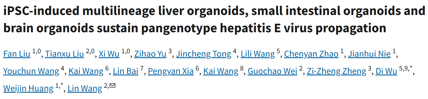

一、研究核心思路:突破传统模型局限,构建多谱系类器官 "病毒研究工厂"
1. 研究背景与痛点
2. 核心研究策略
二、重磅研究成果:三大类器官全面支撑 HEV 感染,破解肝外致病奥秘
成果 1:肝类器官(hLOs)—— 完整支持 HEV 生命周期,重现肝损伤病理
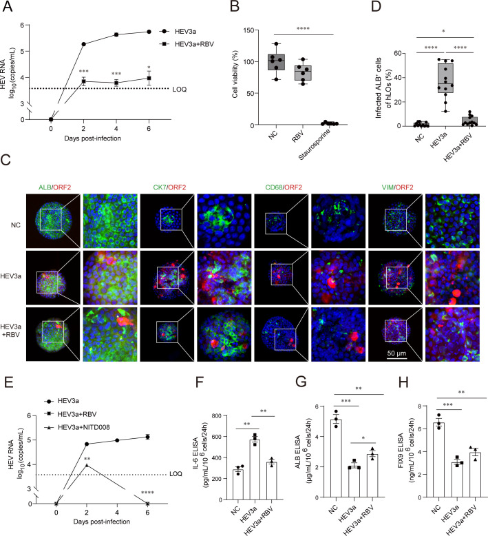 Figure 2.
Infection with HEV impaired hepatic function of hLOs. (A) HEV3a RNA in the supernatants of hLOs. HEV3a+RBV refers to the addition of ribavirin based on HEV3a infection, RBV=500 μM, n=3. (B) Cell viability was determined using the CellTiter-Glo luminescent assay, which quantifies cellular ATP levels as a marker of metabolic activity. Untreated cells served as the NC, while staurosporine was used as the positive control for cytotoxicity induction (RBV=500 μM, staurosporine=10 μM, n=6). (C) Immunofluorescence staining of four hepatic cell types in hLOs after 6 day HEV3a infection. Cultures were stained for HEV ORF2 and ALB, CK7, CD68 and VIM. Scale bars, 50 μm. (D) Quantification of the proportion of HEV infected hepatocyte-like cells in hLOs, n=12. (E) Dynamic changes of HEV3a RNA in hLOs culture supernatant. HEV3a+RBV refers to the addition of ribavirin based on HEV3a infection, RBV=100 μM. HEV3a+NITD008 refers to the addition of NITD008 based on HEV3a infection, NITD008=10 μM, n=3. (F)-(H) ELISA measurement of IL-6 (F), ALB (G), and FIX-9 (H) in hLO culture supernatant after HEV3a infection at day 6, n=3. ALB, albumin; ATP, adenosine triphosphate; FIX, Factor IX; HEV, hepatitis E virus; hLOs, human liver organoids; IL, interleukin; LOQ, limit of quantitation; NC, negative control; ORF, open reading frame; RBV, ribavirin; VIM, vimentin. *P <0.05, **P <0.01, ***P <0.001, ****P <0.0001.
成果 2:小肠类器官(hIOs)—— 揭示 HEV 肠道感染与屏障破坏机制
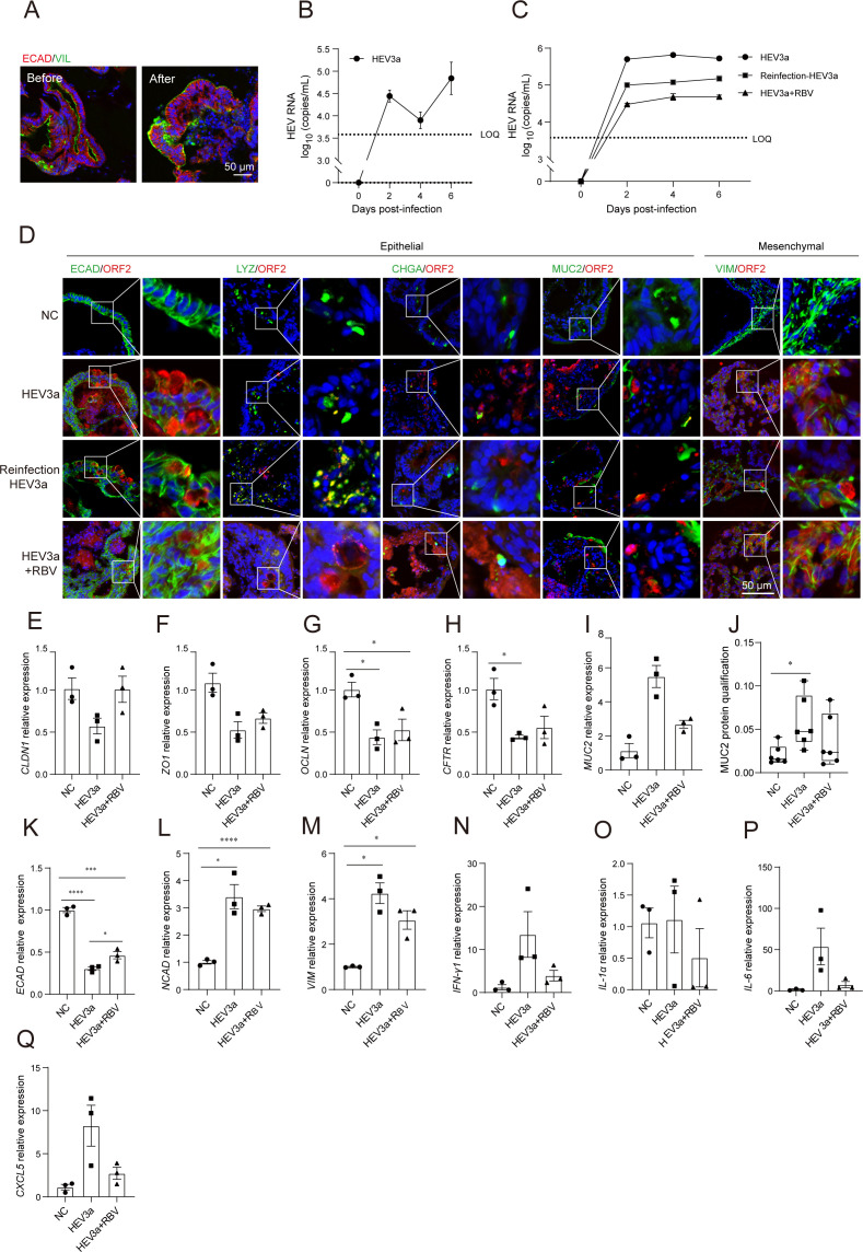 Figure 3.
Human multiple-lineage intestinal organoids support the full life cycle of HEV infection. (A) Immunofluorescence staining of intestinal organoids (hIOs) before and after polarity reversal. Cultures were stained for ECAD and VIL. Scale bars, 50 μm. (B) Dynamic changes of HEV3a RNA in the supernatants of hIOs, n=3. (C) Dynamic changes of HEV3a RNA in hIOs culture supernatant. Reinfection+HEV3a refers to using the supernatant of HEV3a-infected hIOs to reinfect the hIOs, HEV3a+RBV refers to the addition of ribavirin based on HEV3a infection, RBV=500 μM, n=3. (D) Immunofluorescence staining of hIOs infected with HEV for 6 days. Cultures were stained for HEV ORF2 and ECAD, LYZ, CHGA, MUC2 and VIM. Scale bars, 50 μm. (E)-(I) RT-qPCR detection of mRNA expression levels of CLDN1 (E), ZO1 (F), OCLN (G), CFTR (H) and MUC2 (I) in hIOs, n=3. (J) Immunofluorescence quantification of MUC2 expression, n=6. (K)-(Q) RT-qPCR detection of mRNA expression levels of ECAD (K), NCAD (L), VIM (M), IFN-γ1 (N), IL-1α (O), IL-6 (P) and CXCL5 (Q) in hIOs, n=3. CHGA, ahromogranin A; ECAD, E-cadherin; HEV, hepatitis E virus; hIOs, human intestinal organoids; LOQ, limit of quantitation; LYZ, lysozyme; MUC2, mucin 2; mRNA, messenger RNA; NC, negative control; ORF, open reading frame; RBV, ribavirin; RT-qPCR, reverse-transcription quantitative PCR; VIM, vimentin; VIL, villin. *P <0.05, ***P <0.001, ****P <0.0001.
 Figure 4.
Replication of HEV1 and HEV4 in hIOs. (A) Dynamic changes of HEV1 RNA in hIOs culture supernatants. HEV1+RBV refers to the addition of ribavirin based on HEV1 infection, RBV=100 μM, n=3. (B) Dynamic changes of HEV4 RNA in hIOs culture supernatants. HEV4+RBV refers to the addition of ribavirin based on HEV4 infection, RBV=100 μM, n=3. (C) Immunofluorescence staining of hIOs infected with HEV for 6 days. Cultures were stained for HEV ORF2, ECAD and VIM. Scale bars, 50 μm. (D) Quantitative analyses of ECAD+ cells infected with HEV1, n=12. (E) Quantitative analyses of ECAD+ cells infected with HEV4, n=12. ECAD, E-cadherin; HEV, hepatitis E virus; hIOs, human intestinal organoids; LOQ, limit of quantitation; NC, negative control; ORF, open reading frame; RBV, ribavirin; VIM, vimentin. ***P <0.001, ****P <0.0001.
成果 3:肝-肠串联模型 —— 还原 HEV"肠→肝→肠" 自然感染循环
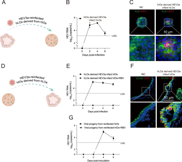 Figure 5.
The hLOs-hIOs system modelled the sequential gut-liver-gut infection cycle of HEV. (A) Schematic diagram of reinfection of liver organoids with supernatant derived from hIOs. (B) The supernatant of hIOs infected with HEV3a was inoculated into hLOs, and HEV RNA was detected in culture supernatants, n=3. (C) Immunofluorescence staining of hLOs infected with HEV for 6 days. Cultures were stained for HEV ORF2 and ALB. Scale bars, 50 μm. (D) Schematic diagram of reinfection of intestine organoids with supernatant derived from hLOs. (E) The supernatant of hLOs infected with HEV3a was inoculated into hIOs, and HEV RNA was detected in culture supernatants. RBV=100 μM, n=3. (F) Immunofluorescence staining of hIOs infected with HEV for 6 days. Cultures were stained for HEV ORF2 and ECAD. Scale bars, 50 μm. (G) Supernatant from HEV-infected hLOs was inoculated into hIOs, which then produced supernatant that was passaged into new hIOs while viral RNA levels in the supernatant were dynamically monitored. RBV=100 μM, n=3. ALB, albumin; ECAD, E-cadherin; HEV, hepatitis E virus; hIOs, human intestinal organoids; hLOs, human liver organoids; LOQ, limit of quantitation; NC, negative control; ORF, open reading frame; RBV, ribavirin.
成果 4:脑类器官(hBOs)—— 证实 HEV 神经嗜性,揭示神经系统损伤机制
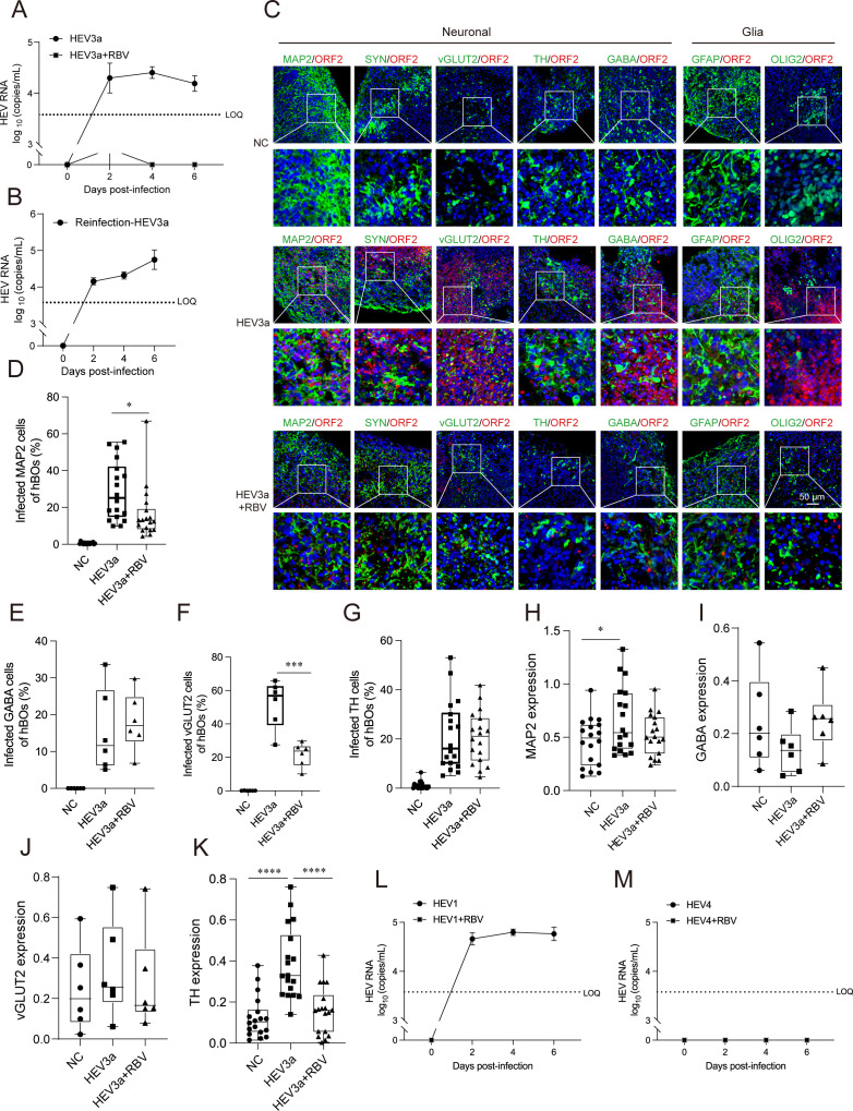 Figure 6.
Replication of HEV3a in hBOs. (A) Dynamic changes of HEV3a RNA in the supernatants of hBOs. HEV3a+RBV refers to the addition of ribavirin based on HEV3a infection, RBV=500 μM, n=3. (B) The supernatant of HEV3a-infected hBOs was used to reinfect the brain organoids, and viral RNA was again detected in the culture supernatant, n=3. (C) Immunofluorescence staining of hBOs infected with HEV3a for 6 days. Cultures were stained for HEV ORF2 and MPA2, SYN, vGLUT2, TH, GABA, GFAP and OLIG2. (D) Quantitative analysis of MAP2+ cells infected with HEV3a, n=18. (E) Quantitative analysis of GABA+ cells infected with HEV3a, n=6. (F) Quantitative analysis of vGLUT2+ cells infected with HEV3a, n=6. (G) Quantitative analysis of TH+ cells infected with HEV3a, n=18. (H) Immunofluorescence quantitation of MAP2 expression, n=18. (I)-(J) Immunofluorescence quantitation of GABA (I) and vGLUT2 (J) expression, n=6. (K) Immunofluorescence quantitation of TH expression, n=18. (L) Dynamic changes of HEV1 RNA in the supernatants of hBOs, HEV1+RBV refers to the addition of ribavirin based on HEV1 infection, RBV=100 μM, n=3. (M) Dynamic changes of HEV4 RNA in the supernatants of hBOs, HEV4+RBV refers to the addition of ribavirin based on HEV4 infection, RBV=100 μM, n=3. GABA, gamma-Aminobutyric acid; GFAP, glial fibrillary acidic protein; HEV, hepatitis E virus; hBOs, human brain organoids; LOQ, limit of quantitation; MAP2, microtubule-associated protein 2; NC, negative control; OLIG2, oligodendrocyte lineage transcription factor 2; ORF, open reading frame; RBV, ribavirin; SYN, synapsin; TH, tyrosine hydroxylase; vGLUT, vesicular glutamate transporter. *P <0.05, ***P<0.001, **** P <0.0001.
三、Absin 核心试剂:精准护航,助力顶刊数据落地
本研究中,Absin 两款明星产品 abs5510778、abs5510777 承担肝损伤核心标志物定量关键任务,为实验结论提供高灵敏、高稳定的数据支撑:
| Absin 产品编号 | 产品名称 | 研究中核心作用 | 对应实验结果 |
|---|---|---|---|
| abs5510778 | 人丙氨酸氨基转移酶(ALT)ELISA 试剂盒 | 精准定量 HEV 感染后肝类器官上清 ALT 水平,量化肝细胞损伤程度 | 检测到 ALT 显著升高,直接证实 HEV 诱导肝细胞损伤 |
| abs5510777 | 人天冬氨酸氨基转移酶(AST)ELISA 试剂盒 | 精准定量 AST 释放,评估肝组织炎症与坏死程度 | AST 水平上升,与临床 HEV 感染肝损伤指标完全吻合 |
Absin 产品核心优势
四、研究科学价值与应用前景
五、Absin 助力 infectious disease 研究,持续赋能生命科学
Absin 始终聚焦病毒学、感染免疫、类器官研究领域,提供 ELISA 试剂盒、抗体、细胞培养试剂、分子生物学试剂等全系列产品,已助力全球 19000+ 篇高水平论文发表,覆盖 Cell、Nature、Gut、Hepatology 等顶刊。
本次 Gut 研究中,abs5510778、abs5510777 以卓越性能助力 HEV 研究突破,再次印证 Absin 试剂 "精准、稳定、可靠" 的硬核实力!

- 手机:13761418683