




- 手机:13761418683
联系人:爱必信
网址:http://www.absin.cn/
地 址:上海市浦东新区新浩路58号18号楼

血浆 PPP2R5C:阿尔茨海默病早期诊断新突破
2026-07-09
阿尔茨海默病(AD)作为全球高发的神经退行性疾病,早期精准诊断一直是临床与科研的核心难题。近期,中南大学湘雅医院团队在Cell Reports Medicine发表重磅研究,首次证实血浆神经元源性 PPP2R5C 可作为 AD 早期诊断潜在生物标志物,并系统阐明其调控 Tau 蛋白的分子机制,为 AD 早筛与机制研究开辟全新方向。
值得关注的是,该研究全程采用Absin 高效 IHC 检测系统(abs957) 完成关键病理验证,为高分成果筑牢实验根基!
文献标题:Neuronal PPP2R5C in plasma is a potential biomarker for early diagnosis of Alzheimer's disease
发表期刊:Cell Rep Med. (IF=10.6)
DOI:https://doi.org/10.1016/j.xcrm.2026.102631
使用 Absin 产品:即用型高效免疫组化二抗试剂盒(货号:abs957)

一、研究核心思路:从血浆外泌体到 AD 早筛标志物,闭环验证突破瓶颈
研究团队围绕AD 早期诊断核心目标,构建临床样本 - 分子机制 - 动物模型的完整研究链条:
标志物发现:分离健康对照、症状前家族性 AD(pre-FAD)、家族性 AD(FAD)血浆神经元源性外泌体(NDEs),通过无标定量蛋白质组筛选到PPP2R5C呈进行性下降趋势;
多队列验证:覆盖散发性 AD、遗忘型轻度认知障碍(aMCI)、FAD 及其他神经退行性疾病队列,确认血浆 PPP2R5C 的早期诊断与鉴别诊断价值;
机制解析:明确 PPP2R5C 通过PP2A 通路 + ULK1 依赖的自噬溶酶体通路双重调控 Tau 蛋白磷酸化与降解,其下降早于 Tau 过度磷酸化;
动物验证:在 Tau P301S 模型鼠中证实,过表达 PPP2R5C 可逆转认知缺陷与 Tau 病理,沉默则加速病变进展。
二、核心研究成果:PPP2R5C——AD 早筛 "理想标志物"
1. 临床诊断价值:早、准、特,突破现有局限
早期预警:血浆 PPP2R5C 在aMCI 阶段即显著降低,早于典型临床症状出现(对应原文Figure 2A);
高诊断效能:区分 AD 与健康对照 AUC=0.8494,区分 aMCI 与健康对照 AUC=0.7360(对应原文Figure 2B);
特异性优异:可有效鉴别 AD 与进行性核上性麻痹(PSP)、额颞叶痴呆(FTD),AUC 分别达 0.8519、0.6622(对应原文Figure 2M、2N);
相关性明确:与 MMSE 评分正相关,与血浆 p-Tau181/217/231 负相关,精准反映 AD 病理进程(对应原文Figure 2C-F)。
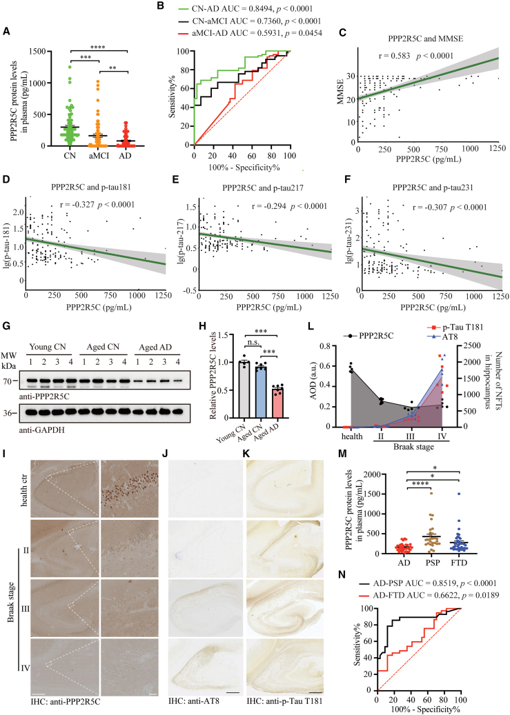 Figure 2. Diagnostic utility of plasma PPP2R5C protein levels in AD
(A) Plasma PPP2R5C protein levels in AD, aMCI, and CN groups by ELISA technology. AD, n = 74. aMCI, n = 76. CN, n = 74.
(B) Evaluation of the diagnostic performance of plasma PPP2R5C protein levels in distinguishing AD from aMCI and CN.
(C–F) Spearman correlation analysis between plasma PPP2R5C levels, Mini-Mental State Examination (MMSE) scores, and other AD biomarkers, including p-Tau T181, p-Tau S231, and p-Tau T217.
(G) Representative western blots for PPP2R5C in brain tissues from young individuals, cognitively normal elderly individuals, and AD patients.
(H) Quantitation of PPP2R5C levels, normalized to young CN (n = 5 or 7).
(I–K) Representative immunohistochemistry images of PPP2R5C (I), AT-8 (J), and p-Tau T181 (K) levels in the hippocampus of healthy controls and Braak-graded AD brain slices. Scale bars: 1 mm in (I, left), 50 μm in (I, right), and 1 mm in (J and K).
(L) Quantitation of PPP2R5C, p-Tau T181, and AT8 levels (sample quantify, health ctr: 5, Braak II: 9, Braak III: 5, and Braak IV: 4).
(M) Difference of plasma PPP2R5C protein levels among AD (n = 34), PSP (n = 28), and FTD (n = 37) patients using ELISA.
(N) ROC curve analysis assessing the discriminative power of plasma PPP2R5C protein levels for distinguishing AD from PSP and FTD.
All the western blot data are representative of three independent experiments. Quantification data are expressed as mean ± SEM (?p < 0.05, ??p < 0.01, ???p < 0.001, and n.s., no statistics with one-way ANOVA with Tukey's multiple comparisons test).
2. 分子机制:双重通路调控 Tau 病理,奠定理论基础
PPP2R5C 直接结合 Tau,通过激活 PP2A促进 Tau 去磷酸化;
同时直接结合 ULK1,启动自噬溶酶体通路加速 Tau 降解;
关键时间窗:PPP2R5C 下降早于 Tau 过度磷酸化,成为 AD 早期病理标志(对应原文Figure 2I-L、7G)。
3. 动物实验:靶向 PPP2R5C 可逆转 AD 样病变
过表达 PPP2R5C 改善 Tau P301S 鼠空间学习记忆,减少 Tau 磷酸化与神经原纤维缠结(对应原文Figure 4);
沉默 PPP2R5C 则加速认知衰退与 Tau 病理,反向验证其保护作用(对应原文Figure 5)。
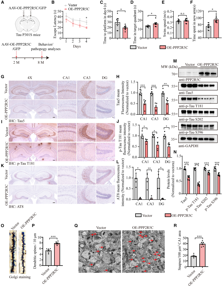 Figure 4. Overexpression of PPP2R5C in Tau P301S mice reduces AD-like pathogenesis and rescues cognitive function
(A) Schematic representation of the experimental design. Two-month-old Tau P301S mice were injected with either AAV-hSyn-EGFP or AAV-hSyn-PPP2R5C-EGFP. Mice were sacrificed 6 months after AAV injection.
(B and C) Morris water maze analysis as escape latency (s) and escape latency on day 4 (s).
(D) Probe trial performance of Morris water maze test.
(E) Swim speed of mice injected with AAVs encoding EGFP or PP2R5C-EGFP (n = 8–10 mice per group).
(F) Time spent in the novel arm in the Y-maze test (n = 8–10 mice per group).
(G, I, and K) Representative immunostaining images of t-Tau, p-Tau T181, and AT8 in the hippocampus of Tau P301S mice injected with AAVs encoding EGFP or PPP2R5C-EGFP. Scale bars: 200 μm for 4× images and 20 μm for magnified images.
(H, J, and L) Quantification of immunoreactivity for t-Tau, p-Tau T181, and AT8 (n = 5 mice per group).
(M and N) Representative western blots showing Tau pathology in mouse brain tissue following PPP2R5C overexpression (n = 6 mice per group).
(O) Golgi staining revealed the dendritic spines in the apical dendritic layer of the CA1 region. Scale bar, 10 μm.
(P) Quantification of spine density (n = 6 mice per group).
(Q) Representative electron microscopy of the synapse structures. Arrows indicate synapses. Scale bar, 1 μm.
(R) Quantification of synaptic density (n = 6 mice per group).
All the western blot data are representative of three independent experiments. Quantification data are expressed as mean ± SEM (?p < 0.05, ??p < 0.01, ???p < 0.001, and n.s., no statistics, Student's ttest).
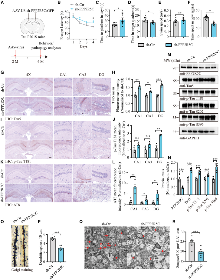 Figure 5. Knockdown of PPP2R5C in Tau P301S mice worsens cognitive dysfunctions
(A) Schematic representation of the experimental design. Two-month-old Tau P301S mice were injected with either AAV-sh-Ctrl-EGFP or AAV-sh-PPP2R5C-EGFP. Mice were sacrificed 4 months after AAV injection.
(B and C) Morris water maze analysis as escape latency (s) and escape latency on day 4 (s).
(D) Probe trial performance of Morris water maze test.
(E) Swim speed of mice injected with AAVs encoding sh-Ctrl-EGFP or sh-PPP2R5C-EGFP (n = 8–10 mice per group).
(F) Time spent in the novel arm in the Y-maze test (n = 8–10 mice per group).
(G, I, and K) Representative immunostaining images of t-Tau, p-Tau T181, and AT8 in the hippocampus of Tau P301S mice injected with AAVs encoding sh-Ctrl-EGFP or sh-PPP2R5C-EGFP.
(H, J, and L) Quantifying immunoreactivity for t-Tau, p-Tau T181, and AT8 (n = 4 mice per group). Scale bars: 200 μm for 4× images and 20 μm for magnified images.
(M and N) Representative western blots showing Tau pathology in mouse brain tissue following PPP2R5C knockdown (n = 6).
(O) Golgi staining revealed the dendritic spines in the apical dendritic layer of the CA1 region. Scale bar, 10 μm.
(P) Quantification of spine density (n = 6 mice per group).
(Q) Electron microscopy of synapses (left) and high magnification of synapses (right). Scale bar: 2 μm in the left panel, 200 μm in the right panel.
(R) Quantification of synaptic density (n = 6 mice per group).
All the western blot data are representative of three independent experiments. Quantification data are expressed as mean ± SEM (?p < 0.05, ??p < 0.01, ???p < 0.001, and n.s., no statistics, Student's ttest).
三、Absin abs957:高分文献的 IHC "硬核支撑"
该研究中,人脑组织、小鼠海马组织的免疫组化(IHC)检测均依赖Absin abs957 高效 IHC 检测系统完成,是验证 PPP2R5C、p-Tau、AT8 表达与定位的核心试剂,对应原文Figure 2I-K、Figure 4G-L、Figure 5G-L等关键图表。
abs957 核心优势,完美适配 AD 病理研究需求
高灵敏度:聚合物信号放大技术,精准捕获低丰度磷酸化 Tau、PPP2R5C信号,避免早期病理遗漏;
低背景干扰:非生物素检测体系,杜绝内源性生物素非特异性染色,人脑 / 鼠脑组织染色背景干净、条带清晰;
操作便捷:鼠兔通用型即用型试剂盒,含封闭液、一抗放大剂、酶标二抗、DAB 显色液全套组分,缩短实验周期,适配大批量临床样本检测;
稳定可靠:适配不同抗原修复条件,重复实验一致性高,支撑多队列、多样本的严谨验证。
在 AD 病理研究中,IHC 是观察 Tau 缠结、神经元蛋白表达的金标准,Absin abs957 凭借优异性能,助力团队清晰呈现PPP2R5C 在 AD 早期的下降趋势、Tau 磷酸化的时空分布,为结论提供直观、可靠的病理证据。
四、研究意义与 Absin 助力
本研究首次将血浆 PPP2R5C确立为 AD 早期诊断的潜在标志物,兼具早期性、特异性、无创性,解决临床血检早筛痛点;同时揭示 PPP2R5C 调控 Tau 的双重机制,为 AD 靶向治疗提供新靶点。
从临床样本验证到动物病理分析,Absin abs957全程护航关键 IHC 实验,以稳定性能、高灵敏度、低背景优势,成为高分成果的 "隐形功臣"。
Absin 助力神经科学研究,一站式解决方案
Absin 专注为神经退行性疾病研究提供高品质试剂,除 abs957 IHC 试剂盒外,还覆盖:
? 神经相关一抗(Tau、p-Tau、ULK1、PP2A 等);
? IHC 配套试剂(抗原修复液、DAB 显色液、封片剂);
? 细胞 / 动物模型培养、蛋白检测全套试剂。
未来,Absin 将持续以优质产品助力 AD 早筛、机制与治疗研究,陪伴科研工作者攻克神经退行性疾病难题!

- 手机:13761418683