




- 手机:13761418683
联系人:爱必信
网址:http://www.absin.cn/
地 址:上海市浦东新区新浩路58号18号楼

顶刊 Nature 子刊揭秘心梗炎症新机制|解锁 Hat1-H3K23succ 表观修饰调控通路
2026-07-20
近期,国际权威期刊Nature Communications在线发表了题为Histone acetyltransferase 1 promotes postinfarction inflammatory response by regulation of monocyte histone succinylation的重磅研究成果,首次揭示组蛋白乙酰转移酶 Hat1 通过调控单核细胞组蛋白琥珀酰化(H3K23succ),驱动心梗后过度炎症反应的全新表观遗传机制,为急性心肌梗死(AMI)的抗炎治疗提供全新靶点。
作为生命科学领域优质试剂耗材提供商,Absin(爱必信)旗下70 μm 细胞过滤器(货号:abs7008) 全程助力本研究关键实验环节,为高分成果保驾护航!
文献标题:Histone acetyltransferase 1 promotes postinfarction inflammatory response by regulation of monocyte histone succinylation
发表期刊:Nature Communications (IF=15.7)
DOI:https://doi.org/10.1038/s41467-025-66721-9
使用 Absin 产品:70um细胞过滤器(200目,白色)(货号:abs7008)

一、研究背景:心梗后炎症 “失控”,表观调控成破题关键
急性心肌梗死(MI)后,单核 / 巨噬细胞过度活化引发的炎症风暴是导致心肌损伤、心室重构的核心诱因。组蛋白琥珀酰化(H3K23succ)作为新型表观修饰,在心血管疾病中的作用尚不明确。
研究团队聚焦Hat1→组蛋白琥珀酰化→单核细胞促炎表型这一核心链条,系统解析心梗早期炎症的表观调控机制,为临床干预提供全新思路。
二、核心研究思路:从临床现象到分子机制,层层递进
1. 临床发现:AMI 患者单核细胞琥珀酰化显著升高
? 纳入48 例 AMI 患者、50 例不稳定型心绞痛(UA)患者,检测血清琥珀酸及单核细胞总琥珀酰化(Pan Ksucc)水平。
? 结果:AMI 组血清琥珀酸、单核细胞 Pan Ksucc 显著升高,且与TNF-α、IL-6、NLR等炎症指标强相关(图 1),提示琥珀酰化参与心梗后炎症调控。
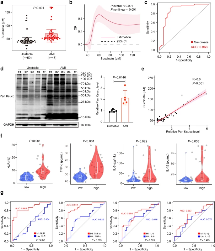
Fig. 1: Upregulated circulating monocyte succinylation positively correlates with inflammatory indicators post-myocardial infarction (MI). a Plasma levels of succinate in unstable angina (UA; n?=?50) and acute myocardial infarction (AMI; n?=?48; two-tailed Mann-Whitney U test). b Restricted cubic spline analysis shows the relationship between succinate concentration and AMI. A spline function term was introduced into the multivariate Logistic regression model and assessed overall effects and nonlinear trends using Wald χ2 test. The solid line represents the adjusted odds ratio (OR), and the shaded area indicates the 95% confidence interval. c Receiver operating characteristic (ROC) curves reveal the diagnostic efficacy of succinate for AMI. d Immunoblot analysis and quantification of Pan Ksucc levels in monocytes from patients with UA and AMI (n?=?5; two-tailed unpaired Student t test). e Correlation of Pan Ksucc levels in human monocytes with plasma succinate levels in patients with AMI (n?=?48; Spearman’s correlation analysis). f The levels of NLR, TNF-α, IL-6, and IL-1β in the plasma of AMI patients with Pan Ksucc levels greater than the median compared with those in patients with Pan Ksucc levels less than or equal to the median (high: n?=?24, low: n?=?24; two-tailed Mann-Whitney U test). g ROC curves reveal the diagnostic efficacy of plasma inflammatory markers for Pan Ksucc levels in monocytes from patients with AMI and UA. Statistical significance between curves was determined using DeLong’s nonparametric test. a, f Data presented as box plots, with 75th and 25th percentiles limits; centre line represents the median; bars represent maximal and minimal values. Results are presented as the mean?±?SD of independent replicates. Pan Ksucc global succinylation, OR odds ratio, AUC area under the curve, NLR neutrophil-to-lymphocyte ratio, TNF-α tumor necrosis factor-α, IL interleukin. Source data are provided as a Source Data file.
2. 动物验证:心梗小鼠单核 / 巨噬细胞 H3K23succ 特异性上调
? 构建小鼠 MI 模型,检测骨髓、外周血单核细胞及心脏浸润巨噬细胞的组蛋白修饰。
? 关键结果:H3K23succ在 MI 后 1 天骨髓单核细胞、3 天外周血单核细胞中显著升高,心脏浸润巨噬细胞同样高表达 Pan Ksucc、H3K23succ(图 2),锁定H3K23succ为核心修饰位点。
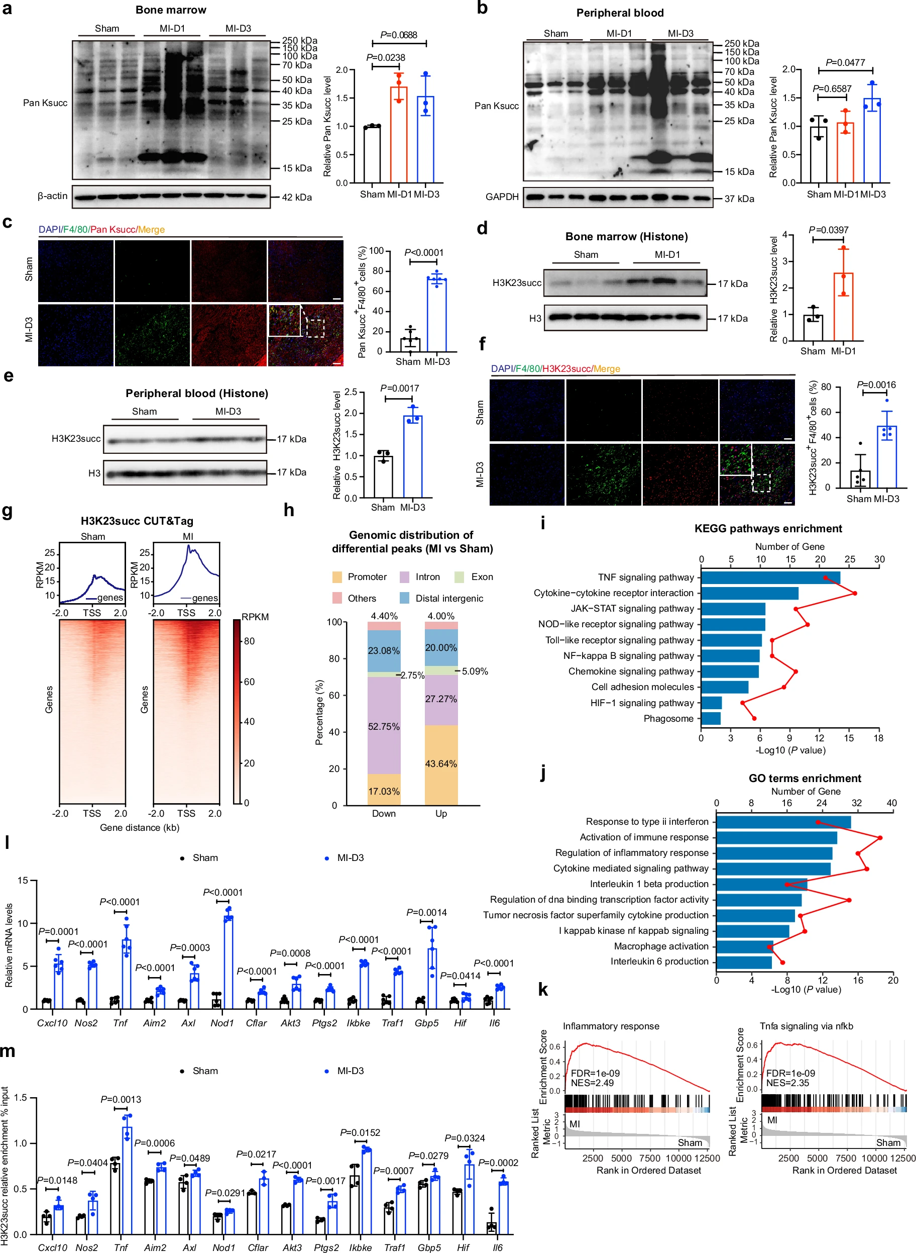
Fig. 2: Histone lysine succinylation is increased in monocytes and activates the transcriptional activity of the inflammatory pathway-related genes post- myocardial infarction (MI). a, b Immunoblot analysis and quantification of Pan Ksucc (global succinylation) level in monocytes sorted from bone marrow (a) and peripheral blood (b) of mice indicated days after MI (n?=?3; one-way ANOVA followed by Bonferroni multiple comparisons test). c Immunofluorescence staining and quantification of the proportion of Pan Ksucc and F4/80 in cardiac tissues on Day 3 with sham operation or after MI (scale bar=50?μm; n?=?6; two-tailed unpaired Student t test). d, e Immunoblots of the levels of H3K23succ (histone) in bone marrow monocytes on Day 1 (d) and circulating monocytes on Day 3 (e) sorted from MI mice (n?=?3; two-tailed unpaired Student t test). The blots were derived from the same experiment and processed in parallel. Total histone H3 level was set as an internal reference. f Immunofluorescence staining and quantification of the proportion of H3K23succ and F4/80 in cardiac tissues on Day 3 with sham operation or after MI (scale bar=50 μm; n?=?5; two-tailed unpaired Student t test). g Heatmaps of H3K23succ binding peaks in circulating monocytes from sham and MI mice. Color intensity reflects the relative read count. Genes exhibiting similar distribution patterns were subjected to clustering analysis using an algorithm, revealing the binding trends of histone succinylation modifications across all genes. h Bar graph showing the genomic distribution of differential H3K23succ peaks (MI vs sham) relative to the translation start site (TSS). i, j KEGG (i) and GO (j) analyses of upregulated genes with increased H3K23succ modification. Statistical significance was assessed using a two-sided hypergeometric test with Benjamini-Hochberg correction for multiple testing. k The GSEA pathways enriched in the MI mice. The indicative genes were enriched for Inflammatory response and Tnf-α signaling via nfκb, as defined by hallmarks. l The mRNA levels of H3K23succ-enriched inflammatory genes in sham and MI mice (n?=?6; two-tailed unpaired Student t test). m ChIP–qPCR analysis of H3K23succ enrichment at the promoters of inflammatory genes in sham and MI mice (n?=?4; two-tailed unpaired Student t test). Results are presented as the mean?±?SD of independent replicates. DAPI 4?,6-diamidino-2-phenylindole, GO Gene Ontology, KEGG Kyoto Encyclopedia of Genes and Genomes, GSEA gene set enrichment analysis, NES normalized enrichment score, ChIP chromatin immunoprecipitation, PCR polymerase chain reaction. Source data are provided as a Source Data file.
3. 机制深挖:Hat1 作为琥珀酰转移酶催化 H3K23succ,驱动促炎基因转录
? 筛选发现Hat1在 MI 后心脏巨噬细胞、循环单核细胞中特异性高表达(图 3),且与 H3K23succ 时空表达高度一致。
? 功能实验:Hat1 敲除(KO)/ 敲低(KD)显著降低 H3K23succ 水平,减少促炎巨噬细胞浸润、缩小心梗面积、改善心功能(图 4、5)。
? 分子机制:Hat1 通过I243 位点催化 H3K23succ,增强促炎基因(Cxcl10、Tnf、Nos2等)启动子区域染色质可及性,激活炎症通路(图 6、7、8)。
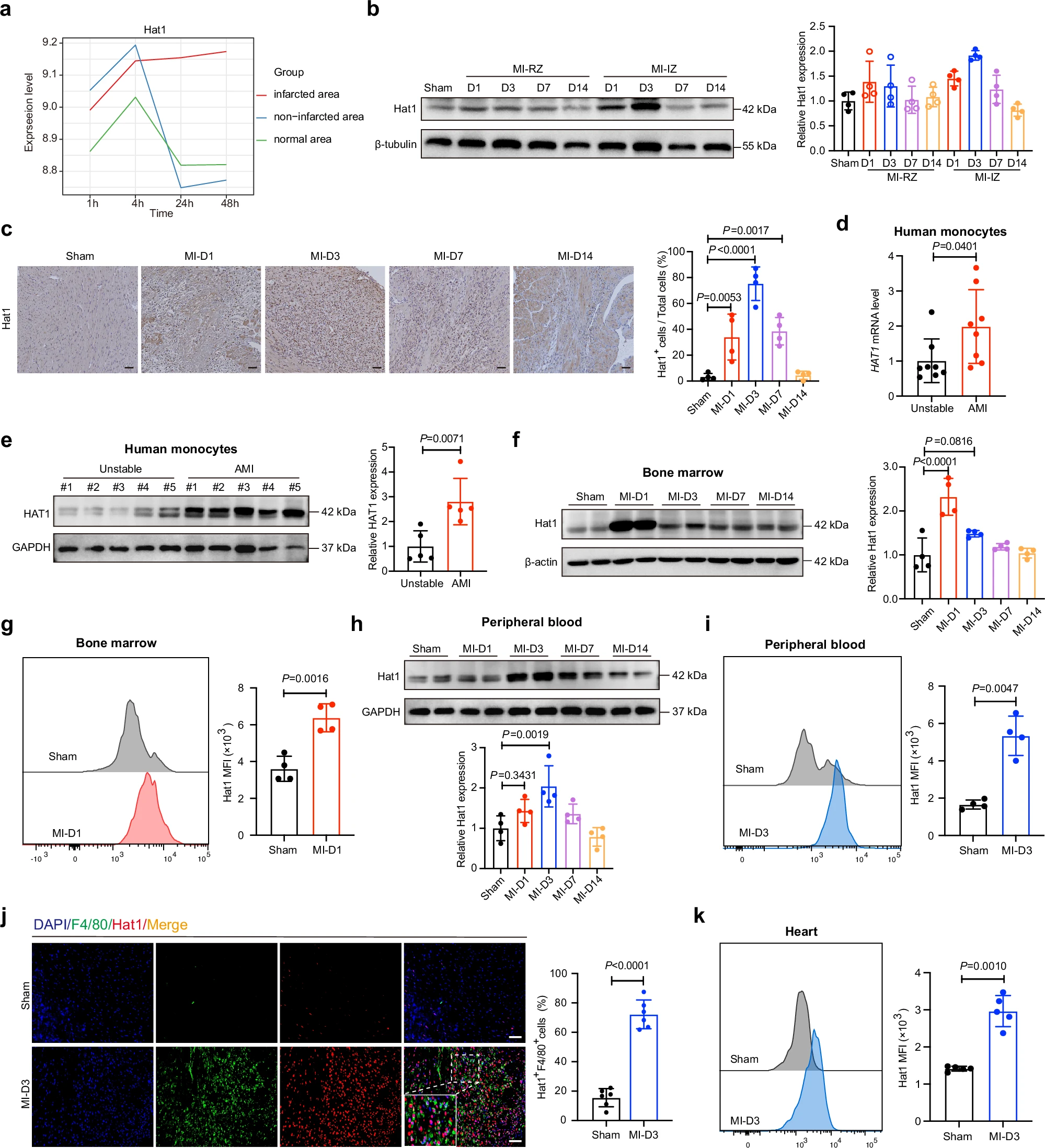
Fig. 3: Hat1 is specifically elevated in monocytes after myocardial infarction (MI). a Line graph showing the expression and distribution of Hat1 at different time points in MI mouse heart tissues, as revealed through reanalysis of transcriptome database (GSE775). b Immunoblot analysis of Hat1 expression in infarct zone (IZ) and remote zone (RZ) of post-MI hearts and sham controls at the indicated days (n?=?4). c Representative immunohistochemical analyses of Hat1 in sham-operation and MI mice at the indicated days after MI (scale bar?=?50?μm; n?=?5). Hat1 expression was quantified and compared (n?=?4; one-way ANOVA followed by Bonferroni multiple comparisons test). d Quantitative PCR analysis of HAT1 mRNA expression in the peripheral circulating monocytes of patients with UA and AMI (n?=?8; two-tailed unpaired Student t test). e Representative Western blotting and quantification of Hat1 expression in circulating monocytes from patients with UA or AMI (n?=?5; two-tailed unpaired Student t test). f Immunoblot analysis and quantification of Hat1 expression in bone marrow monocytes from MI mice at the indicated days (n?=?4; one-way ANOVA followed by Bonferroni multiple comparisons test). g Flow cytometry analysis and quantification of Hat1 expression in the bone marrow monocytes on Day 1 after MI (n?=?4 per group; two-tailed unpaired Student t test). h Representative Western blotting and quantification of Hat1 expression in circulating monocytes from MI mice at the indicated days (n?=?4; one-way ANOVA followed by Bonferroni multiple comparisons test). i Flow cytometry analysis and quantification of Hat1 expression in the blood monocytes on Day 3 post-MI (n?=?4 per group; two-tailed unpaired Student t test). j Dual immunofluorescence staining and quantification of the proportion of Hat1 and F4/80 in murine heart on Day 3 after MI (scale bar=50 μm; n?=?6; two-tailed unpaired Student t test). k Flow cytometry analysis and quantification of Hat1 expression in the heart on Day 3 after MI (n?=?5; two-tailed unpaired Student t test). Results are presented as the mean?±?SD of independent replicates. Hat1 histone acetyltransferase 1, UA unstable angina, AMI acute myocardial infarction, DAPI 4?,6-diamidino-2-phenylindole, PCR polymerase chain reaction, MFI mean fluorescence intensity. Source data are provided as a Source Data file.
4. 转化价值:抑制 Hat1 可改善心肌缺血 / 再灌注(I/R)损伤
? 巨噬细胞特异性敲低 Hat1,显著减轻 I/R 模型心肌炎症、减小梗死面积,证实Hat1 是 MI 潜在治疗靶点。
三、核心研究成果(3 大创新突破)
1. 首次证实:组蛋白琥珀酰化(H3K23succ)是心梗后单核细胞促炎活化的关键表观标记。
2. 全新机制:Hat1 兼具乙酰转移酶与琥珀酰转移酶活性,通过 I243 位点催化 H3K23succ,调控促炎基因转录。
3. 转化潜力:靶向抑制 Hat1 可缓解心梗后过度炎症,为 AMI 抗炎治疗提供新策略。
四、Absin abs7008:高分文章的 “硬核支撑”
本研究中,Absin 70 μm 细胞过滤器(abs7008) 用于小鼠心脏组织单细胞悬液制备,是流式细胞术、细胞分选等关键实验的基础耗材,对应原文方法学部分「Flow cytometric analysis」 及图 5、图 6 等流式相关实验。
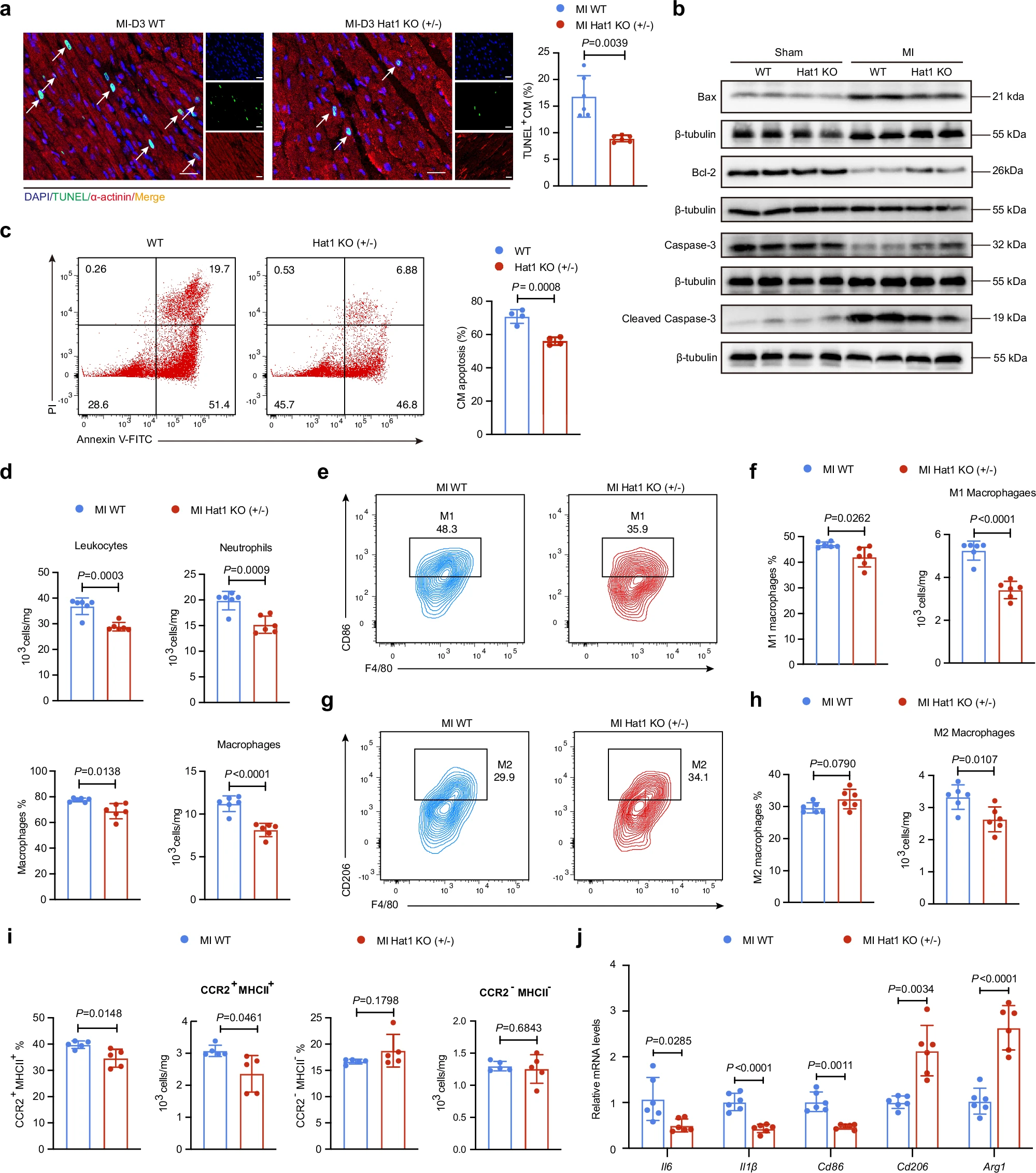
Fig. 5: Loss of Hat1 attenuates cardiomyocyte apoptosis and suppresses macrophage-mediated inflammation in the myocardial infarction (MI) tissue. a TUNEL and α-actinin co-staining in the border regions of WT and Hat1 KO mouse hearts on Day 1 after MI. Nuclear staining with DAPI (scale bar=50 μm; n?=?6; two-tailed unpaired Student t test). b Immunoblot analysis and quantification of apoptosis-related proteins including Bax, Bcl-2, Caspase-3, and cleaved Caspase-3 in peri-infarct areas of WT and Hat1 KO from infarcted hearts on Day 3 post-MI (n?=?6; two-way ANOVA followed by Bonferroni multiple comparisons test). c Flow cytometry analysis of cardiomyocyte apoptosis following co-culture with LPS/IFN-γ-stimulated WT versus Hat1KO BMDMs (n?=?4; two-tailed unpaired Student t test). d Flow cytometric analysis of leukocytes (CD45+), neutrophils (CD45+CD11b+Ly6G+) and macrophages (CD45+CD11b+Ly6G-F4/80+CD64+) in WT and Hat1 KO infarct tissues after MI 3 days (n?=?6; two-tailed unpaired Student t test). e, f Flow cytometric analysis of pro-inflammatory (M1) macrophages (CD45?CD11b?Ly6G? F4/80?CD64?CD86?) in infarcted heart tissues from WT and Hat1 KO mice at 3 days post-MI (n?=?6; two-tailed unpaired Student t test). g, h Flow cytometric analysis of reparative (M2) macrophages (CD45?CD11b?Ly6G?F4/80?CD64?CD206?) in infarcted heart tissues from WT and Hat1 KO mice at 3 days post-MI (n?=?6; two-tailed unpaired Student t test). i Flow cytometric analysis of monocyte-derived (CD45+CD11b+Ly6G-F4/80+CD64+CCR2+MHCII+) and tissue-resident (CD45+ CD11b+Ly6G-F4/80+CD64+CCR2-MHCII-) macrophage subsets in infarcted heart tissues from WT and Hat1 KO mice at 3 days post-MI (n?=?5; two-tailed unpaired Student t test). j The mRNA expression levels of pro-inflammatory and reparative genes in the infarcted heart tissues of WT and Hat1 KO mice on Day 3 post-MI (n?=?6; two-tailed unpaired Student t test). Results are presented as the mean?±?SD of independent replicates. WT wild type, Hat1 histone acetyltransferase 1, TUNEL terminal deoxynucleotidyl transferase-mediated deoxyuridine triphosphate nick-end labeling, DAPI 4?,6-diamidino-2-phenylindole. Source data are provided as a Source Data file.
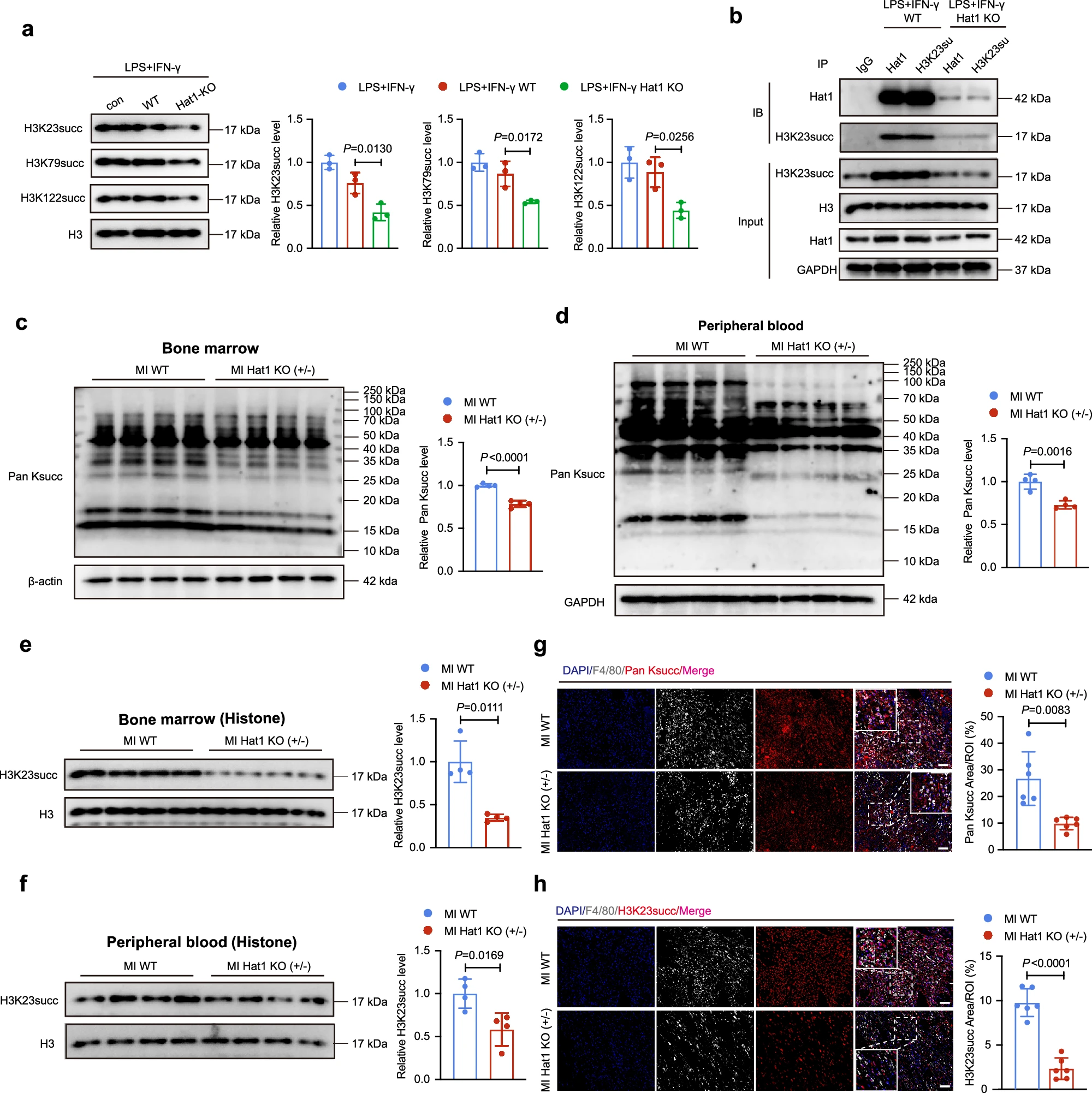
Fig. 6: Hat1 may function as a succinyltransferase and regulate elevated H3K23succ levels post-myocardial infarction (MI). a The H3K23succ, H3K79succ, and H3K122succ immunoblots of WT or Hat1 KO BMDMs treated with LPS and IFN-γ for 24?h (n?=?3; one-way ANOVA followed by Bonferroni multiple comparisons test). The blots were derived from the same experiment and processed in parallel. Total histone H3 level was set as an internal reference. b Immunoblot analysis of Hat1 and H3K23succ in protein complexes immunoprecipitated with anti-Hat1 or anti-H3K23succ antibody from lysates of WT or Hat1 KO BMDMs with LPS/IFN-γ stimulation (n?=?3). c, d Immunoblots of the expression levels of Pan Ksucc in bone marrow (c) and circulating monocytes (d) sorted from WT and Hat1 KO mice post-MI (n?=?4; two-tailed unpaired Student t test). e, f Immunoblots of the expression levels of H3K23succ in bone marrow (e) and circulating monocytes (f) sorted from WT and Hat1 KO mice post-MI (n?=?4; two-tailed unpaired Student t test). The blots were derived from the same experiment and processed in parallel. Total histone H3 level was set as an internal reference. g, h Dual immunofluorescence staining and quantification of the proportion for Pan Ksucc (g) or H3K23succ (h) with F4/80 in the ischemic heart tissues of WT and Hat1 mice on Day 3 after MI (n?=?6; Scale bar=50?μm; two-tailed unpaired Student t test). Results are presented as the mean?±?SD of independent replicates. WT wild type, Hat1 histone acetyltransferase 1, LPS lipopolysaccharide, IFN-γ interferon, BMDMs bone marrow-derived macrophages; DAPI 4?,6-diamidino-2-phenylindole. Source data are provided as a Source Data file.
abs7008 核心优势(适配高分实验需求)
? 精准孔径:70 μm 尼龙滤网,高效过滤组织碎片、细胞团块,获得高活性、均一性单细胞悬液,避免流式仪器堵塞。
? 无菌保障:伽马射线灭菌,无 DNase/RNase、无热源、无内毒素,满足原代细胞、免疫细胞等高敏感实验要求。
? 便捷设计:适配标准 50 mL 离心管,带手柄易操作,独立包装防污染,提升实验效率与数据可靠性。
原文应用场景
研究团队处理小鼠心脏组织时,经胶原酶消化后,使用 abs7008 过滤细胞悬液,去除组织残渣与细胞团,获得纯净的心脏免疫细胞(CD45 + 白细胞、F4/80 + 巨噬细胞等),为后续流式分析、CUT&Tag、ATAC-seq 等高通量实验提供高质量单细胞样本,保障数据准确性与重复性。
五、Absin:助力生命科学研究,赋能高分文章
Absin 始终专注于为科研工作者提供高品质、高稳定性的试剂与耗材,产品覆盖细胞生物学、分子生物学、免疫学等多领域,已助力全球科研团队发表Nature、Cell、Science等顶刊文章数百篇。
本次 abs7008 在 Nature 子刊研究中的成功应用,再次印证 Absin 产品的硬核品质与科研适配性。未来,Absin 将持续深耕生命科学领域,为心血管疾病、肿瘤、炎症等研究提供更全面的解决方案,助力更多科研成果落地!

- 手机:13761418683