




- 手机:13761418683
联系人:爱必信
网址:http://www.absin.cn/
地 址:上海市浦东新区新浩路58号18号楼

突破免疫治疗耐药:GBP2 相分离驱动铁死亡,为黑色素瘤治疗赋能
2026-07-22
2025 年 12 月,《Nature Communications》发表的一项重磅研究揭示了 GBP2 在黑色素瘤免疫治疗中的关键调控机制。该研究由武汉大学中南医院团队完成,全程使用爱必信(absin)#abs955 GBP2 抗体,为机制验证提供了核心工具支撑,为克服 PD-L1 免疫治疗耐药提供了全新靶点与策略。
文献标题:GBP2 condensates promote ferroptosis to sensitize anti-PD-L1 immunotherapy in melanoma
发表期刊:Nature Communications (IF=15.7)
DOI:https://doi.org/10.1038/s41467-025-67690-9
使用 Absin 产品:免疫(共)沉淀(IP/CoIP)试剂盒(货号:abs955)

一、研究背景:免疫治疗耐药的未解难题
免疫检查点阻断(ICB)疗法(如抗 PD-L1 抗体)已成为黑色素瘤治疗的核心手段,但超过 50% 患者会出现原发或获得性耐药。其核心瓶颈在于肿瘤微环境(TME)的免疫抑制状态,尤其是干扰素 -γ(IFN-γ)信号通路受损,导致 CD8?T 细胞杀伤功能减弱。
近年研究发现,铁死亡(ferroptosis) 可增强肿瘤免疫原性,与 ICB 具有显著协同效应。但 IFN-γ 如何调控铁死亡、能否通过靶向该通路逆转耐药,仍缺乏清晰机制。本研究以此为切入点,聚焦 IFN-γ 诱导的关键分子 GBP2,探索其在 “铁死亡 - 免疫激活” 轴中的作用。
二、研究思路:从大数据到机制的精准锁定
研究团队采用 “生信筛选 + 体内外验证 + 机制深挖 + 临床关联” 的递进式思路,层层破解 GBP2 的功能:
1. WGCNA 生信筛选:分析黑色素瘤队列,锁定与 IFN-γ 应答、CTL 杀伤活性高度相关的GBP2 基因;
2. 临床样本验证:证实 GBP2 高表达与患者良好预后、ICB 应答率正相关;
3. 体内外功能实验:构建 GBP2 敲除 / 过表达细胞系与动物模型,验证其对铁死亡和 ICB 疗效的调控作用;
4. 机制解析:聚焦 GBP2 相分离,明确其通过 “GBP2-SHP1-STAT1-SLC7A11” 轴调控铁死亡;
5. 临床转化:验证 GBP2/ SLC7A11 可作为 ICB 疗效预测标志物。
三、核心研究成果:GBP2—— 免疫治疗的 “黄金靶点”
1. GBP2 是 IFN-γ 驱动铁死亡的关键放大器
研究证实,GBP2 是IFN-γ 诱导的核心功能分子,可显著增强 CD8?T 细胞介导的铁死亡。
? 体外实验:GBP2 过表达显著升高脂质 ROS(铁死亡标志物),敲除则完全逆转该效应(对应原文图 2G、2H);
? 体内实验:GBP2 过表达联合抗 PD-L1 可显著抑制肿瘤生长,敲除则疗效丧失(对应原文图 2C、2D)。
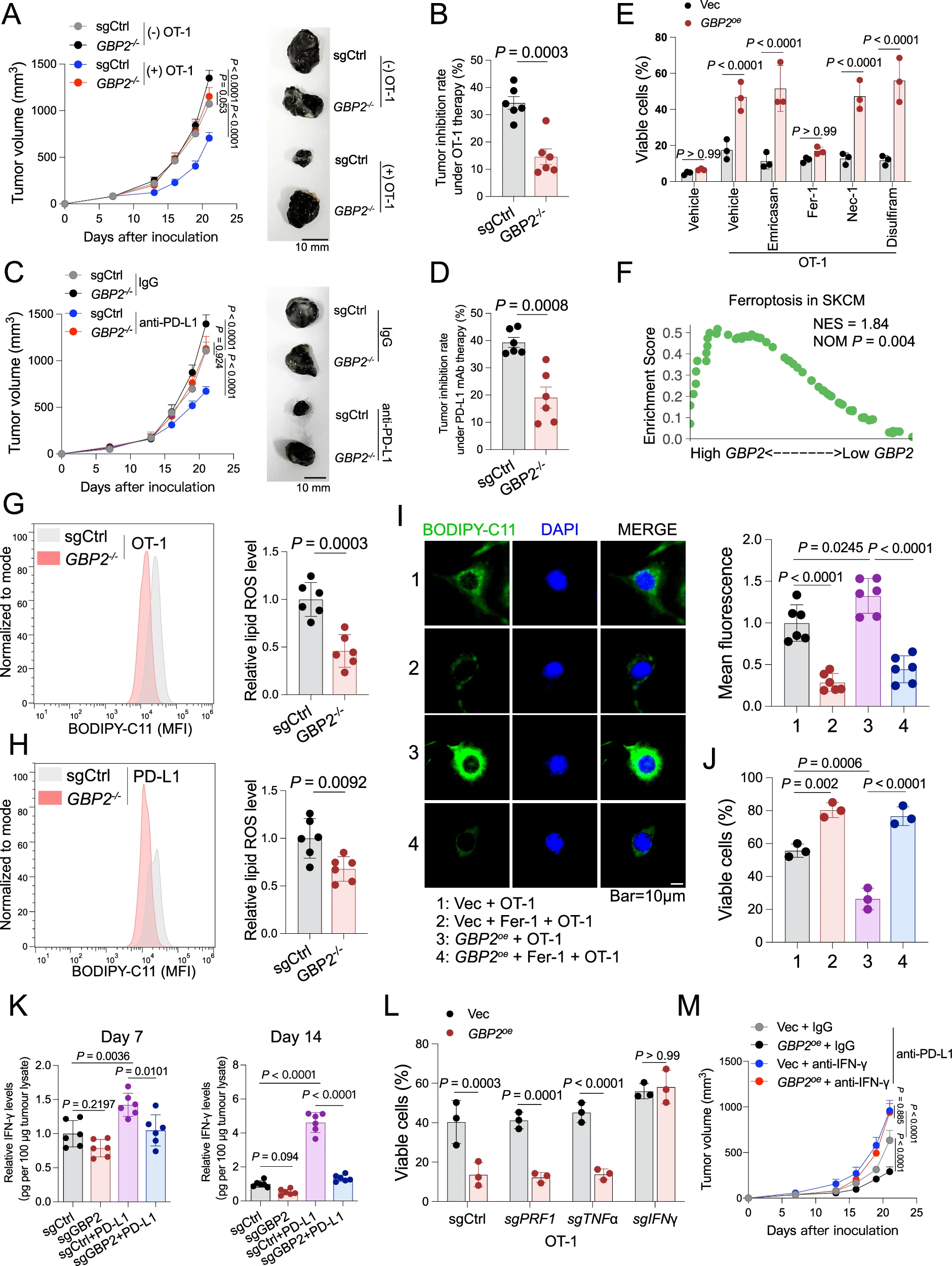
Fig2 Knockout of GBP2 blunted the efficacy of ICB and inhibited the CTL-induced ferroptosis.
A Tumor growth in mice subcutaneously injected with sgCtrl or GBP2?/? B16-OVA cells and treated with OT 1 T cells or PBS every 7 days. B Tumor inhibition rate of GBP2?/? relative to sgCtrl tumors under OT-1 treatment (n?=?6). C Tumor growth in mice bearing sgCtrl or GBP2?/?B16-OVA tumors treated with anti–PD L1 (200?μg/mouse) or IgG every 3 days. D Tumor inhibition rate of GBP2?/? versus sgCtrl tumors following anti–PD-L1 treatment (n?=?6). E Control or GBP2-overexpressing tumor cells were co-cultured with OT-1 cells (E:T?=?1:1, 24?h) with or without emricasan (20?μM), ferrostatin-1 (Fer-1, 10?μM), necrostatin-1 (Nec-1, 20?μM), or disulfiram (20?μM) (n?=?3). F GSEA of ferroptosis between high- and low-GBP2 patients according to the SKCM cohort. Lipid ROS in B16-OVA tumors treated with OT-1 (G) or anti–PD-L1 (H), quantified in CD45? cells by flow cytometry. Representative lipid ROS images (I) and viability (J) of Vec or GBP2oe B16-OVA cells co-cultured with OT-1 (E:T?=?5:1, 24?h), followed by Fer-1 (10?μM) for 24?h. K IFN-γ levels in tumor lysates from anti–PD-L1 or IgG-treated mice measured by ELISA. L Viability of Vec or GBP2oe cells after co-culture (24?h, E:T?=?1:1) with OT-1 T cells transduced with sgCtrl, sgIFNγ, sgTNFα, or sgPRF1 (n?=?3). M Vec or GBP2oe B16-OVA cells were subcutaneously implanted into C57BL/6 mice. When tumors reached ~125?mm3, both Vec and GBP2oe groups were respectively treated with either anti-IFN-γ or control IgG antibodies every 3 days. Tumor-bearing mice from both groups received anti-PD-L1 therapy every 3 days. All p value?<?0.05 as statistic difference. n?=?3 independent experiments (E, J, L); n?=?6 independent experiments (A–D, G–I, K, M). The data are represented as mean?±?SD. p value by unpaired two-tailed t-test (B, D, G, H) or one-way ANOVA (I–K) or two-way ANOVA (A, C, E, L, M) or two-sided permutation test (F). Source data are provided as a Source Data file.
2. GBP2 通过相分离 “扣押” SHP1,激活 STAT1 通路
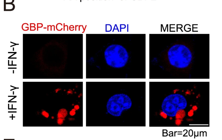
这是本研究最突破性的发现:IFN-γ 诱导 GBP2 发生液 - 液相分离(LLPS),形成动态凝聚体。
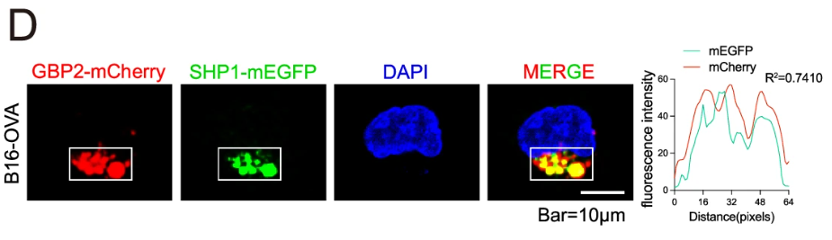
? 机制:GBP2 通过其无序区(IDR,aa436-579)形成凝聚体,特异性结合并扣押磷酸酶 SHP1,阻止 SHP1 去磷酸化 STAT1;
? 效应:持续激活的 STAT1 抑制铁死亡抑制蛋白 SLC7A11 转录,触发铁死亡(对应原文图 4、5);
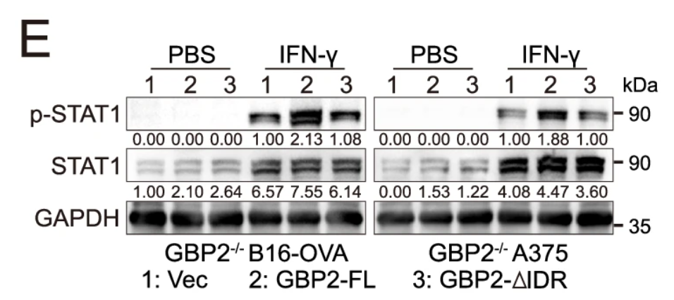
? 验证:删除 GBP2 的 IDR 区域,凝聚体无法形成,SHP1 不能被扣押,STAT1 激活受阻,铁死亡完全抑制(对应原文图 5K、5L)。
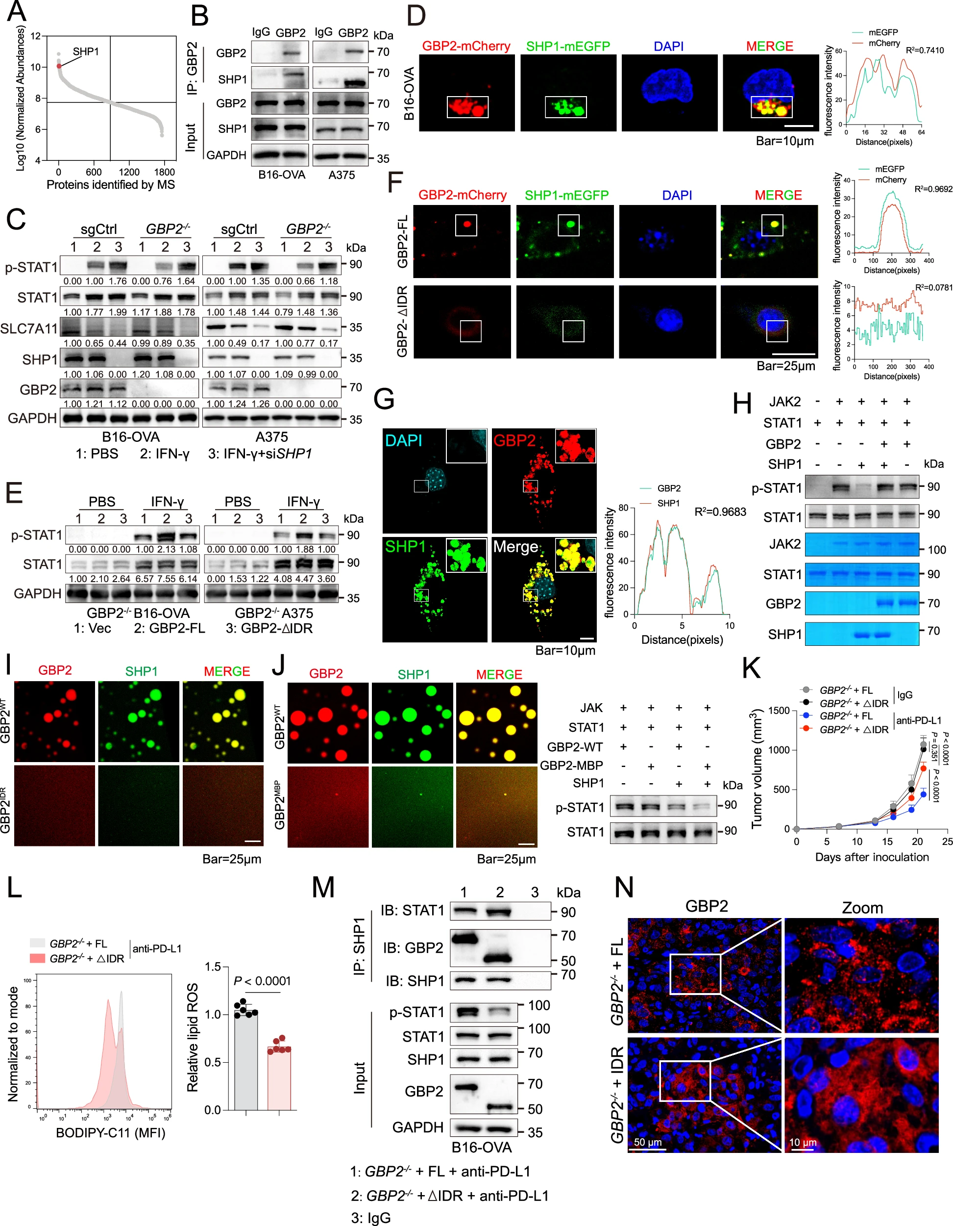
Fig5 GBP2 sequesters SHP1 via IDR-mediated phase separation to augment ferroptosis.
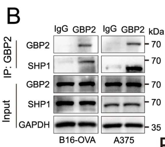
A B16-OVA cells treated with IFN-γ (10?ng/mL, 12?h) were immunoprecipitated with anti-GBP2 for mass spectrometry (MS); SHP1 was selected. B IP assay shows endogenous GBP2 interact with SHP1 in B16-OVA and A375 cells. C sgCtrl and GBP2?/? of B16-OVA and A375 cells were treated with IFN-γ (10?ng/mL, 12?h), followed by SHP1 knockdown using siRNA for 24?h; cell lysates were analyzed by western blot. D Representative live-cell imaging of B16-OVA cells expressing GBP2-mCherry stimulated with 10?ng/ml IFN-γ for 6?h, and transfected with SHP1-mEGFP plasmid for 36?h. The line plots show GBP2-mCherry and SHP1-mEGFP colocalization of areas. E Western blot analysis of B16-OVA GBP2?/? and A375 GBP2?/? cells treated with 10?ng/ml IFN-γ for 12?h, followed by transfection with vector, GBP2-FL, or GBP2 ΔIDR plasmids for 24?h. F, G Representative live-cell imaging and high-resolution confocal demonstrated colocalization of GBP2-mCherry with SHP1-mEGFP, but not GBP2ΔIDR, upon IFN-γ stimulation. H In vitro kinase assays revealed enhanced STAT1 phosphorylation in the presence of GBP2 condensates. I, J In vitro phase separation assays compared condensate formation between GBP2-WT, GBP2ΔIDR, and GBP2-MBP, confirming IDR-dependent condensate activity and STAT1 phosphorylation enhancement. K GBP2?/? B16-OVA cells expressing GBP2-FL or GBP2ΔIDR (1?×?106) were subcutaneously injected into C57BL/6 mice and treated with anti–PD-L1 antibody (200?μg/mouse) or IgG every 3 days. L Lipid ROS in CD45? tumor cells were quantified by flow cytometry. M Tumors from GBP2-FL and GBP2ΔIDR groups were immunoprecipitated with anti-SHP1 and analyzed by western blot. N Representative immunofluorescence images of GBP2 in GBP2-FL and GBP2ΔIDR tumors treated with anti–PD-L1 (n?=?3). All p value?<?0.05 as statistic difference. n?=?3 independent experiments (B–J, M, N); n?=?6 independent experiments (K, L). The data are represented as mean?±?SD. p value by unpaired two-tailed t-test (L) or two-way ANOVA (K). Source data are provided as a Source Data file.
3. GBP2 增强肿瘤免疫原性,促进 CD8?T 细胞浸润
铁死亡的肿瘤细胞释放HMGB1等损伤相关分子(DAMPs),促进树突状细胞(DC)成熟,增强 CD8?T 细胞浸润与活化。
? GBP2 高表达肿瘤中 CD8?T 细胞浸润显著增加(对应原文图 6G);
? 临床样本证实:GBP2 高表达患者肿瘤组织中 CD8?T 细胞浸润水平更高,预后更好(对应原文图 7I)。
4. GBP2 可作为 ICB 疗效预测标志物
多中心临床队列验证:
? GBP2 高表达 + SLC7A11 低表达的黑色素瘤患者,抗 PD-L1 治疗应答率最高、生存期最长(对应原文图 7D、7F);
? 该标志物在肺癌、尿路上皮癌中同样具有预测价值。
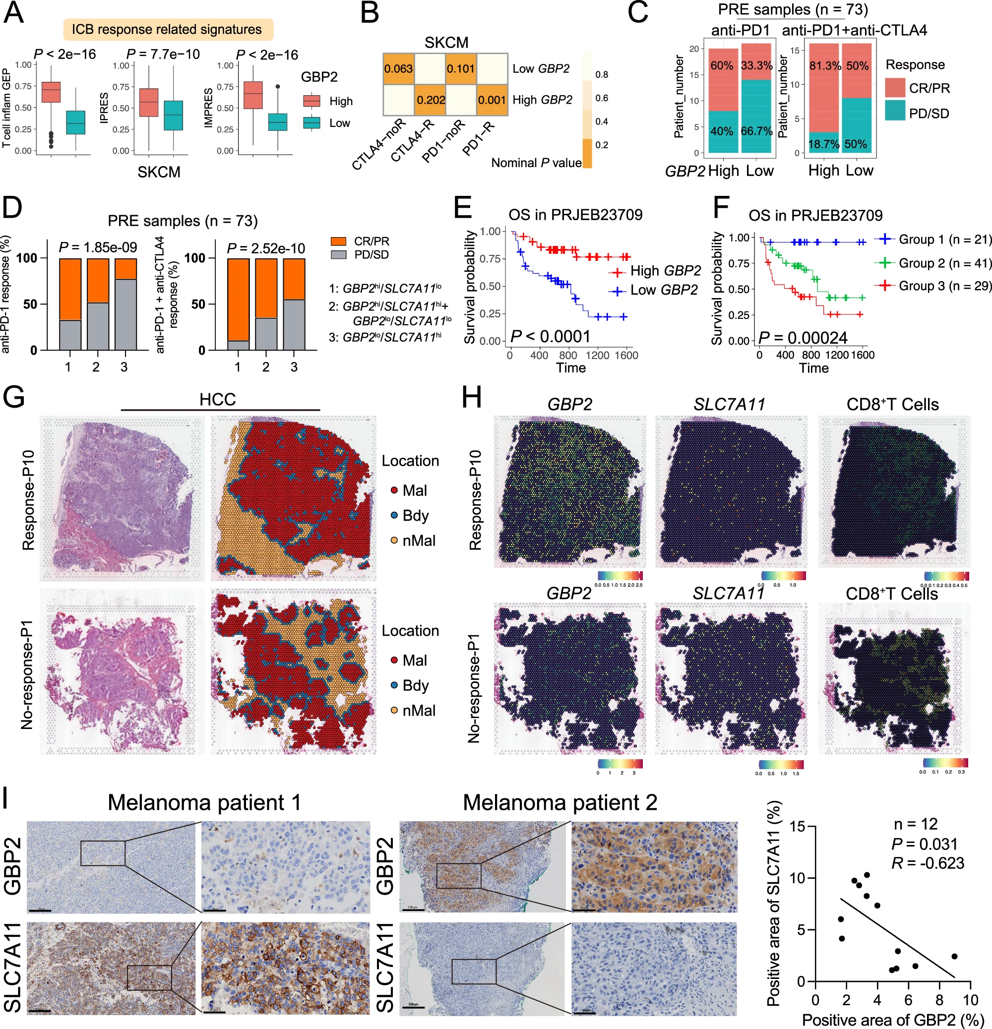
Fig7 GBP2 acts as an immunotherapeutic target in patients with cancer.
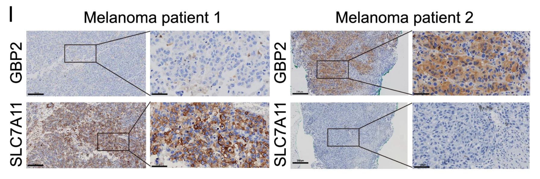
A Box plots showing expression of ICB response-related signatures between high and low GBP2 group in the SKCM cohort (n?=?470). IMPRES: Minimum (high vs. low: 0.064 vs. 0.001); Median (high vs. low: 0.668 vs. 0.332); Maximum (high vs. low: 1.0 vs. 0.751). IPRES: Minimum (high vs. low: 0.0 vs. 0.0); Median (high vs. low: 0.57 vs. 0.419); Maximum (high vs. low: 1.0 vs. 1.0). T cell-inflam GEP: Minimum (high vs. low: 0.047 vs. 0.0); Median (high vs. low: 0.707 vs. 0.316); Maximum (high vs. low: 0.941 vs. 0.732). B Submap analysis demonstrates high GBP2 group in SKCM samples closely align with the PD1-response (PD1-R) group defined in the melanoma cohort. C Treatment response rates to anti-PD-1 or combination immunotherapy in pre-therapy (PRE) patients stratified in high or low GBP2 expression groups. SD stable disease, PD progressive disease, CR complete response, PR partial response. D PRE patients were stratified into three groups based on SLC7A11 and GBP2 expression, with corresponding immunotherapy response rates shown. Kaplan–Meier survival analysis for GBP2-high vs. -low groups (E), and for the three SLC7A11/GBP2-based groups (F). G Spatial transcriptomics of two hepatocellular carcinoma sections (one responder, one non-responder) identified morphology-aligned clusters, annotated as malignant, boundary, or non-malignant regions. H The spatial feature plot that visualizes the proportion of the CD8+T cells and spatial expression patterns of GBP2 and SLC7A11 in spatial spots (n?=?2). I The tissue sections from 12 patients with melanoma were immunohistochemically stained with anti-GBP2 or anti-SLC7A11 antibody. The correlation between the expression of GBP2 and SLC7A11 is shown (right). Scale bars, 200?μm. All p value?<?0.05 as statistic difference. p value by unpaired two-tailed Wilcoxon test (A) or two-sided permutation test (B) or Pearson’s chi-square test (D) or Log-rank test (E, F) or the correlation coefficient and two-tailed p value were calculated using Pearson’s correlation analysis (I). Source data are provided as a Source Data file.
四、爱必信(absin)#abs955 抗体:研究的核心工具
本研究全程使用爱必信(absin)#abs955 GBP2 抗体,支撑了从分子机制到临床验证的全链条实验,其高特异性、高灵敏度成为关键保障。
1. 核心应用场景(对应原文图片)
? Western Blot(WB):精准检测细胞 / 组织中 GBP2 表达及 STAT1、SLC7A11 等通路蛋白变化(对应原文图 3G、5E);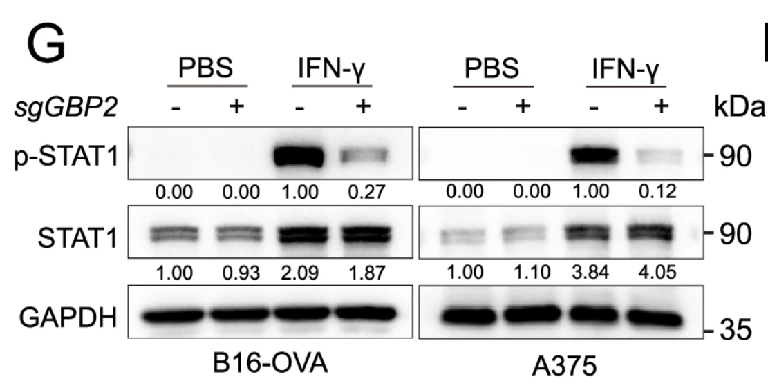
? 免疫荧光(IF):清晰显示 GBP2 在细胞内的凝聚体结构及与 SHP1 的共定位(对应原文图 4B、5D);
? 免疫组化(IHC):验证临床样本中 GBP2 表达水平及与 CD8?T 细胞的空间分布(对应原文图 7I);
? 免疫共沉淀(Co-IP):证实 GBP2 与 SHP1 的直接相互作用(对应原文图 5B)。
2. 产品核心优势
? 高特异性:仅识别天然 / 重组 GBP2,无交叉反应,保障机制结论可靠性;
? 多应用兼容:适配 WB、IF、IHC、Co-IP 等多种实验,满足多维度验证需求;
? 性能稳定:批次间一致性强,助力体内外实验重复性与可靠性。
五、研究意义与转化价值
1. 机制创新:首次揭示GBP2 相分离是连接 IFN-γ、铁死亡与肿瘤免疫的核心桥梁,填补了免疫治疗耐药机制的空白;
2. 靶点价值:GBP2 可作为黑色素瘤联合免疫治疗的全新靶点,通过靶向 GBP2 相分离,逆转 ICB 耐药;
3. 临床应用:GBP2/SLC7A 表达谱可作为 ICB 疗效预测标志物,指导精准用药;
4. 工具认可:爱必信 #abs955 GBP2 抗体在顶级研究中的成功应用,印证了其在生命科学研究中的可靠性与权威性。

- 手机:13761418683