




- 手机:13761418683
联系人:爱必信
网址:http://www.absin.cn/
地 址:上海市浦东新区新浩路58号18号楼

文献解析:《TFPI2 promotes NK cell–mediated glioblastoma killing through adhesion and checkpoint control》
2026-07-31
一、文献综述
(一)基础文献信息
? 文章标题:TFPI2 promotes NK cell–mediated glioblastoma killing through adhesion and checkpoint control
? 期刊:PNAS(美国国家科学院院刊,综合性顶刊,2026 IF≈12.7)
? 卷期页码:2026, Vol.123, No.25, e2529973123
? DOI:https://doi.org/10.1073/pnas.2529973123
? 发表时间:2026 年 6 月 18 日
(二)核心结论
? 上游诱导机制:NK 细胞与胶质母细胞瘤(GBM)接触后分泌 IL1β、TNFα,共同激活 GBM 细胞经典 / 非经典 NF-κB 通路,特异性诱导TFPI2表达;该诱导效应仅存在于 GBM 细胞(U87MG、U138、A172),HeLa、A549 等非脑肿瘤细胞无响应。
? 肿瘤细胞自主抑癌通路:TFPI2 抑制转录因子 POU2F2,下调周期蛋白 CCND1,阻滞细胞 S 期,直接抑制 GBM 增殖、3D 球体形成。
? 双重免疫增强轴(两大核心机制)
① TFPI2-LGALS1-ICAM1 黏附轴:TFPI2 与 LGALS1 直接结合,抑制 LGALS 泛素化蛋白酶体降解,稳定 LGALS1 蛋白,持续上调黏附分子 ICAM1,强化 NK - 肿瘤免疫突触,提升 NK 细胞黏附、浸润与杀伤;
② TFPI2-SIGLEC15 检查点轴:TFPI2 选择性抑制免疫检查点 SIGLEC15(不调控 PD-L1),解除 SIGLEC15 对 NK 细胞的抑制,恢复 NK IFNγ 分泌与细胞毒活性。
? 体内分层验证
GL261 小鼠颅内模型(炎症可诱导 Tfpi2):Tfpi2 敲低加速肿瘤生长、脑内 NK 浸润显著减少,NK 过继治疗完全失效;清除内源 NK 后无叠加促瘤效应,证明 TFPI 抑瘤完全依赖 NK 细胞。
CT-2A 小鼠 GBM:IL1β/TNFα 刺激无法上调 Tfpi2,敲低无肿瘤表型,证实 TFPI 通路存在肿瘤异质性。
NCG 人源颅内异种移植:U87MG-TFPI2 敲除小鼠输注人 PB-NK 后,肿瘤无明显抑制,验证人体系保守机制。
? 临床转化意义:TFPI 可作为 GBM 免疫分型标志物;通过 STING 激动剂、去甲基化药物提升肿瘤 TFPI 表达,联合过继 NK 细胞、SIGLEC15 阻断剂,改善 GBM 免疫治疗疗效。
(三)本研究使用 Absin(爱必信)产品及作用
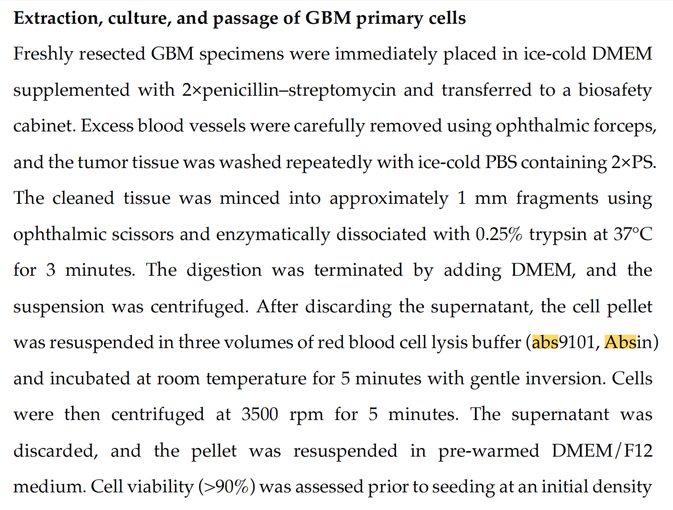
使用爱必信abs9101 红细胞裂解液,是免疫细胞分离的关键前处理试剂:
1. 人外周血 NK 制备:健康人外周血经 Ficoll 梯度分离 PBMC 后,使用 abs9101 裂解残留红细胞,去除血红蛋白、红细胞碎片杂质,保证 PBMC 纯度,用于后续 NK 扩增与体外共培养;
2. 小鼠脑肿瘤浸润淋巴细胞(TIL)提取:颅内 GL261/CT-2A 肿瘤组织酶解制备单细胞悬液,加入 abs9101 清除组织内红细胞,消除流式背景干扰,保证 CD45+CD3-NK1.1+ NK 分群准确;
3. 样本保护优势:abs9101 选择性裂解无核红细胞,不损伤 NK、T 等有核免疫细胞,细胞活性稳定,满足流式染色、体外杀伤、体内过继回输全部后续实验要求。
二、研究领域背景介绍与本文科学切入点
(一) 领域研究现状
1. 胶质母细胞瘤(GBM)是成人恶性最高原发性脑肿瘤,标准手术 + 替莫唑胺放化疗预后极差,5 年生存率不足 10%;血脑屏障、高度免疫抑制肿瘤微环境极大限制 CAR-T、免疫检查点单药疗效。
2. NK 细胞优势:无需 MHC 抗原递呈、移植物抗宿主风险低,是实体瘤过继免疫优选,但 GBM 多重逃逸机制削弱 NK 细胞功能:
① 分泌 TGFβ 等抑制因子、下调 NKG2D 配体;
② 低表达 ICAM1,破坏 NK - 肿瘤细胞黏附;
③ 高表达 SIGLEC15 新型抑制检查点,独立于 PD-L1 通路抑制 NK 细胞毒。
3. 研究空白:既往研究多改造 NK 细胞本身,缺乏肿瘤细胞响应 NK 攻击的反馈调控分子研究;TFPI2 功能存在争议:多数肿瘤为抑癌基因,但 GBM 干细胞中可促干性,其在 NK-GBM 互作中的功能完全未知;SIGLEC15 上游肿瘤内调控靶点未被报道。
4. 临床痛点:GBM 存在明显免疫异质性,部分患者完全不响应免疫治疗,缺少分层标志物。
(二)本文创新切入点
1. 视角创新:聚焦 NK 刺激后 GBM 细胞产生的反馈调控分子 TFPI2,挖掘肿瘤自身双向调控环路(抑制增殖 + 重塑免疫微环境);
2. 机制创新:同时解析 TFPI2 调控细胞周期、细胞黏附、新型免疫检查点三条独立通路,搭建 TFPI2-LGALS1-ICAM1、TFPI2-SIGLEC15 两大功能轴;
3. 异质性解释:基于人 / 鼠多细胞系、体内模型,阐明仅炎症敏感型 GBM 可激活 TFPI 通路,为患者免疫分层提供依据;
4. 实验技术支撑:依托 abs9101 完成人 / 鼠 NK 细胞纯化,建立标准化体外、体内免疫互作体系,保障所有免疫功能实验数据可靠。
三、作者整体研究思路总结
全文遵循临床生信筛选→体外诱导通路解析→细胞自主增殖机制→免疫调控分子互作→体外 2D/3D 功能验证→人 / 鼠多层体内模型→临床转化推论完整闭环:
1. 临床队列挖掘:TCGA、CGGA 人 GBM 队列区分高 / 低活化 NK 浸润组,筛选差异基因 TFPI2;临床组织 IHC 证实 TFPI2 与 CD56+ NK 浸润正相关;
2. 上游通路解析:NK 分泌 IL1β/TNFα 激活 NF-κB 诱导 TFPI2,区分直接接触 / 细胞因子的诱导作用;
3. 增殖抑制机制:构建 TFPI2 敲低 / 回补细胞,明确 POU2F2-CCND1 周期轴;
4. 免疫黏附机制:IP-MS 筛选 TFPI2 结合蛋白 LGALS1,通过泛素实验阐明 ICAM1 上调机制;
5. 检查点调控:转录组筛选出 SIGLEC15 为 TFPI 特异性抑制靶点,体外回补 / 双敲验证功能;
6. 体外功能:2D LDH 杀伤、3D 肿瘤球体共培养、NK 黏附实验,全部依赖 abs9101 纯化的人 / 鼠 NK 细胞完成;
7. 体内分层验证:免疫健全 GL261、免疫冷肿瘤 CT2A、NCG 人源异种移植三套动物模型,流式检测 TIL 均使用 abs9101 处理组织单细胞;
8. 转化推导:提出 TFPI 作为预后分层标志物,炎症诱导联合 NK 过继治疗新策略。
四、分模块详细研究思路、实验结果
模块 1:临床生信 + 细胞初筛,TFPI2 为 NK 特异性诱导 GBM 分子
研究思路
TCGA/CGGA 生信筛选高 NK 浸润差异基因;人 GBM 细胞、患者原代细胞、对照肿瘤细胞(HeLa/A549)NK 刺激验证 TFPI2 表达;临床组织 IHC 关联 TFPI 与 NK 浸润。
实验结果
1. TCGA、CGGA 高活化 NK 组 TFPI2 mRNA 显著上调,Venn 交集锁定候选分子(图 1A、SI Fig S1H);
2. NK92 时间梯度刺激 U87MG/U138/A172,TFPI2 蛋白持续上调;宫颈癌、肺癌细胞无诱导(图 1C-E、SI Fig S2C-D);
3. 4 例患者原代 GBM 细胞 NK 处理后 TFPI2 升高(图 1G);
4. 人 GBM 组织 IHC:TFPI2 表达与 CD56+ NK 浸润显著正相关(图 1H-J)。
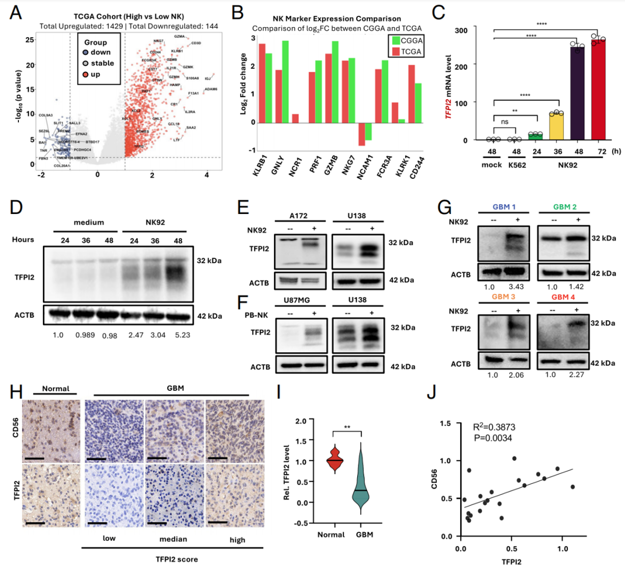
Fig. 1. (A) Volcano plots displaying differentially expressed genes (DEGs) between high-activated (n = 45) and low-activated (n = 53) NK infiltration groups in the TCGA GBM cohort. (B) The log2 Fold change (Log2 FC) of 10 selected NK cell signature genes in the high- activated NK infiltration group (TCGA, n = 45; CGGA, n = 20) relative to the low-activated NK infiltration group (TCGA, n = 53; CGGA, n = 26). (C) U87MG cells were cocultured with NK92 cells (E:T ratio=3:1) or K562 cells (same cell count ratio as NK92: U87MG) for indicated hours. RNA was analyzed by RT- qPCR, normalized with GAPDH. One- way ANOVA; n = 3. (D) U87MG cells were cocultured with NK92 at E:T ratio=3:1 for indicated hours. Protein was analyzed by Western blotting. The quantitated ratio of TFPI2 to ACTB from three independent analyses was shown at the bottom. (E) TFPI2 protein expression was detected by Western blotting in A172 and U138 GBM cells following NK92 stimulation (E:T ratio=3:1, 24 h). (F) TFPI2 protein expression was detected in U87MG and U138 cells stimulated with PB-NK (E:T ratio=1:1, 8 h). (G) TFPI2 protein expression in primary GBM cells from four patients (numbered as 1 to 4) after NK92 cell coculturing (E:T ratio= 3:1, 24 h) was detected by Western blotting. The quantitated ratio of TFPI2 to ACTB was shown at the bottom. (H) CD56+ cell infiltration and the level of TFPI2 in GBM or normal brain tissues. (Representative IHC images; Scale bar, 50 μm.) TFPI2 expression level was scored as low, median, and high based on the tertile distribution (see Materials and Methods for details). (I) Quantification of TFPI2 and CD56 levels in GBM or normal brain tissues (unpaired t test; Normal Brain Tissues, n = 4; GBM, n = 20). (J) Correlation analysis of CD56 and TFPI2 measurement in GBM (n = 20). Data in all quantitative panels are displayed as mean ± SD, *P < 0.05, **P < 0.01, ***P < 0.001, ****P < 0.0001.
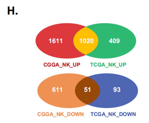
(H) Venn diagram of upregulated and downregulated genes from TCGA and CGGA GBM cohorts.
模块 2:NK 通过 IL1β/TNFα-NF-κB 通路诱导 TFPI2 转录
研究思路
Transwell 区分直接接触 / 可溶性因子;RNA-seq 富集 NF-κB 通路;抑制剂、启动子截短、RELB 结合位点突变验证转录调控;单独 / 联合细胞因子刺激确认上游信号分子。
实验结果
1. NK 直接接触 + 上清因子均可诱导 TFPI,直接接触效应更强(图 2A);
2. NK 刺激后 GBM 转录组 NF-κB 通路显著富集(图 2B、SI Fig S3B-D);
3. NF-κB 抑制剂 BMS、BOT64 完全阻断 TFPI2 上调(图 2C);
4. TFPI2 启动子 - 254~+100bp 为核心激活区,RELB 结合基序突变丧失诱导活性(图 2D-F);
5. NK 刺激先诱导 IL1β,再上调 TFPI;IL1B 敲低联合 TNFα 中和显著抑制 TFPI 表达(图 2G-J)。
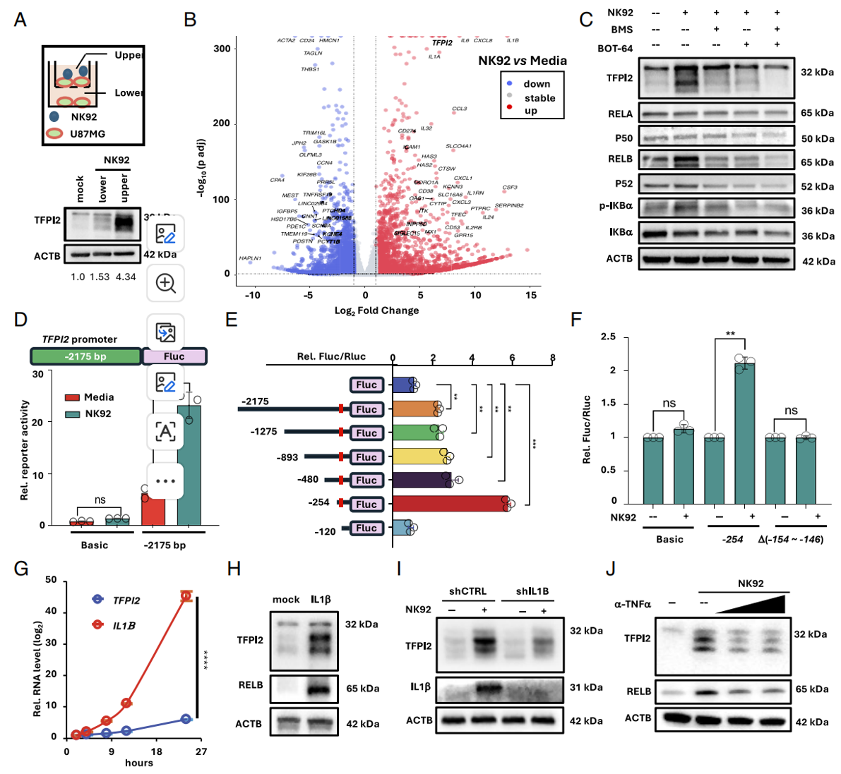
Fig. 2. NK cells induce TFPI2 expression through activation of the NF-κB signaling pathway. (A) TFPI2 protein expression in U87MG cells from upper vs. lower chambers in a Trans- well coculturing with NK92 cells (E:T ratio = 3:1, 24 h). The quantitated ratio of TFPI2 and ACTB of three independent experiments was shown at the bottom. (B) Differential gene expression by volcano plot between U87MG cells with or without NK92 stimulation. (C) Expression of TFPI2 and NF- κB pathway components in U87MG cells pretreated with BOT64 or BMS- 345541 (both at 10 μM as final concentration, 8 h) and then stimulated with NK92 cells (E:T ratio = 3:1) for 10 h. (D) U87MG cells were transfected with pGL3- basic vector or TFPI2 promoter-driven Firefly luciferase (Fluc) vector (pGL3- TFPI2). 48 h later, cells were stimulated with NK92 cells (E:T ratio = 3:1) for 8 h and then collected for luciferase activity measurement (unpaired t test; n = 3). (E) Truncated TFPI2 promoter constructs were transfected in U87MG cells and then stimulated with NK92 cells (one-way ANOVA, n = 3). (F) Luciferase activity measurement in U87MG cells transfected with truncated promoter regions indicated followed by NK92 stimulation (E:T ratio = 3:1, 8 h) (unpaired t test; n = 3). (G) Time- course (2, 4, 8,12, 24 h) of IL1β and TFPI2 mRNA induction in U87MG cells upon NK92 stimulation (E:T ratio = 3:1) (two-way ANOVA, n = 3). (H) TFPI2 and RELB protein expression in U87MG cells treated with recombinant human IL1β (40 ng/mL, 24 h). (I) TFPI2 and IL1β protein levels were detected by Western blotting in U87MG (IL1B knockdown or control) upon NK92 stimulation (E:T ratio = 3:1, 24 h). (J) TFPI2 induction was measured in U87MG cells upon NK92 treatment (E:T ratio = 3:1, 24 h) in the presence of anti- TNFα neutralizing antibody (Infliximab@ 0, 10, 20 μg/mL) vs. isotype control. Data in all quantitative panels are displayed as mean ± SD, **P < 0.01, ***P < 0.001, ****P < 0.0001, ns, not significant.
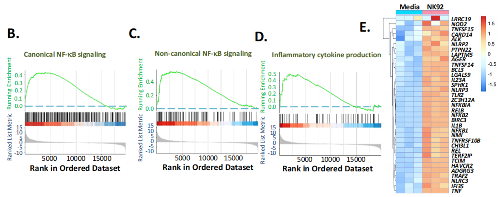
(B-D) Enrichment of NF-kB and inflammatory response pathways from Figure 2B (B-D) and in NK high-activated group of TCGA (F) and CGGA (G) GBM cohorts.
模块 3:TFPI2 通过 POU2F2-CCND1 轴抑制 GBM 细胞增殖
研究思路
构建 TFPI2 稳定敲低、回补细胞;CCK8、EdU、3D 球体、流式细胞周期检测增殖表型;转录组筛选周期基因,双敲 TFPI2+POU2F2 进行挽救验证。
实验结果
1. TFPI2 敲低提升细胞活力、增大 3D 球体、EdU 阳性增殖细胞增多,回补 TFPI 逆转表型;
2. TFPI2 缺失上调 POU2F2 与周期蛋白 CCND1;双敲消除 S 期增殖异常;
3. 外源 TFPI 蛋白剂量依赖性下调 POU2F2/CCND1。
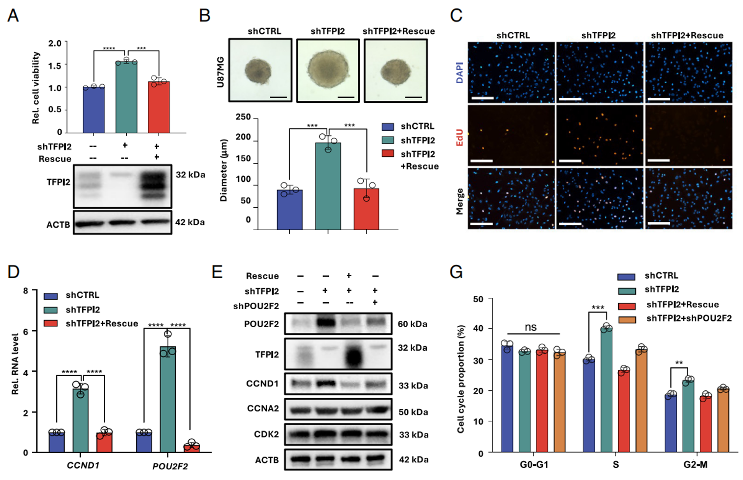
Fig. 3. TFPI2 suppresses GBM proliferation through the POU2F2–CCND1 axis. (A) TFPI2 protein levels and cell viability in U87MG cells with TFPI2 knockdown and its rescue after 48 h culturing were tested by Western blotting and CCK-8 respectively (one-way ANOVA, n = 3). (B) Tumor spheroid formation for 7 d in U87MG cells with TFPI2 knockdown and rescue. (Representative images; Scale bar, 200 μm; one-way ANOVA, n = 3.) (C) EdU incorporation in U87MG cells with TFPI2 knockdown and rescue. (Scale bar, 200 μm.) (D) POU2F2 and CCND1 mRNA levels in U87MG cells with TFPI2 knockdown and rescue (one-way ANOVA, n = 3) was measured by RT-qPCR, normalized with GAPDH. (E) Protein expression of POU2F2, CCND1, CCNA2, and CDK2 in U87MG cells under TFPI2 and POU2F2 modulation were detected by Western blotting. (F and G) Cell cycle distribution in U87MG cells with TFPI2 knockdown and TFPI2&POU2F2 double knockdown was analyzed via Propidium Iodide (PI)-staining followed by FACS analysis and quantification (G, one-way ANOVA, n = 3). Data in all quantitative panels are displayed as mean ± SD, **P < 0.01, ***P < 0.001, ****P < 0.0001, ns, not significant.
模块 4:TFPI2 稳定 LGALS1 上调 ICAM1,促进 NK - 肿瘤黏附杀伤
研究思路
TFPI2 敲低后 NK 杀伤、黏附能力下降;转录组锁定 ICAM1;Co-IP 质谱筛选互作 LGALS1;泛素化实验阐明分子机制;LGALS1 敲低回补验证轴功能。
实验结果
1. TFPI2 敲低显著削弱 2D、3D 球体 NK 杀伤,NK 细胞在肿瘤球表面黏附数量大幅降低;
2. TFPI2 缺失下调 ICAM1,过表达 ICAM1 可恢复 TFPI 敲除细胞对 NK 敏感性;
3. TFPI 与 LGALS1 胞内共定位、直接结合;TFPI2 缺失促进 LGALS1 泛素化降解,MG132 可挽救 LGALS1 蛋白;
4. LGALS1 敲低直接下调 ICAM1,重现 TFPI2 缺失表型。
Absin abs9101 使用说明
本模块人原代 PB-NK 细胞分离扩增环节必须使用 abs9101:健康人外周血分离 PBMC 后,abs9101 裂解红细胞,去除杂质,获得高活性 NK 细胞用于共培养、荧光黏附成像实验,是本模块体外功能实验的基础样本制备试剂。
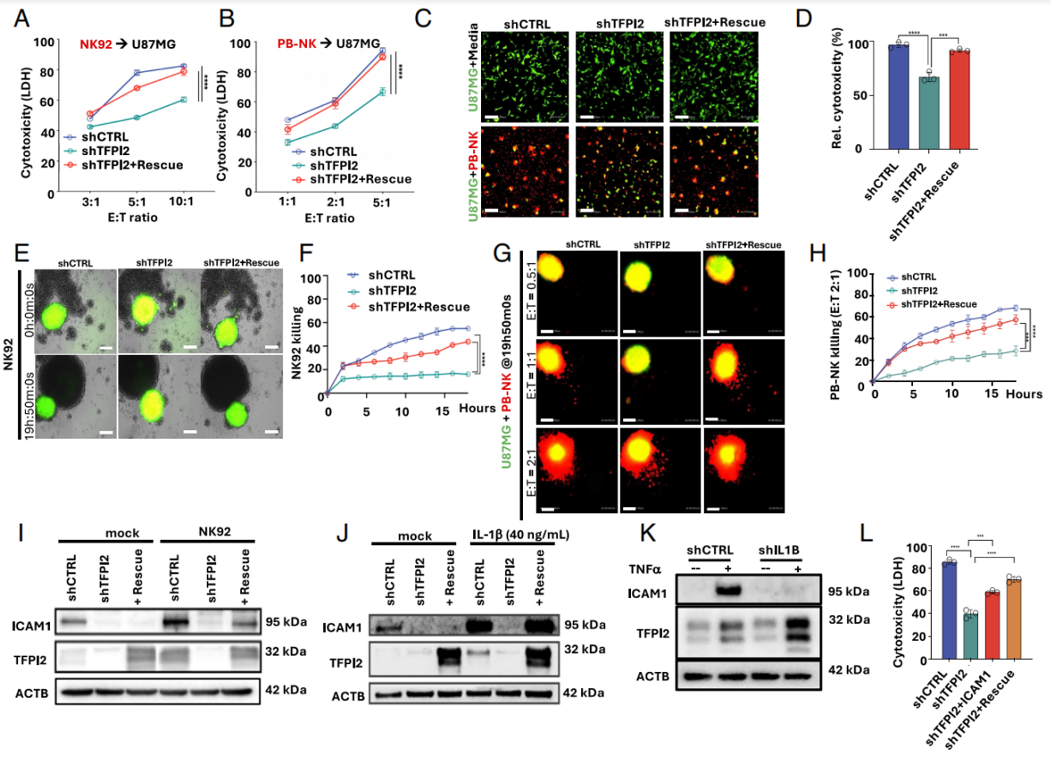
Fig. 4. TFPI2 enhances NK cell–mediated cytotoxicity by supporting ICAM1 expression. (A and B) Cytotoxicity of NK92 cells (24 h coculturing) and PB-NK cells (6 h coculturing) against TFPI2 knockdown-U87MG cells at indicated E:T ratios was measured by LDH assay (two-way ANOVA, n = 3). (C and D) Representative images and quantification for PB-NK-mediated killing of EGFP-U87MG cells with TFPI2 knockdown at an E:T ratio of 5:1 for 6 h. (Scale bar, 200 μm; one-way ANOVA, n = 3; green, EGFP-U87MG; red, live cell tracker stained-PB-NK.) The cytotoxicity was measured by collecting EGFP signals and normalized against the signal in shCTRL cells. (E and F) Representative images and quantification for NK92-mediated killing (E:T ratio of 5:1) of 3D-cultured EGFP-U87MG spheroids at indicated time points. (Scale bar, 200 μm; two-way ANOVA, n = 3; green, EGFP-U87MG; bright field, NK92.) NK92 killing was measured in the same way as in (D). (G and H) Representative images and quantification for PB-NK-mediated killing of EGFP-U87MG spheroids with TFPI2 expression at indicated E:T ratios and incubation time. (Scale bar, 200 μm; Red, live cell tracker stained-PB-NK; Green, U87MG; two-way ANOVA, n = 3.) PB-NK killing was measured in the same way as in (D). (I) ICAM1 protein expression in U87MG under TFPI2 modulation with NK92 stimulation (E:T ratio = 3:1, 24 h). (J) ICAM1 protein expression in U87MG under TFPI2 modulation with or without IL1β stimulation (40 ng/mL, 24 h). (K) ICAM1 and TFPI2 protein levels in IL1B knockdown U87MG cells treated with recombinant TNFα (40 ng/mL, 24 h). (L) U87MG cells indicated were infected with ICAM1-encoding lentivectors for stable cell line establishment and challenged by NK92 cells at an E:T ratio of 5:1 for 24 h (one-way ANOVA, n = 3). Cytotoxicity was measured by LDH assay. Data in all quantitative panels are displayed as mean ± SD, ***P < 0.001, ****P < 0.0001.
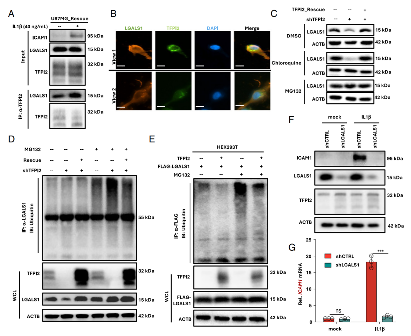
Fig. 5. LGALS1 required for optimal ICAM1 expression and is stabilized by TFPI2 interaction. (A) Enhanced interaction between endogenous TFPI2 and LGALS1 in U87MG cells upon IL1β (40 ng/mL, 24 h) stimulation. (B) Colocalization of TFPI2 and LGALS1 in U87MG cells. (Scale bar, 5 μm.) (C) LGALS1 protein level was detected in TFPI2-knockdown U87MG cells treated with proteasome inhibitor MG132 (10 μM, 6 h) or lysosomal inhibitor chloroquine (30 μM, 24 h). (D) Polyubiquitination of LGALS1 in U87MG cells with TFPI2 knockdown. Total LGALS1 proteins were immunoprecipitated by anti-LGALS1 antibody, and Ub-LGALS1 was detected by anti-Ubiquitin antibody. The band at 55 kDa represents IgG heavy chain. WCL, whole cell lysates. (E) Ubiquitination of LGALS1 in HEK293T cells with TFPI2 overexpression. Total FLAG-tagged LGALS1 proteins were immunoprecipitated by anti-FLAG antibody, and Ub-LGALS1 was detected by antiUbiquitin antibody. (F and G) ICAM1 protein and mRNA expression in U87MG cells upon IL1β stimulation (40 ng/mL, 24 h) and LGALS1 knockdown (unpaired t test; n = 3). Data in all quantitative panels are displayed as mean ± SD, ***P < 0.001, ns, not significant.
模块 5:TFPI2 选择性抑制 SIGLEC15,解除 NK 免疫抑制
研究思路
TFPI2 敲低后全免疫检查点筛选,仅 SIGLEC15 特异性上调;外源 SIGLEC15 处理抑制 NK 杀伤、IFNγ 分泌;TFPI2+SIGLEC15 双敲实现功能挽救。
实验结果
1. TFPI2 缺失仅上调 SIGLEC15,PD-L1 表达无变化;
2. 外源 SIGLEC15 处理显著降低 NK 介导肿瘤裂解、IFNγ 分泌;
3. 同时敲除 TFPI2 与 SIGLEC15,可恢复 NK IFNγ 分泌与细胞毒能力。
Absin abs9101 使用说明
体外 PB-NK 细胞均经 abs9101 裂红纯化后扩增,用于 LDH 杀伤、IFNγ ELISA 检测。
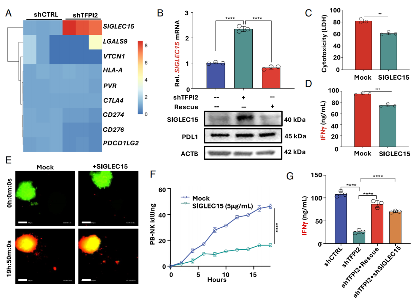 Fig. 6. TFPI2 promotes NK cell activity by selectively suppressing the immune checkpoint SIGLEC15. (A) Heatmap of expression of immune checkpoint molecules in control and TFPI2-knockdown U87MG cells from SI Appendix, Fig. S5G. (B) mRNA and protein expression of SIGLEC15 in TFPI2-knockdown and rescue U87MG cells (one-way ANOVA, n = 3). (C) U87MG cells were treated with recombinant SIGLEC15 (5 μg/mL) and NK92 cells (E:T ratio = 5:1) for 24 h, then were analyzed by LDH assay (unpaired t test; n = 3). (D) IFNγ secretion in (C) was measured by ELISA (unpaired t test; n = 3). (E, F) Representative images and quantification of live cell tracker stained-PB-NK-mediated killing of 3D-cultured EGFP-U87MG spheroids treated with recombinant SIGLEC15 at 5 μg/mL as final concentration for 19 h 50 min. (Scale bar, 200 μm; two-way ANOVA, n = 3.) The PB-NK killing was measured in the same way as in Fig. 4D. (G) IFNγ secretion by ELISA in coculturing supernatants of NK92 cells (E:T ratio = 5:1, 24 h) with U87MG cells under single or dual knockdown conditions indicated (one-way ANOVA, n = 3). Data in all quantitative panels are displayed as mean ± SD, **P < 0.01, ***P < 0.001, ****P < 0.0001.
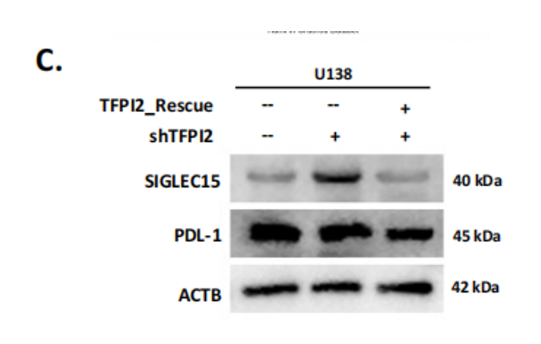 (C) Protein expression of SIGLEC15, PD-L1 in TFPI2-knockdown and rescue U138MG cells.
模块 6:多层体内动物模型验证 TFPI2 体内抑瘤依赖 NK
研究思路
三套颅内肿瘤模型:免疫健全 GL261、免疫冷肿瘤 CT2A、NCG 人源 U87 异种移植;设置 NK 清除、人 PB-NK 过继分组;小鼠脑组织消化后提取 TIL,流式检测 NK 浸润数量。
实验结果
1. GL261 模型:Tfpi2 敲低肿瘤荧光负荷升高、生存期缩短;NK 清除后无额外促瘤,脑内 CD45+CD3-NK1.1+ NK 浸润显著减少;
2. CT2A 细胞对 IL1β/TNFα 无响应,Tfpi2 敲低不改变肿瘤生长;
3. NCG 小鼠输注 abs9101 纯化人 PB-NK,对照组肿瘤明显缩小,TFPI2 敲除组无治疗获益。
Absin abs9101 使用说明(核心应用场景)
1. 小鼠肿瘤 TIL 提取:颅内 GL261/CT2A 脑组织酶解为单细胞悬液,加入 abs9101 裂解组织内红细胞,去除血红蛋白对流式荧光的干扰,保证 NK 细胞分群准确,用于定量肿瘤浸润 NK 比例;
2. 人源过继 NK 制备:健康人外周血 PBMC 分离后,abs9101 去除红细胞,纯化 NK 细胞扩增,尾静脉回输 NCG 小鼠,是体内人源免疫重建的必备前处理试剂;
3. 所有小鼠脾脏、外周血流式样本预处理统一使用 abs9101 标准化裂红流程,保证多组动物实验样本处理一致性,降低实验误差。
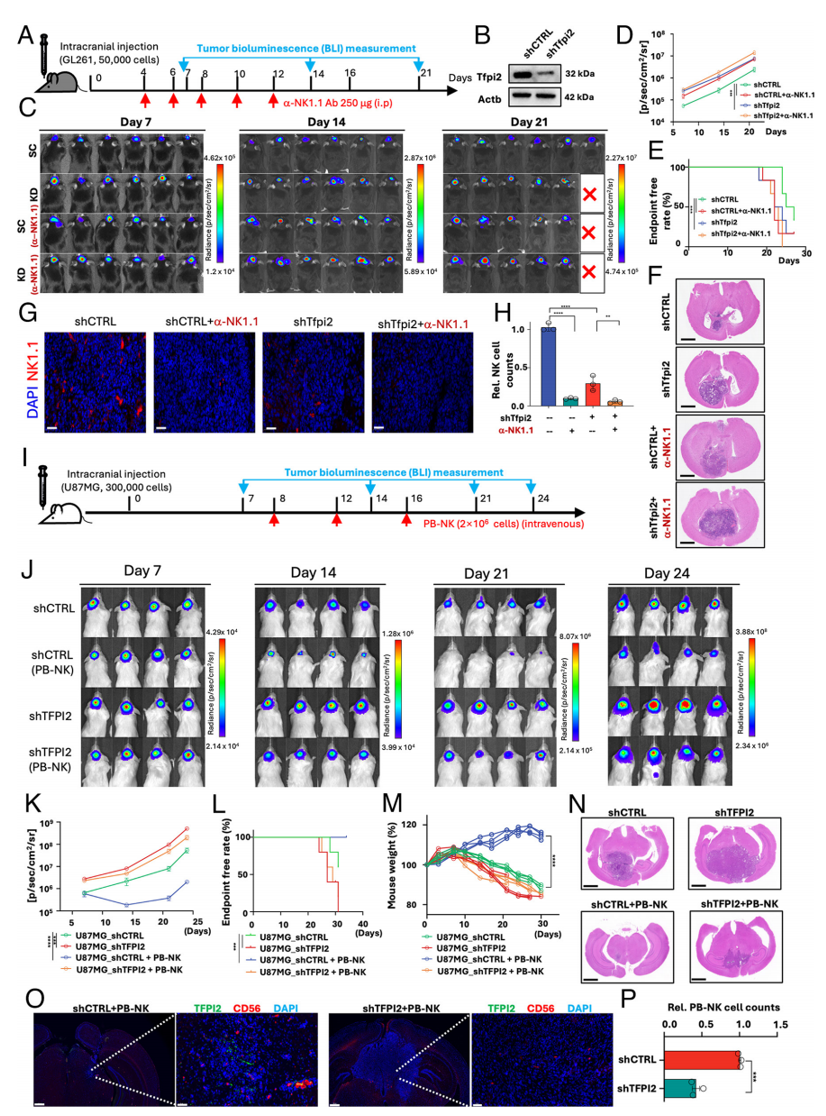 Fig. 7. TFPI2 promotes NK-mediated GBM killing in vivo. (A) Experimental timeline for intracranial tumor implantation and anti-NK1.1 antibody-mediated NK cell depletion in C57BL/6 mice. (B) Tfpi2 knockdown in mouse GBM GL261-Luc cells. (C) Representative bioluminescence images of mice bearing intracranial GL261-Luc tumors with or without Tfpi2 knockdown and anti-NK1.1 antibody treatment. (D) Quantification (Two-way ANOVA, n = 6) of bioluminescence flux from (C). (E) Endpoint-free rate of mice over a 27-d monitoring period. (F) Representative H&E-stained brain sections from each group at endpoint. (Scale bar,2 mm.) (G and H) Representative images and quantification for immunofluorescence staining of NK cells (red, NK1.1) in brain sections. NK cell counts were determined by calculating red staining using ImageJ. (Scale bar, 50 μm; oneway ANOVA, n = 3 for H.) (I) Experimental timeline for intracranial implantation of human U87MG-Luc cells and adoptive transfer of human primary NK cells (PB-NK) in NCG mice. (J) Representative bioluminescence images of mice after tumor implantation and NK cell transfer (days indicated). (K) Quantification of bioluminescence flux from (J) (Two-way ANOVA, n = 4). (L) Endpoint-free rate of mice over a 32-d monitoring period. (M) Body weight changes of mice during the experiment (Two-way ANOVA, n = 4). (N) Representative H&E-stained brain sections from each group at endpoint. (Scale bar, 2 mm.) (O and P) Representative images and quantification for immunofluorescence staining of human NK cells (red, CD56) and TFPI2 (green) in brain sections. PB-NK cell counts were measured as in (H). (Scale bar, 50 μm; unpaired t test, n = 3.) Data in all quantitative panels are displayed as mean ± SD, **P < 0.01, ***P < 0.001, ****P < 0.0001.
五、Absin abs9101 红细胞裂解液整体作用总结
Absin abs9101 是贯穿本研究全部原代免疫细胞体外、体内实验的标准化前处理核心试剂,为全文 NK 细胞相关功能数据提供基础样本保障,整体作用分为 3 个层面:
1. 人原代 NK 制备支撑:用于健康人外周血 PBMC 分离后裂解红细胞,去除细胞碎片与血红蛋白杂质,完整保留 NK 细胞活性,满足体外共培养、3D 球体杀伤、细胞因子分泌检测全部体外功能实验;
2. 小鼠肿瘤免疫流式标准化:颅内胶质瘤组织、小鼠外周 / 脾脏单细胞悬液统一采用 abs9101 裂红,消除红细胞造成的流式背景噪音,精准定量脑内浸润 NK 细胞比例,支撑 GL261/CT2A 模型异质性对比实验;
3. 体内过继细胞质控:经 abs9101 纯化的人 NK 细胞活性稳定,回输 NC 免疫缺陷小鼠后可稳定重建人抗肿瘤免疫体系,保证体内疗效对比实验可重复;
4. 实验稳定性优势:abs9101 配方温和,仅裂解无核红细胞,不损伤 NK、淋巴细胞等目标免疫细胞,兼顾细胞培养、流式、体内回输多场景,统一人 / 鼠样本前处理方案,减少实验变量,是本研究免疫表型、杀伤功能、体内生存期等关键结论的基础耗材支撑。

- 手机:13761418683