




- 手机:13761418683
联系人:爱必信
网址:http://www.absin.cn/
地 址:上海市浦东新区新浩路58号18号楼

文献深度解析:《PRMT3 通过调控犬尿氨酸代谢介导 NSCLC 放疗抵抗与免疫抑制》
2026-07-31
期刊信息:CANCER RESEARCH,2026,86 (2):421-437,肿瘤领域权威 A + 期刊,AACR 官方旗舰刊(IF=22.6)
完整文章标题:PRMT3 Drives IDO1-Dependent Radioresistance and Immunosuppression by Promoting Kynurenine Metabolism in Non–Small Cell Lung Cancer
DOI:https://doi.org/ 10.1158/0008-5472.CAN-24-4162
全文核心结论:肿瘤高表达 PRMT3 可精氨酸甲基化修饰转录因子 TFAP2A(R363 位点),提升 TFAP2A 蛋白稳定性、核定位与二聚化能力,进而转录激活色氨酸代谢限速酶 IDO1,大量生成犬尿氨酸(Kyn);一方面 Kyn 直接诱导 NSCLC 细胞放疗抵抗,另一方面 Kyn 激活 CD8?T 细胞 AhR 通路造成 T 细胞耗竭、肿瘤免疫抑制;联合 PRMT3 抑制剂 SGC707 与 IDO1 抑制剂 1-MT 可同步逆转放疗耐药、恢复抗肿瘤免疫,PRMT3/IDO1 双标志物可预测 NSCLC 患者放疗疗效。
使用爱必信产品:本研究选用abs955 免疫共沉淀(Co-IP)试剂盒与abs50028 四色多重免疫荧光 IHC 染色试剂盒两款产品参与实验。其中abs955 免疫共沉淀(Co-IP)试剂盒直接验证PRMT3 与 TFAP2A 存在细胞内蛋白相互作用,排除 PRMT3 与 IDO1 直接结合;结合 4D-DIA 质谱,筛选 PRMT3 全基因组互作蛋白,锁定转录因子 TFAP2A 为下游关键效应分子,搭建全文分子通路骨架。abs50028 四色多重免疫荧光 IHC 染色试剂盒直观建立 “PRMT3 高表达 —CD8?T 浸润减少” 空间关联;TSA 信号放大技术解决低丰度免疫荧光信号弱问题,精准定量肿瘤微环境免疫抑制表型。
一、研究领域背景介绍
1. 非小细胞肺癌(NSCLC)放疗耐药临床痛点
NSCLC 占肺癌 85% 以上,放疗是局部晚期、不可手术 NSCLC 核心根治手段,但放疗抵抗是治疗失败、肿瘤复发转移首要诱因。现有耐药机制包含 DNA 损伤修复异常、肿瘤代谢重编程、免疫抑制微环境三大维度,临床仍缺乏精准靶点与联合增敏方案。 色氨酸 - 犬尿氨酸(Trp-Kyn)代谢是肿瘤核心免疫抑制通路:限速酶 IDO1 催化 Trp 生成 Kyn,Kyn 激活 CD8?T 细胞芳香烃受体(AhR),诱导 T 细胞耗竭、PD-1 上调,形成 “放疗抵抗 + 免疫逃逸” 双重抑制表型。既往 IDO1 单药抑制剂临床三期失败,核心原因为未明确 IDO1 上游转录调控轴,缺少分层标志物与联合靶点。
2. PRMT 蛋白家族科研现状
蛋白精氨酸甲基转移酶(PRMT)介导蛋白精氨酸残基甲基化,属于关键表观翻译后修饰酶,调控转录因子稳定性、核转位、二聚化。近年 PRMT 家族被证实广泛参与肿瘤放化疗抵抗、免疫重塑,但PRMT3 在 NSCLC 放疗中的功能与上下游调控网络完全未知,是本研究核心创新切入点。
二、文献综述、作者完整研究思路与分模块实验解析
(一)文献综述(领域空白与科学问题)
1. 已知研究:IDO1-Kyn-AHR 通路介导肿瘤放疗抵抗与 T 细胞耗竭;PRMT 家族调控肿瘤治疗敏感性;放疗会上调 IDO1、重塑肿瘤代谢。
2. 领域空白: ① 哪种 PRMT 亚型特异性调控 NSCLC 放疗抵抗尚不明确; ② IDO1 转录上游甲基化调控轴未被解析; ③ PRMT3 是否同步调控肿瘤固有放疗抵抗 + 肿瘤免疫抑制双重表型; ④ PRMT3/IDO1 双抑制联合放疗、免疫治疗的体内协同效应无体内 PDX 验证。
3. 本文科学问题:肿瘤内源 PRMT3 是否通过表观修饰转录因子调控 IDO1 依赖的犬尿氨酸代谢,同时介导 NSCLC 放疗抵抗与 CD8?T 细胞免疫抑制,能否作为放疗增敏联合靶点?
(二)整体研究思路总览(5 大递进式研究模块)
1. 临床队列筛选:临床 NSCLC 放疗应答 / 无应答样本筛选放疗关键调控分子 PRMT3;
2. 细胞 / 类器官 / PDX 表型验证:体内外证实 PRMT3 促进 NSCLC 放疗抵抗;
3. 代谢组锁定下游效应通路:PRMT3 依赖 Trp-Kyn 代谢发挥放疗保护作用;
4. 分子机制解析(Absin Co-IP 核心支撑):PRMT3 甲基化 TFAP2A,稳定 TFAP2A 并激活 IDO1 转录;
5. 肿瘤免疫微环境机制 + 体内联合治疗验证:Kyn 激活 T 细胞 AhR 通路诱导耗竭;PRMT3 抑制剂 + IDO1 抑制剂联合放疗 / 抗 PD-L1 可逆转耐药、恢复抗肿瘤免疫。
三、分模块详细研究思路、实验、结果、配套产品
模块 1:临床样本 + 细胞 / 类器官 / PDX 证实 PRMT3 促进放疗抵抗
1. 研究逻辑:113 例 NSCLC 放疗患者分组筛选差异 PRMT 亚型,体外细胞、患者类器官、PDX 动物验证 PRMT3 放疗抵抗表型。
2. 核心实验:患者 qRT/IHC、TCGA 生信、克隆形成、迁移增殖、ROS、凋亡、γH2AX 荧光、PDX 与类器官给药。
3. 关键结果:放疗无应答组 PRMT3 显著高表达;PRMT3 上调降低放疗敏感性;抑制剂 SGC707 联合放疗抑制体内肿瘤生长。
4. 配套试剂:普通 qPCR、IHC、细胞荧光试剂
5. 小结:PRMT3 是 NSCLC 放疗不良预后标志物与放疗抵抗驱动基因。
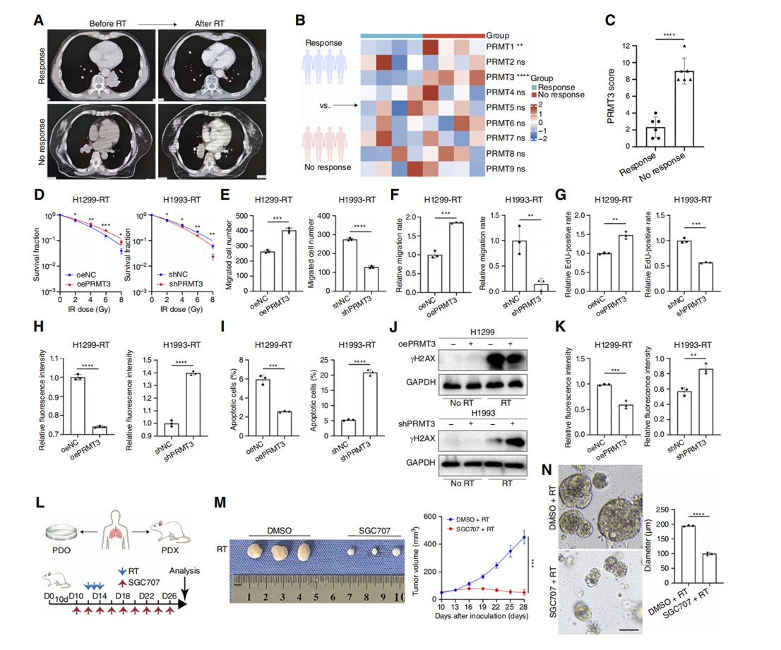
Figure 1. PRMT3 promotes radiotherapy resistance in NSCLC.
A, Tumor response to radiotherapy (RT) was classified into response and nonresponse groups based on changes in tumor size after treatment. B, The mRNA expression levels of nine PRMT family genes were quantified in both response and nonresponse groups using RT-PCR, with GAPDH serving as a loading control (n ? 4). NSCLC tissues were collected from patients prior to any treatment. C, PRMT3 expression was significantly elevated in the nonresponse group (n ? 6). D, Overexpression of PRMT3 enhanced the clonogenic survival of NSCLC cells after radiotherapy (0–8 Gy), whereas PRMT3 knockout reduced clonogenic survival (n ? 3). E–G, Overexpression of PRMT3 promoted the migration and proliferation of NSCLC cells after radiotherapy (4 Gy; n ? 3). H, Overexpression of PRMT3 inhibited the production of ROS in cells after radiotherapy (4 Gy; n ? 3). I, Knockdown of PRMT3 promoted apoptosis in NSCLC cells 24 hours after radiotherapy (6 Gy; n ? 3). J, NSCLC cells from the designated group were subjected to 6 Gy irradiation and subsequently cultured for 1 hour under standard conditions, followed by Western blot analysis using γH2AX antibodies. K, IF confocal microscopy was used to examine the regulatory effect of PRMT3 expression on γH2AX formation after radiotherapy (6 Gy; n ? 3). L, PDX tumors were treated with the PRMT3 inhibitor SGC707 (10 mg/kg, every 2 days via intraperitoneal injection) and/or radiotherapy (8Gy/day, three consecutive days). D, day; PDO, patient derived tumor organoid. M, Tumor growth curve was plotted (n ? 3 mice per group). N, Corresponding patient-derived tumor organoids were treated with SGC707 (10 μmol/L) and/or radiotherapy (8 Gy, once). Scale bar, 100 μm. Data represent the mean ± SD. Differences were tested using two-way ANOVA test (B) and unpaired two-sided Student t test (C–I, K, M, and N). ns, not significant; *, P < 0.05; **, P < 0.01; ***, P < 0.001; ****, P < 0.0001.
模块 2:代谢组证明 PRMT3 功能依赖色氨酸 - 犬尿氨酸通路
1. 研究逻辑:非靶向代谢组筛选 PRMT3 调控代谢物,色氨酸缺失培养基回补验证 Kyn 是功能必需介质。
2. 核心实验:4D-DIA 蛋白组、非靶向代谢组、Trp/Kyn ELISA、无 Trp 培养基细胞功能实验。
3. 关键结果:PRMT3 显著上调 Kyn;去除色氨酸后 PRMT3 无法诱导放疗抵抗。
4. 配套试剂:代谢组试剂盒、Trp/Kyn ELISA 试剂盒
5. 小结:PRMT3 通过上调犬尿氨酸代谢介导 NSCLC 放疗耐受。
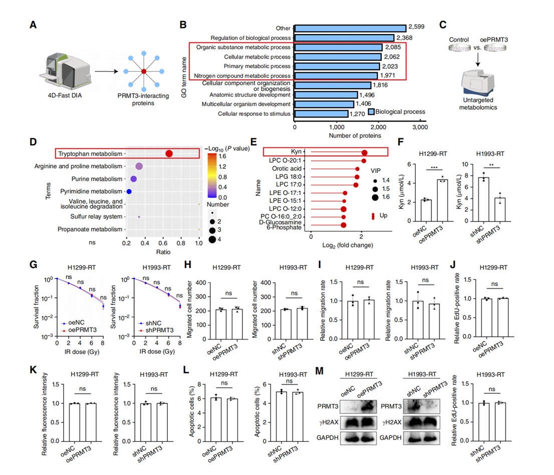
Figure 2. Kyn metabolism is a key mediator of PRMT3 function in NSCLC.
A, 4D-Fast DIA qualitative proteomics was utilized to identify PRMT3-interacting proteins from co-IP experiments. B, Gene Ontology (GO) enrichment analysis was performed on the identified PRMT3-interacting proteins. C, Nontargeted metabolomics analysis was conducted on specific NSCLC cell groups. D, Kyoto Encyclopedia of Genes and Genomes enrichment analysis was applied to the nontargeted metabolomics data. E, Elevated levels of Kyn were detected in PRMT3-overexpressing cells compared with controls. Up, upregulated. F, The Kyn concentration in the culture supernatant of specific NSCLC cells after radiotherapy (RT; 4 Gy) was measured to assess Kyn metabolism (n ? 3). G–M, The impact of PRMT3 on NSCLC cell clonogenic survival (0–8 Gy; n ? 3; G), migration (4 Gy; n ? 3; H), wound-healing (4 Gy; n ? 3; I), and proliferation (4 Gy; n ? 3; J) after radiotherapy was evaluated in Trp-depleted culture conditions. Additionally, ROS production (6 Gy; n ? 3; K), apoptotic cell ratio (6 Gy; n ? 3; L), and γH2AX expression (6 Gy; M) were also assessed in Trp-depleted NSCLC cells following radiotherapy. Data represent the mean ± SD. Differences were tested using unpaired two-sided Student t test (F–L). ns, not significant; **, P < 0.01; ***, P < 0.001.
模块 3:核心分子机制 ——PRMT3 甲基化 TFAP2A 激活 IDO1
1. 研究逻辑:通过 Absin abs955 Co-IP 捕获 PRMT3 互作蛋白 TFAP2A,逐层验证甲基化修饰调控 IDO1 转录完整通路。
2. 核心实验:Co-IP、质谱、ChIP、双荧光素酶、TFAP2A R363 突变、蛋白半衰期、核质分离、TFAP2A 二聚实验、PRMT3 酶活突变。
3. 关键结果:
1. 图 5C、5D:abs955 Co-IP 验证 PRMT3 与 TFAP2A 存在内源相互作用;
2. 图 5L、5O:abs955 富集产物 WB 证明 PRMT3 介导 TFAP2A 发生 ADMA 甲基化;
3. 图 6 全子图、图 7 全子图:甲基化修饰延长 TFAP2A 半衰期、促进核转位与二聚,R363 位点突变完全阻断调控效应。
4. 配套产品:Absin abs955 免疫共沉淀试剂盒、ChIP 试剂盒。
5. 小结:PRMT3 依赖 TFAP2A R363 甲基化促进 IDO1 转录,驱动 Kyn 大量生成。
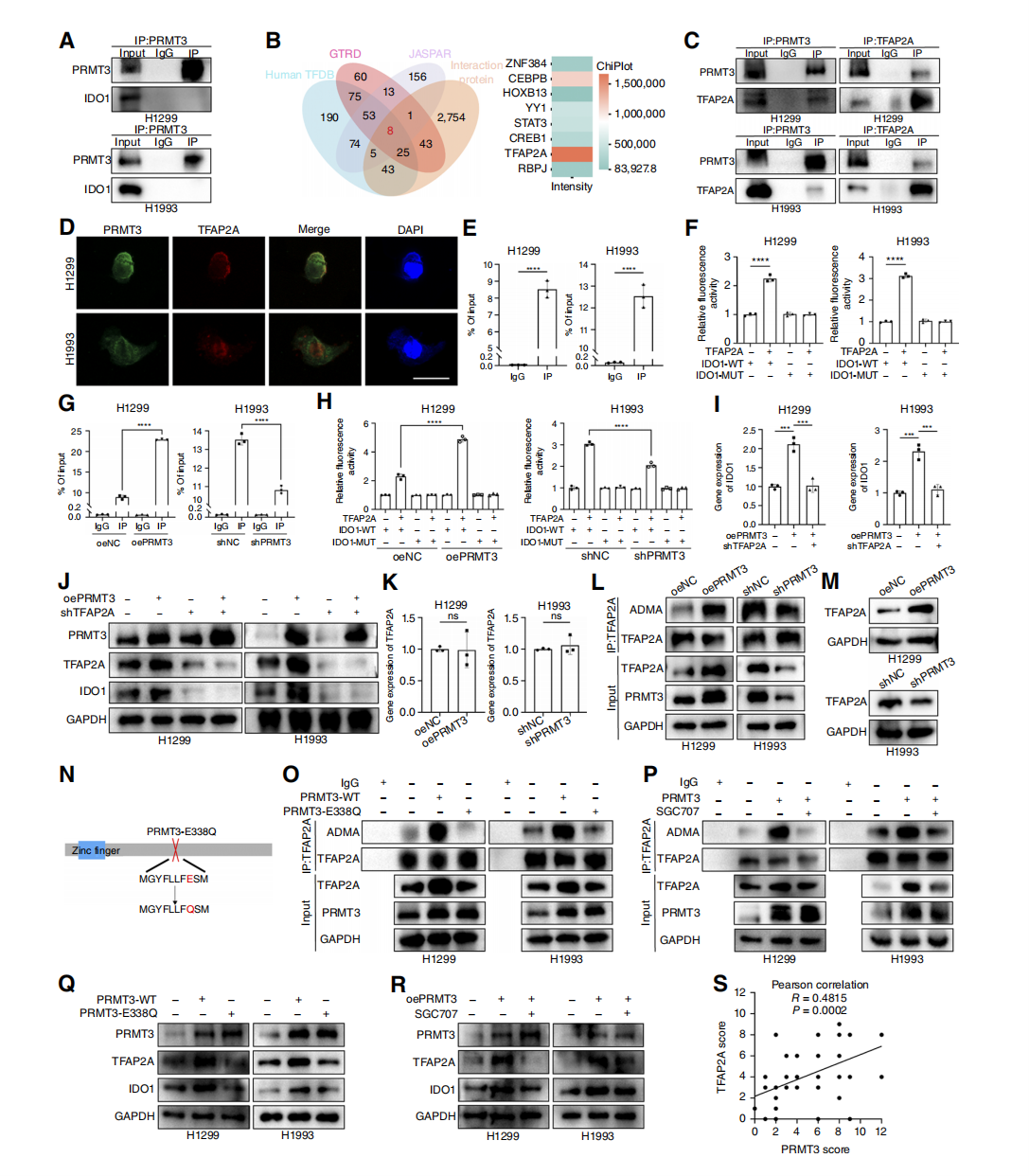
Figure 5. PRMT3 regulates IDO1 transcription through TFAP2A.
A, Co-IP experiments showed that PRMT3 does not directly interact with IDO1. B, Predicted transcription factors of IDO1 intersected with PRMT3-interacting proteins. C and D, PRMT3 interacts with TFAP2A (C), and they colocalize (D). Scale bar, 20 μm. E, ChIP assays validated the binding of TFAP2A to the IDO1 promoter (n ? 3). F, Luciferase assays indicated that TFAP2A is a transcription factor for IDO1 (n ? 3). G, PRMT3 overexpression promoted the binding of TFAP2A to the IDO1 promoter (n ? 3). H and I, PRMT3 knockdown inhibited TFAP2A binding to the IDO1 promoter, whereas TFAP2A knockdown rescued the PRMT3-mediated upregulation of IDO1 (n ? 3). J, Overexpression of PRMT3 did not increase IDO1 expression in the absence of TFAP2A. K, PRMT3 did not affect TFAP2A mRNA levels (n ? 3). L and M, Overexpression of PRMT3 promoted ADMA generation in TFAP2A (L), whereas PRMT3 influenced TFAP2A protein levels (M). N, Construction of PRMT3 enzyme inactivation mutant. O, Overexpression of PRMT3 with enzyme inactivation did not increase ADMA production in TFAP2A. P, Observation of the effect of treating cells with PRMT3 inhibitor on ADMA production in TFAP2A. Q, Western blot is used to detect the regulatory effect of enzyme inactivation mutants on TFAP2A protein levels. R, PRMT3 inhibitors can rescue the upregulation of TFAP2A protein levels caused by overexpression of PRMT3. S, There was a positive correlation between PRMT3 and TFAP2A expression in the NSCLC cohort. Data represent the mean ± SD. Differences were tested using unpaired two-sided Student t test (E and K) and one-way ANOVA test (F–I). S, The correlation was determined using Pearson correlation test. *, P < 0.05; **, P < 0.01; ***, P < 0.001; ****, P < 0.0001.
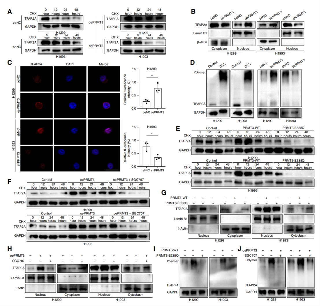
Figure 6. PRMT3-mediated methylation extends TFAP2A protein stability.
A and B, PRMT3 overexpression prolonged TFAP2A half-life and promoted nuclear uptake. C, IF revealed that PRMT3 regulates TFAP2A nuclear localization (n ? 3). Scale bar, 20 μm. D, Disuccinimidyl suberate (DSS) assays showed that PRMT3 knockdown inhibited TFAP2A dimerization. E–J, PRMT3 enzyme-inactivated mutants did not affect TFAP2A half-life (E), nuclear uptake (G), and dimer formation (I). The PRMT3 inhibitor SGC707 rescued the effects of PRMT3 overexpression on TFAP2A half-life (F), nuclear localization (H), and dimer formation (J). Data represent the mean ± SD. Differences were tested using unpaired two-sided Student t test (C). *, P < 0.05; **, P < 0.01. CHX, cycloheximide.
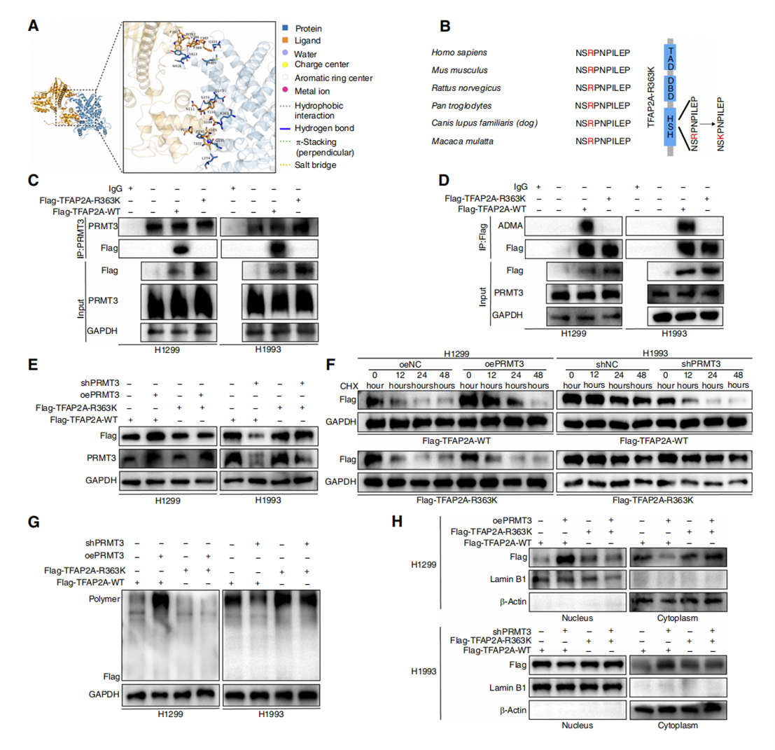
Figure 7. PRMT3 methylates TFAP2A at R363.
A, Molecular docking simulations were conducted to predict the potential methylation sites of TFAP2A. B, In mammals, the amino acid sequence surrounding the R363 site of TFAP2A is highly conserved. A mutant with the substitution R363K was constructed. DBD, DNA-binding domain; TAD, transactivation domain. C, PRMT3 was unable to directly interact with the mutant proteins. D–H, The mutant proteins were incapable of generating ADMA (D), and PRMT3 could not regulate their protein levels (E), half-life (F), dimerization (G), or nuclear localization (H). CHX, cycloheximide.
模块 4:Kyn-AHR 通路诱导 CD8?T 细胞耗竭,形成免疫抑制微环境
1. 研究逻辑:依托 Absin abs50028 多重荧光试剂盒原位观测肿瘤 CD8?T 浸润,体外共培养验证 Kyn-AHR 介导 T 细胞耗竭。
2. 核心实验:TCGA 免疫浸润分析、Absin 四色多重荧光 IHC、T 细胞 - 肿瘤共培养、流式检测耗竭标志物、IDO1 抑制剂回补。
3. 关键结果:
1. 图 4C、4D(多重荧光切片图):abs50028 试剂盒染色结果直观显示 PRMT3 高表达区域 CD8?T、Granzyme B 浸润显著减少;
2. 图 4E–L 流式与 qPCR 证明 Kyn 激活 T 细胞 AhR、上调 PD-1,抑制杀伤功能。
4. 配套产品:Absin abs50028 四色多重荧光免疫组化试剂盒、流式抗体。
5. 小结:PRMT3-IDO1-Kyn 轴激活 CD8?T 细胞 AhR 通路,诱导 T 细胞耗竭,构建免疫抑制微环境。
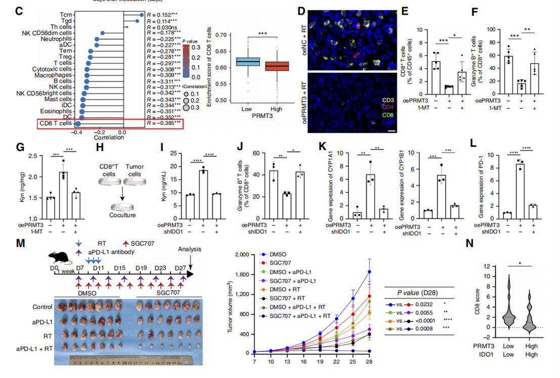
Figure 4. PRMT3 influences NSCLC by modulating the tumor-immune microenvironment.
A, A subcutaneous xenograft model was constructed in immunodeficient nude mice. Compared with immune-competent mice, PRMT3 overexpression did not exhibit the same tumor-promoting effects in the nude mice (n ? 7). RT, radiotherapy. B, Pan-cancer analysis from TCGA revealed that PRMT3 affects the tumor-immune microenvironment. C, High PRMT3 expression in patients with NSCLC was associated with reduced CD8+ T-cell infiltration. D, IF confirmed PRMT3’s inhibitory effect on CD8+ T cells. Scale bar, 20 μm. E and F, Flow cytometry analysis of mouse tumors was performed (n ? 5). G, ELISA demonstrated that PRMT3’s effect on Kyn metabolism could be rescued by 1-MT (n ? 3). H–J, Coculturing CD8+ T cells extracted from mouse spleen with LLC cells (H) showed that PRMT3-overexpressing tumor cells upregulated Kyn metabolism (I) and reduced granzyme B expression in CD8+ T cells (J). K and L, qRT-PCR analysis of CYP1A1, CYP1B1 (K), and PD-1 (L) expression in CD8+ T cells (n ? 3). M, PRMT3 inhibitor enhanced the efficacy of radiotherapy combined with immunotherapy in NSCLC (n ? 7). aPD-L1, anti–PD-L1; D, day. N, IHC was used to stain relevant markers in the NSCLC patient cohort and assess their correlation. Data represent the mean ± SD. Differences were tested using one-way ANOVA test (A and E–M), Wilcoxon rank-sum test (C), and unpaired two-sided Student t test (N). The correlation was determined using Spearman correlation test (B and C). *, P < 0.05; **, P < 0.01; ***, P < 0.001; ****, P < 0.0001.
模块 5:体内联合治疗与临床分层验证双靶点临床价值【对应:图 8 A、B、C、D、E、F、G】
1. 研究逻辑:多组荷瘤小鼠验证 PRMT3 抑制剂 + IDO 抑制剂联合放疗 / 免疫治疗的抑瘤效果;临床队列按 PRMT3/IDO1 表达分层分析放疗应答与生存。
2. 核心实验:LLC 皮下瘤、PDX 给药、肿瘤生长曲线、生存分析、临床样本 IHC 分层统计、CD8 清除对照。
3. 关键结果:双抑制剂联合放疗抑瘤效果最优;PRMT3high/IDO1high 患者放疗有效率低、生存期短。
4. 配套试剂:小动物放疗设备、免疫检查点中和抗体。
全文通路总结
肿瘤 PRMT3 上调→甲基化 TFAP2A(R363)→TFAP2A 稳定性 / 核转位 / 二聚增强→IDO1 转录升高→Kyn 大量蓄积: ① 肿瘤细胞内 Kyn 促进 DNA 损伤修复、抑制凋亡,产生放疗抵抗; ② Kyn 释放至微环境激活 CD8?T 细胞 AhR,上调 PD-1、降低颗粒酶 B,诱发 T 细胞耗竭; 联合抑制 PRMT3 与 IDO1 可同步逆转放疗耐药、重建抗肿瘤免疫。
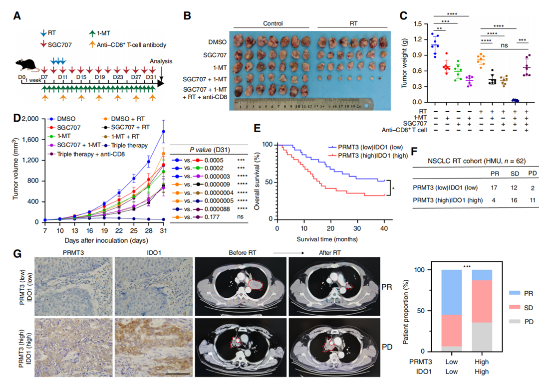
Figure 8. Targeting the PRMT3–IDO1 axis sensitizes radiotherapy in NSCLC.
A–D, Mouse xenograft models demonstrated that the combination of PRMT3 and IDO1 inhibitors enhanced the sensitization of radiotherapy (RT). Tumor growth, weight, and survival were compared among different groups (n ? 7). D, day. E, Patient survival data were stratified by different expression levels of PRMT3 and IDO1. F and G, The therapeutic efficacy was assessed in an NSCLC patient cohort. HMU, Harbin Medical University; SD, stable disease. Data represent the mean ± SD. Differences were tested using one-way ANOVA test (C and D), log-rank test (E), and Pearson χ2 test (F). ns, not significant; **, P < 0.01; ***, P < 0.001; ****, P < 0.0001.
四、爱必信(Absin)产品整体作用总结
本研究分子互作机制、肿瘤免疫原位成像两大核心创新结论完全依赖 Absin 两款核心试剂实现,整体科研价值分为四层:
1. 机制挖掘基础工具(abs955 Co-IP 试剂盒) 突破常规单蛋白检测局限,稳定捕获细胞内 PRMT3-TFAP2A 天然蛋白复合物,精准检测 TFAP2A 精氨酸甲基化修饰,直接锁定 IDO1 上游表观调控轴,搭建全文分子通路核心骨架;试剂盒低非特异性结合、蛋白完整保留特性,适配 4D-DIA 质谱联合实验,支撑翻译后修饰机制创新。
2. 肿瘤微环境原位可视化金标准(abs50028 四色多重荧光 IHC 试剂盒) 解决传统单标 IHC 无法同步观测 “肿瘤驱动蛋白 + 杀伤性 T 细胞” 空间共定位难题,TSA 信号放大技术实现低丰度免疫标志物清晰成像,为临床样本、动物模型提供直观原位证据,建立 PRMT3 表达与免疫抑制表型直接关联,支撑免疫微环境机制结论。
3. 适配高分肿瘤期刊标准化实验体系 两款产品操作流程标准化、实验可重复性高,完全匹配 Cancer Research 等顶级肿瘤学杂志实验质控要求,数据可信度满足临床转化类 SCI 发文标准,是本文通路创新性、临床转化价值论证的关键试剂支撑。
4. 临床转化研究支撑 依托 Absin 多重荧光试剂盒可对大批量 NSCLC 临床肿瘤切片进行多指标分层染色,快速完成 PRMT3/IDO1 双标志物预后分层验证,为 PRMT3-IDO1 联合靶点临床转化、患者分层治疗方案提供稳定检测技术平台。
综上,Absin 产品贯穿本文从 “蛋白互作分子机制” 到 “组织原位免疫表型” 的全链条关键实验,是解析 PRMT3 介导放疗抵抗与免疫抑制双重生物学效应不可或缺的核心科研工具。

- 手机:13761418683