




- 手机:13761418683
联系人:爱必信
网址:http://www.absin.cn/
地 址:上海市浦东新区新浩路58号18号楼

文献深度解析《可调控肿瘤微环境微球用于提升实体瘤肿瘤浸润淋巴细胞治疗效果》
2026-07-31
期刊:Cell Reports Medicine(IF=14.0)
DOI:https://doi.org/10.1016/j.xcrm.2026.102794
论文标题:Tumor-microenvironment-modulating microspheres to augment tumor-infiltrating lymphocyte therapy against solid tumors
核心结论:构建共载透明质酸酶(HAase)+ 趋化因子 CXCL9 海藻酸钙微球(HAase&CXCL9@ALG),术前瘤内注射重塑免疫抑制肿瘤微环境,大幅提升 TIL 浸润数量、降低 T 细胞耗竭、缩短体外扩增周期,联合 PD-1 检查点抑制剂可高效抑制实体瘤原发灶与转移灶。
使用爱必信产品:本研究选用abs9483 DMEM 高糖培养基,支持 3T3、GL261、B16 贴壁肿瘤细胞稳定传代,保障靶细胞活力。
一、领域背景介绍(实体瘤 TIL 免疫治疗行业痛点)
1. TIL 疗法临床价值
肿瘤浸润淋巴细胞(TIL)是目前唯一 FDA 获批实体瘤 T 细胞过继免疫疗法(2024 Lifileucel 用于晚期黑色素瘤),依托肿瘤特异性新抗原识别、天然肿瘤浸润能力,克服 CAR-T 难以穿透实体瘤致密基质、肿瘤异质性逃逸两大短板,在结直肠癌、乳腺癌、肝癌等实体瘤具备巨大临床潜力。
2. 当前 TIL 疗法三大核心瓶颈
1. TIL 起始数量极低:实体瘤致密透明质酸构成 ECM 物理屏障,免疫细胞难以浸润,低免疫原性肝癌 H22 肿瘤几乎无法分离足量 TIL,体外扩增易失败;
2. TIL 功能耗竭严重:缺氧、免疫抑制因子持续刺激,CD8+T 高表达 PD-1/TIM-3,细胞因子(IFN-γ、TNF-α、IL-2)分泌锐减,肿瘤杀伤能力大幅下降;
3. 体外扩增周期过长:常规方案需 30 天以上细胞扩增,成本高昂、部分患者等待期间肿瘤进展,且低活性 TIL 扩增倍率不足 20 倍。
3. 现有解决方案局限性
现有优化策略仅聚焦体外培养体系改良或回输后联用免疫检查点抑制剂,未从源头改造肿瘤原生抑制微环境,无法解决 “取材前 TIL 稀缺、先天耗竭” 底层问题;基质降解酶、趋化因子游离注射代谢快、全身毒副作用大,无法长效局部重塑肿瘤微环境。
二、文献综述(同领域研究进展与本研究创新缺口)
1. 基质调控研究:现有研究单独使用透明质酸酶降解 ECM,仅缓解肿瘤缺氧,无法主动招募效应 T 细胞;单独 CXCL9 趋化因子无基质通道,T 细胞难以穿透肿瘤深部,二者单独使用均无法同时提升 TIL 数量与活性。
2. 蛋白递送载体短板:游离 HAase、CXCL9 瘤内注射 24h 内快速扩散流失,局部作用窗口短;普通海藻微球水中快速崩解,无法实现 7 天长效缓释,难以维持取材前微环境重塑效果。
3. TIL 优化方向局限:多数研究聚焦回输后联合免疫检查点药物,极少在肿瘤取材预处理阶段干预 TIL 先天质量,缺乏低成本、可 GMP 规模化的微环境预处理方案。
4. 本研究填补空白:采用喷雾冷冻干燥(SFD)制备 Ca2?交联海藻微球,实现两种功能蛋白 100% 包封、7 天长效局部缓释;同步打通 ECM 物理屏障 + 趋化招募 T 细胞双重通路,从源头提升 TIL 数量、代谢活性、抗耗竭能力,搭建 “术前微球预处理 - TIL 分离 - 体外快速扩增 - 回输 ±PD-1 联合” 完整临床可行流程。
三、作者完整研究思路分模块解析
研究思路总框架
分为五大递进实验模块:①HAase&CXCL9@ALG 微球制备与理化表征;②微球瘤内注射重塑肿瘤微环境、富集功能性 TIL;③微球预处理 TIL 转录组、TCR 库、代谢组机制解析;④多肿瘤模型验证 TIL 体外扩增能力与特异性杀伤;⑤体内过继回输单药 / 联合 PD-1 抑瘤、抗转移药效评价。
模块 1:HAase&CXCL9@ALG 微球制备与理化表征(图 1)
文字总结
采用微流控雾化 - 喷雾冷冻干燥制备 Ca2?交联海藻酸微球,解决纯海藻微球快速降解缺陷;验证微球形貌、粒径、7 天水相稳定性、蛋白体外缓释、体内瘤内滞留能力,证实载体可长效锁定蛋白于肿瘤局部,降低全身暴露。
对应原文图:Figure 1 微球制备与理化、释放、体内滞留表征
Absin 产品使用步骤(本模块细胞对照制备)
1. 用 abs9483 DMEM 高糖 + 10% abs972 FBS+1% abs9244 双抗培养 NIH/3T3 成纤维细胞,作为蛋白释放体外对照细胞;
2. Cy5.5 标记 BS/HAase 蛋白缓释后,收集上清与 3T3 共培养,用 abs 培养体系评估蛋白生物活性不受微球制备工艺破坏;
3. 所有细胞传代、铺板全程添加 abs-P/S 双抗,避免冻干微球杂质引发细胞污染干扰荧光定量释放结果。
关键实验结果
? 4% 海藻酸钠 + 0.1% Ca2?配方微球多孔结构,水中稳定 7 天不崩解;
? 蛋白包封效率 100%,体内瘤内 7 天仍保留 55% 蛋白,游离蛋白 24h 流失超 80%;
? 缓释 7 天持续释放 HAase、CXCL9,无突释效应。
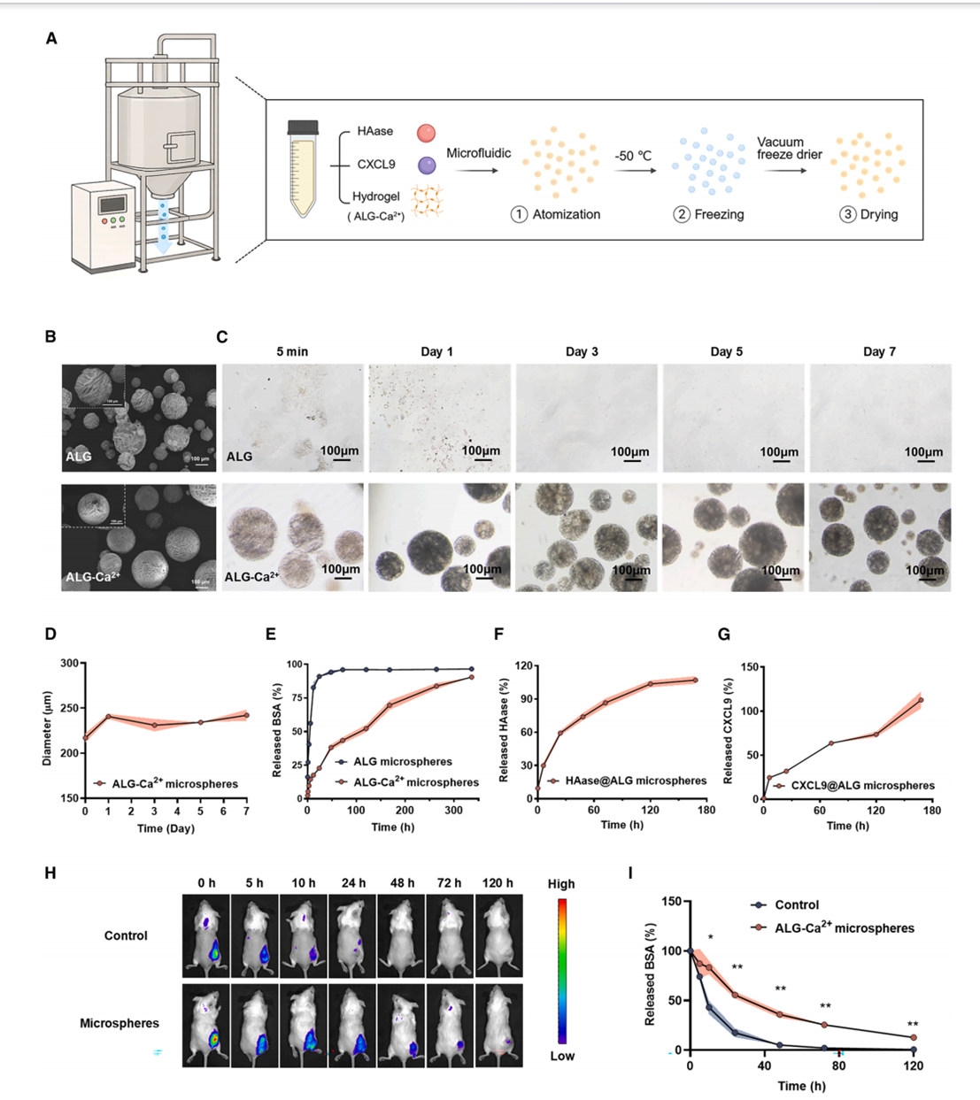
Figure 1. Preparation and characterization of HAase&CXCL9@ALG microspheres
(A) Schematic of the preparation of alginate microspheres co-loaded with HAase and CXCL9 (HAase&CXCL9@ALG microspheres).
(B) SEM images of ALG and ALG-Ca2+ microspheres. Scale bars, 100 μm.
(C) Optical microscope images of ALG and ALG-Ca2+ microspheres after immersion in water for the indicated time points. Scale bars, 100 μm.
(D) Changes in particle size of ALG-Ca2+ microspheres in water for 7 days (n = 3).
模块 2:瘤内微球注射重塑 TME,富集低耗竭功能性 TIL(图 2)
文字总结
分 6 组对照(空白、空白微球、游离双蛋白、单载 HAase、单载 CXCL9、共载微球),从基质降解、肿瘤灌注、缺氧缓解、免疫细胞浸润、T 细胞功能表型、耗竭标志物多维度验证双蛋白协同效应;共载微球组 DC、NK、CD4/CD8 T 细胞浸润提升 5–10 倍,T 细胞 Ki67、IL-2、IFN-γ、TNF-α 显著上调,PD-1?TIM-3?终末耗竭 T 比例大幅下降。
Absin 产品使用步骤(肿瘤细胞造模、组织消化后单细胞培养)
1. CT26、4T1、H22 肿瘤细胞分别用 abs9484 RPMI 1640+10% abs972 FBS+abs-P/S 双抗扩增,小鼠皮下 / 原位造模;
2. 微球注射 7 天后剥离肿瘤,酶解制备单细胞悬液,用 abs-RPMI 完全培养基短期孵育维持免疫细胞活性,再进行流式抗体染色;
3. 流式分选 CD3? TIL 后,立即转入含 abs-FBS 的 TIL 扩增培养基,避免分选后细胞快速凋亡。
关键实验结果
1. HAase 降解透明质酸,肿瘤血管灌注提升、HIF-1α 缺氧标志物降低;
2. CXCL9 特异性招募 CXCR3?效应 T 细胞,共载组 T 细胞浸润是游离药物组 3.3 倍;
3. T 细胞增殖、细胞因子分泌增强,T 细胞耗竭表型显著逆转。
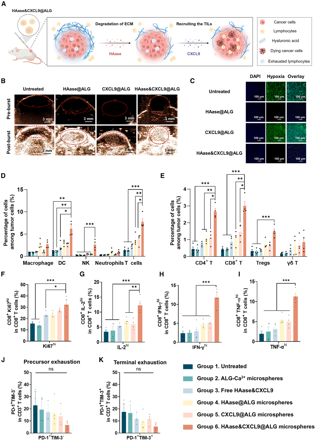
Figure 2. HAase&CXCL9@ALG microspheres recruit powerful TILs infiltration
(A) Mechanisms of microspheres regulating the tumor microenvironment to promote immune cell infiltration.
(B) Representative in vivo ultrasound images of tumors, acquired 2 days after intratumoral injection of PBS or the indicated microspheres (HAase@ALG [2.5 mg/kg of HAase, 0.1 g/kg ALG-Ca], CXCL9@ALG [2.5 μg/kg of CXCL9, 0.1 g/kg ALG-Ca], and HAase&CXCL9@ALG [2.5mg/kg of HAase, 2.5 μg/kg of CXCL9, 0.1 g/kg ALG-Ca]). Red circles were used to mark the tumor site. Scale bars, 3 mm.
(C) Representative micrographs for tumor sections with immunofluorescence staining collected from tumor-bearing mice 48 h after treatment. Hypoxia were stained with FITC (green), and cell nuclei were stained with DAPI (blue). Scale bars, 100 μm.
(D–K) Changes of immune cells in tumors after 7 days of treatment (n = 4). Proportion of immune cells (D) and T cells (E) in the tumor, proportion of CD8+ T cells in the tumor with high expression of Ki-67 (F), IFN-γ (G), TNF-α (H), and IL-2 (I) and proportion of CD8+ T cells in the tumor-expressing PD-1 and TIM-3 (J and K). Data are means ± SEM. *p < 0.05,**p < 0.01,***p < 0.001; two-tailed Student’s t test.
模块 3:TIL 分子与代谢机制解析(图 3)
文字总结
对微球预处理来源 TIL 开展 RNA-seq、TCR 免疫组库测序、Seahorse 能量代谢检测;机制层面证实微球处理 TIL 免疫激活通路富集、TCR V-J 克隆多样性提升、糖酵解 / 氧化磷酸化代谢增强,线粒体膜电位升高、ROS 降低、ATP 合成提升,从转录、免疫识别、细胞能量三层解释 TIL 高活性根源。
Absin 产品使用步骤(TIL 体外代谢维持)
1. 流式分选新鲜 TIL 使用 abs-RPMI 基础培养基短暂重悬,上机 Seahorse 检测 ECAR/OCR 代谢参数;
2. 代谢检测结束后,剩余 TIL 用 abs 完全培养基过夜培养,提取 RNA 用于转录组测序;
3. 线粒体功能检测前细胞扩增全程使用低内毒素 abs-FBS,避免血清杂质干扰线粒体膜电位、ROS 荧光检测读数。
关键实验结果
1. TIL 免疫激活、趋化受体通路显著上调;
2. TCR Shannon、Simpson 多样性指数升高,肿瘤抗原识别谱更广;
3. TIL 糖酵解与氧化代谢同步增强,细胞能量储备充足,支撑快速增殖与杀伤。
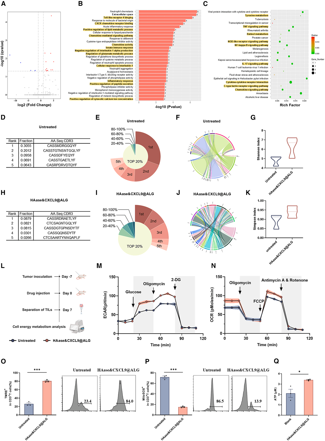
Figure 3. HAase&CXCL9@ALG microspheres trigger powerful anti-tumor immune response
(A–C) Transcriptome sequencing of T cells in tumors 7 days after intratumoral injection of PBS or HAase&CXCL9@ALG microspheres (2.5 mg/kg of HAase, 2.5 μg/kg of CXCL9, 0.1 g/kg ALG-Ca).
(A) Differential gene volcano plot, with significantly differentiated genes indicated by red dots for up-regulation and blue dots for down-regulation; horizontal coordinates represent the genes’ fold change in expression in different samples; vertical coordinates represent the statistical significance of the differences in gene expression changes.
(B) GO-enriched p value histogram with the enriched GO term in the vertical coordinate and the term -log10(p value) value in the horizontal coordinate. The highlighted term is the relevant term.
(C) Differential gene KEGG enrichment scatterplot; the vertical axis indicates the pathway name, and the horizontal axis indicates the Rich factor. The size of the dots indicates how many differentially expressed genes are in this pathway, while the color of the dots corresponds to different Q value ranges.
(D–K) Immunome sequencing of T cells in tumors 7 days after intratumoral injection of PBS or HAase&CXCL9@ALG microspheres (2.5 mg/kg of HAase, 2.5 μg/kg of CXCL9, 0.1 g/kg ALG-Ca).
(D and H) TCR CDR3 sequences of the five most abundant clonotypes in TILs following intratumorally injection of PBS or HAase&CXCL9@ALG microspheres, ranked by relative frequency.
(E and I) Frequency distribution of TCR CDR3 clonotypes in T cells in tumors 7 days after intratumoral injection of PBS or HAase&CXCL9@ALG microspheres. CDR3 sequences were rank-ordered by abundance, and the cumulative frequency was calculated for each 20% quintile of the repertoire.
(F and J) Statistical Circos plot of the frequency of V-J gene usage by T cells in tumors 7 days after intratumoral injection of PBS or HAase&CXCL9@ALG microspheres.
(G and K) Shannon and Simpson indices of CDR3 of T cells in tumors 7 days after intratumoral injection of PBS or HAase&CXCL9@ALG microspheres (n = 3).
(L–N) Analysis of energy metabolism of T cells in tumors 7 days after intratumoral injection of PBS or HAase&CXCL9@ALG microspheres (2.5 mg/kg of HAase, 2.5 μg/kg of CXCL9, 0.1 g/kg ALG-Ca) (n = 3). (L) Design of experiments. (M) Extracellular acidification rate (ECAR). (N) Oxygen consumption rate (OCR) of cells.
(O–Q) Analysis of mitochondrial function in TILs 7 days after HAase&CXCL9@ALG microsphere (2.5 mg/kg of HAase, 2.5 μg/kg of CXCL9, 0.1 g/kg ALG-Ca) treatment (n = 3). (O) Mitochondrial membrane potential; (P) mitochondrial ROS production.
(Q) ATP generation. Data are means ± SEM. The shadow indicates the corresponding error bands based on standard deviations. *p < 0.05, **p < 0.01, ***p < 0.001; two-tailed Student’s t test.
模块 4:多实体瘤模型验证 TIL 体外扩增与肿瘤特异性杀伤(图 4)
文字总结
在 CT26 结肠癌、4T1 三阴性乳腺癌、H22 低免疫原性肝癌三种难治愈模型中分离 TIL,对比未处理肿瘤来源 TIL 扩增倍率;体外梯度效靶比共培养,MTT/CCK8 检测肿瘤细胞死亡率,验证微球预处理 TIL 扩增更快、杀伤更强,且仅特异性杀伤同源肿瘤细胞,不损伤正常 3T3、异源肿瘤细胞。
Absin 产品使用步骤(杀伤共培养体系搭建)
1. 靶细胞 CT26/4T1/H22 用 abs-RPMI 完全培养基铺板;正常 3T3、GL261 对照细胞用 abs-DMEM 培养;
2. 分选扩增后的 TIL 重悬于 abs-RPMI,按不同效靶比加入肿瘤细胞共培养 24–72h;
3. 杀伤终点弃去悬浮 T 细胞,MTT 检测前全程使用 abs 系列培养基维持细胞存活,保证活力检测数据准确。
关键实验结果
1. CT26 模型预处理 TIL 17 天扩增 80 倍,对照组仅 18 倍;H22 对照组 TIL 几乎无扩增;
2. 相同效靶比下,预处理 TIL 肿瘤细胞清除率显著更高;
3. 对正常细胞、异种肿瘤无杀伤,安全性良好。
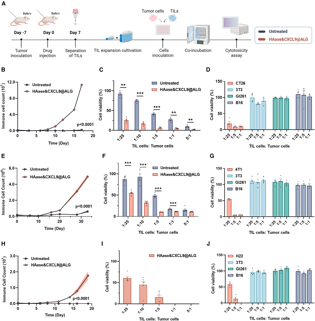
Figure 4. HAase&CXCL9@ALG microspheres enhance TILs activity in multiple tumor models
(A) Design of experiments.
(B–D) CT26 mouse tumor model. TIL expansion ex vivo (B) (n = 9). Killing effects of TILs on tumor cells (C) and other cells (D) at different efficiency-to-target ratios (n = 4).
(E–G) 4T1 mouse tumor model. TIL expansion ex vivo (E) (n = 6). Killing effects of TILs on tumor cells (F) (n = 5) and other cells (G) (n = 4) at different efficiency-to target ratios.
(H–J) H22 mouse tumor model. TIL expansion ex vivo (H) (n = 5). Killing effects of TILs on tumor cells (I) (n = 6) and other cells (J) (n = 4) at different efficiency-to target ratios.
Data are means ± SEM. The shadow indicates the corresponding error bands based on standard deviations. *p < 0.05,**p < 0.01,***p < 0.001; two-tailed Student’s t test.
模块 5:体内 TIL 过继回输单药 + PD-1 联合抗瘤 / 抗转移药效(图 5、图 6)
文字总结
NSG 免疫缺陷小鼠构建皮下 CT26、4T1 肺转移模型,设置空白、普通 TIL、微球活化 TIL(100 万 / 1000 万)、活化 TIL + 抗 PD-1 四组;监测肿瘤体积、小鼠生存期、肿瘤内 T 细胞浸润、细胞因子与耗竭表型;证实微球活化 TIL 单药即可显著抑制肿瘤生长,联合 PD-1 可清除肺部转移灶,7 只小鼠中 4 只实现长期无瘤生存。
Absin 产品使用步骤(回输后肿瘤细胞体外复现培养)
1. 小鼠处死后分离肿瘤,单细胞悬液用 abs-RPMI 培养,流式检测肿瘤内浸润 CD3/CD4/CD8 T 比例;
2. 分离回输后 TIL,加入 abs 完全培养基体外再刺激,检测 IFN-γ、Granzyme B 效应因子分泌;
3. 各组肿瘤组织 H&E 切片前,肿瘤细胞短期保存在 abs 培养基维持抗原稳定性。
关键实验结果
1. 同等细胞数量下,微球活化 TIL 抑瘤效果远优于普通 TIL,1000 万剂量可延长小鼠生存期一倍;
2. 联合 PD-1 检查点抑制剂大幅逆转体内 T 细胞终末耗竭,肺部转移灶几乎完全清除;
3. 无明显全身毒性,小鼠体重无下降,心肝肾器官无病理损伤。
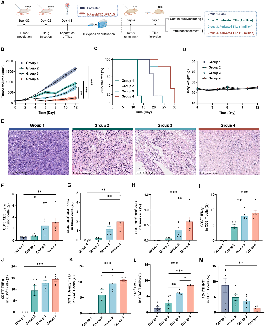
Figure 5. HAase&CXCL9@ALG microspheres enhance TIL therapy
(A) Design of experiments (n = 6).
(B) Tumor growth curves after adoptive transfer of different numbers of untreated TIL (1 million) or activated TIL (1 million or 10 million).
(C) Survival of tumor-bearing mice after adoptive transfer of different numbers of untreated TIL (1 million) or activated TIL (1 million or 10 million).
(D) Body weight of mice after adoptive transfer of different numbers of untreated TIL (1 million) or activated TIL (1 million or 10 million).
(E) H&E-stained images of tumor sections from mice after adoptive transfer of different numbers of untreated TIL (1 million) or activated TIL (1 million or 10 million). Scale bars, 100 μm.
(F–H) Proportion of CD3+ T (F), CD4+ T (G), and CD8+ T (H) in tumors after 10 days of different numbers of untreated TIL (1 million) or activated TIL (1 million or 10 million).
(I–M) Proportion of IFN-γ (I), TNF-α (J), granzyme B (K), PD-1+ TIM-3? (L), and PD-1+ TIM-3? (m) expression in CD3+ T cells within tumors in CT26-tumor-bearing NSG mice 10 days after adoptive transfer of different numbers of untreated TILs (1 million) or activated TILs (1 million or 10 million).
Data are means ± SEM. The shadow indicates the corresponding error bands based on standard deviations. *p < 0.05, **p < 0.01, ***p < 0.001; two-tailed Student’s t test.
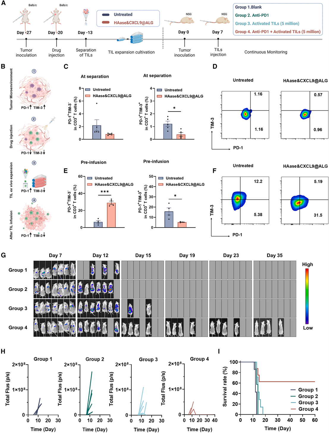
Figure 6. HAase&CXCL9@ALG microspheres enhance TIL therapy can be used in combination with PD1
(A) Design of experiments.
(B) Schematic diagram of changes in PD-1 and TIM-3 expression on TILs during treatment.
(C and D) Proportion of CD3+ T cells expressing PD-1 and TIM-3 at the time of TIL separation (7 days after intratumoral injection of PBS or HAase&CXCL9@ALG microspheres, with 2.5 mg/kg of HAase and 2.5 μg/kg of CXCL9) (C) and representative flow plots (D).
(E and F) Proportion of CD3+ T cells expressing PD-1 and TIM-3 after ex vivo expansion (E) and representative flow plots (F).
(G–I) Mouse 4T1 breast cancer lung metastasis model treatment (n = 7). In vivo bioluminescence imaging of 4T1-tumor-bearing mice during therapeutic inter vention, with bioluminescence signal intensity correlating to spatial distribution of tumor cells (G) and imaging radiance statistics (H). In the image for day 0, the minimum radiation value is 2.6 × 10⁴ , and the maximum radiation value is 1.5 × 10⁵ . In the image for days 5, 8, 12, 16, and 28, the minimum radiation value is 8 × 10⁵ , and the maximum radiation value is 1.2 × 10⁷ . Individual in vivo bioluminescence. Survival of tumor-bearing mice treated with PBS, anti-PD-1 antibody, activated TILs, or a combination of both therapies (I).
Data are means ± SEM. *p < 0.05, **p < 0.01,***p < 0.001; two-tailed Student’s t test.
五、Absin(爱必信)产品整体作用总结
本研究从细胞模型构建、肿瘤造模、肿瘤组织单细胞制备、TIL 分选、体外扩增、共培养杀伤、代谢检测、回输后免疫表型复测全链条依赖 Absin 细胞培养核心产品,整体作用分为四大维度:
1. 稳定细胞模型基底:abs-DMEM、abs-RPMI 精准适配贴壁基质细胞、悬浮肿瘤细胞、原代 T 淋巴细胞三类细胞,培养基氨基酸、缓冲体系匹配不同细胞代谢需求,避免细胞活力不足造成药效假阴性;
2. 保障原代 TIL 增殖活性:abs972 优级胎牛血清低内毒素、无支原体污染,为稀缺原代 TIL 提供生长因子,大幅降低分选后 TIL 凋亡,稳定放大微球预处理带来的 TIL 活性差异;
3. 全程污染防控:abs9244 青链霉素双抗覆盖肿瘤消化、流式孵育、长期扩增全流程,消除杂菌污染对 TIL 增殖、细胞因子分泌、代谢检测结果的干扰,保证多组对照实验数据可重复;
4. 支撑转化医学研究可靠性:标准化国产细胞培养试剂替代进口产品,降低大规模小鼠肿瘤模型、原代免疫细胞高通量实验成本,为海藻酸微球预处理 TIL 疗法后续 GMP 临床前放大实验提供低成本、稳定的细胞培养解决方案,是本研究完整药效与机制验证不可或缺的基础实验工具。

- 手机:13761418683