




- 手机:13761418683
联系人:爱必信
网址:http://www.absin.cn/
地 址:上海市浦东新区新浩路58号18号楼

- 期刊:Advanced Science(Wiley 顶级综合交叉期刊,2026 最新 IF=14.1,中科院一区 TOP,综合 Q1 分区)
- DOI:10.1002/advs.202520963
- 正式标题:CD28-Targeted Enzyme-Responsive Conformation-Switching Peptide Self-Assembly for Selective T-Cell Acute Lymphoblastic Leukemia (T-ALL) Therapy
-
发表时间:2026 年 3 月接收、在线刊发
- 靶点创新:首次提出 CD28 作为 T-ALL 高特异性治疗靶点,规避 CD3/CD5/CD7 靶向药物杀伤正常 T 细胞的脱靶毒性;T-ALL 细胞 CD28 异常高表达,正常 T 细胞在肿瘤微环境 CD28 表达耗竭,天然存在靶向窗口。
- 多肽分子设计:模块化磷酸化多肽 SAp-CD28,三功能结构:CD28 靶向序列 SPMLVAYD、D 型氨基酸磷酸化自组装基序 G?F?F?pY、NBD 荧光示踪基团;静脉给药水溶性优异。
- 酶促构象转换机制:肿瘤高表达碱性磷酸酶(ALP)介导多肽去磷酸化,实现α- 螺旋→β- 折叠 /β- 转角构象重排,CD28 结合亲和力提升 36 倍;原位形成纳米寡聚纤维。
- 三重细胞杀伤通路 ① 抑制 PI3K-Akt 存活通路:总 Akt 下调>54%,诱发氧化应激、ROS 蓄积; ② 阻断 PLCγ 钙信号通路:总 PLCγ 下降 48%,磷酸化 PLCγ 比例下降 80%,胞内钙超载,激活钙蛋白酶 calpain,细胞骨架崩解; ③ 纳米纤维入核物理破坏:多肽内吞逃逸溶酶后入核,持续组装长纤维撕裂核膜,诱导坏死性细胞死亡。
- 体内药效:Jurkat 异种移植瘤模型,SAp-CD28 单药抑瘤率 78.09%;与阿糖胞苷联用抑瘤率 92.9%,实现肿瘤近乎消退;小鼠脏器无病理损伤,生物安全性优异。
- 通用性:该酶响应构象自组装多肽策略可拓展至其他 CD28 高表达血液肿瘤。
- 特异性识别 CST 来源兔源一抗(总 Akt、p-Akt、总 PLCγ1、p-PLCγ1),精准结合一抗恒定区;
- 偶联 HRP 辣根过氧化物酶,通过化学发光显影清晰输出蛋白条带(对应原文图 5A WB 条带图);
- 信噪比高、非特异性背景极低,实现总蛋白与磷酸化蛋白定量差异可视化,直接证明 SAp-CD28 对两条 CD28 下游关键通路的抑制作用;
- 配套灰度定量支撑统计学分析,为转录组差异基因提供蛋白水平金标准验证。
-
T-ALL 临床治疗痛点
T 细胞急性淋巴细胞白血病(T-ALL)是高侵袭血液恶性肿瘤,占儿童 ALL 15%、成人 ALL 25%;成人患者复发率高、化疗耐药、预后极差。现有靶向靶点存在固有缺陷:
- CD3 靶向:正常 T 细胞同步清除,T 细胞自相残杀;
- CD5/CD7 靶向:对正常免疫细胞强毒性,免疫抑制严重;
- 缺乏仅在白血病细胞特异性高表达、正常 T 细胞低表达的安全靶点,临床亟需全新靶向体系。
-
CD28 靶点研究空白
CD28 是 T 细胞活化核心共刺激受体;现有免疫治疗多靶向 PD-1/CTLA-4,极少开发 CD28 靶向药物。已有临床样本证实 T-ALL 细胞 CD28 异常过表达,肿瘤微环境内正常 T 细胞 CD28 表达耗竭,存在天然靶向差异窗口,但无 CD28 特异性纳米多肽药物开发报道。
-
酶原位自组装多肽技术现状
酶指导多肽胞内自组装是近年纳米药物热点,可通过肿瘤特异性酶触发原位纳米结构生成,提升选择性杀伤;但既往多肽缺少特异性肿瘤细胞受体靶向模块,无法精准富集 T-ALL,限制体内疗效。
-
现有研究双重局限,引出本文创新切入点
现有 T-ALL 药物脱靶毒性大、CD28 靶点未被多肽纳米药开发、酶自组装多肽缺乏肿瘤特异性靶向;本文同时解决靶点特异性、多肽酶响应组装、多重细胞毒性机制三大瓶颈,构建 CD28 靶向酶构象开关多肽纳米治疗新策略。
- 靶点筛选:文献 + 细胞流式证实 Jurkat(T-ALL)高 CD28、高 ALP,正常 CD3+T / 肝细胞低表达,锁定 CD28 为特异性靶点;
- 多肽分子设计 + 对照构建:主分子 SAp-CD28,同步设计 3 组缺失型对照(SA-CD28p、SAp、CD28p)排除单一组分干扰;引入乱序肽 SAp-CD28-Scr 验证靶向序列必要性;
- 体外理化表征:TEM、FTIR、圆二色谱 CD、MST 亲和力实验,证明 ALP 诱导去磷酸化→α→β 构象转换、CD28 结合大幅提升;
- 细胞层面功能验证:共聚焦观察细胞摄取、内吞途径、溶酶逃逸、核损伤;MTT 特异性毒性;流式 Annexin V 明确坏死为主死亡方式;钙荧光、鬼笔环肽染色验证钙超载与骨架破坏;
- 组学初筛机制:RNA-seq 转录组差异基因 + KEGG 富集,锁定 PI3K-Akt、PLCγ 钙信号两条核心失调通路;
- 蛋白水平通路验证(核心 Absin 试剂应用环节):Western Blot 检测总 / 磷酸化 Akt、PLCγ1,定量验证通路抑制,从蛋白层面解释细胞坏死机制;
- 体内动物药效:Jurkat 裸鼠移植瘤,单药 / 阿糖胞苷联合给药,监测肿瘤体积、体重、血清氧化标志物 8-OHdG、肿瘤 Ki67 增殖、脏器病理;
- 机制汇总与策略拓展:整合信号通路紊乱 + 胞内纳米纤维物理破核双重杀伤机制,提出可通用的 CD28 靶向酶响应多肽设计范式。
- 未酶解前所有多肽水溶性好,无丁达尔效应;ALP 处理仅 SAp-CD28 形成稳定纳米纤维凝胶;
- TEM:SAp-CD28 酶解后形成 4.15nm 交织纳米纤维;
- FTIR + 圆二色谱 CD:ALP 处理后 α 螺旋含量下降 96%,β 折叠 80.3%、β 转角 17.9%;
- MST 亲和力:酶解后 SAp-CD28 KD 由 293.8μM 降至 8.1μM,结合提升 36 倍,对照多肽亲和力无明显改善。
- 图 1:多肽结构与杀伤机制示意图;
- 图 2:丁达尔、HPLC 去磷酸化动力学、TEM 电镜、FTIR 红外、CD 圆二色谱、MST 结合曲线。
- SAp-CD28 特异性富集 Jurkat,可逃逸溶酶入核,造成染色质浓缩、核膜破裂;对照多肽几乎无胞内摄取;
- 摄取依赖微管、动力蛋白、巨胞饮通路;
- 16μM SAp-CD28 处理 Jurkat 细胞活力仅 20%,正常 T / 肝细胞活力>85%,选择性极佳;
- 细胞死亡以坏死为主(78.03% 坏死细胞)。
- 总 Akt 蛋白下调>54%,p-Akt 活化水平显著降低,PI3K-Akt 存活通路阻断;
- 总 PLCγ1 下降 48%,磷酸化 PLCγ/ 总 PLCγ 比值下降 80%,经典钙动员通路被抑制;
- 胞内钙离子荧光强度提升 1.4 倍,F - 肌动蛋白骨架碎片化,证实钙蛋白酶介导骨架崩溃。
- SAp-CD28 单药抑瘤 78.09%,联用组抑瘤 92.9%,肿瘤体积小幅消退;
- 各组小鼠体重无显著下降,无系统毒性;
- 联用组肿瘤坏死区域>50%,Ki67 增殖标记降至 20%;主要脏器无组织病变。
- 数据可靠性保障:适配商用兔源磷酸化 / 总蛋白一抗,特异性强、非特异杂带少,WB 条带清晰可定量,保证 Akt、PLCγ 通路抑制结论的统计学可信度;
- 机制逻辑串联关键:承接转录组高通量筛选结果,从分子蛋白层面解释多肽诱导氧化应激、钙超载、细胞坏死的上游信号紊乱,搭建 “多肽结合 CD28→通路抑制→多重细胞死亡” 完整分子机制链条;
- 文章顶级期刊发表基础:Advanced Science对蛋白免疫印迹数据成像质量、定量重复性要求严苛,Absin 二抗稳定发光、批间差异小,满足顶刊图片标准化要求;
- 通用性科研工具价值:该 Absin 二抗可广泛用于肿瘤信号通路(PI3K/Akt、PLC / 钙信号)WB 验证,为血液肿瘤、多肽纳米药物、靶向小分子机制研究提供标准化免疫学检测方案。
文献深度解析《CD28-Targeted Enzyme-Responsive Conformation-Switching Peptide Self-Assembly for Selective T-ALL Therapy》
2026-08-03
本研究Western Blot 关键通路验证实验全程使用 Absin HRP 标记二抗 abs20039、abs20040,是解析 Akt、PLCγ 磷酸化信号通路的核心试剂:
本研究遵循靶点挖掘→分子模块化设计→体外理化表征→细胞水平靶向 / 毒性 / 机制验证→转录组高通量通路筛选→蛋白 WB 通路确证(Absin 二抗支撑)→体内异种移植药效 + 安全性评价→联合化疗增效完整闭环思路:
1.实验思路 构建三功能多肽 SAp-CD28 及三组对照,验证磷酸化位点、靶向序列、组装基序各自功能;通过 ALP 酶处理模拟肿瘤微环境,多手段检测溶解度、组装形貌、二级结构、CD28 结合亲和力。
2.实验结果
3.对应原图
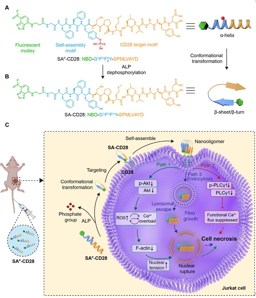 FIGURE 1
Schematic of peptide molecular design and its tumor-killing mechanism. (A) Chemical structure and secondary conformation of SApCD28. (B) Chemical structure and secondary conformation of SA-CD28, generated via alkaline phosphatase-mediated dephosphorylation of SAp-CD28. (C) Schematic illustration of SAp-CD28 inducing nuclear rupture through dysregulation of CD28 downstream signaling and physical damage, and its synergy with the chemotherapeutic agent cytarabine in cancer treatment.
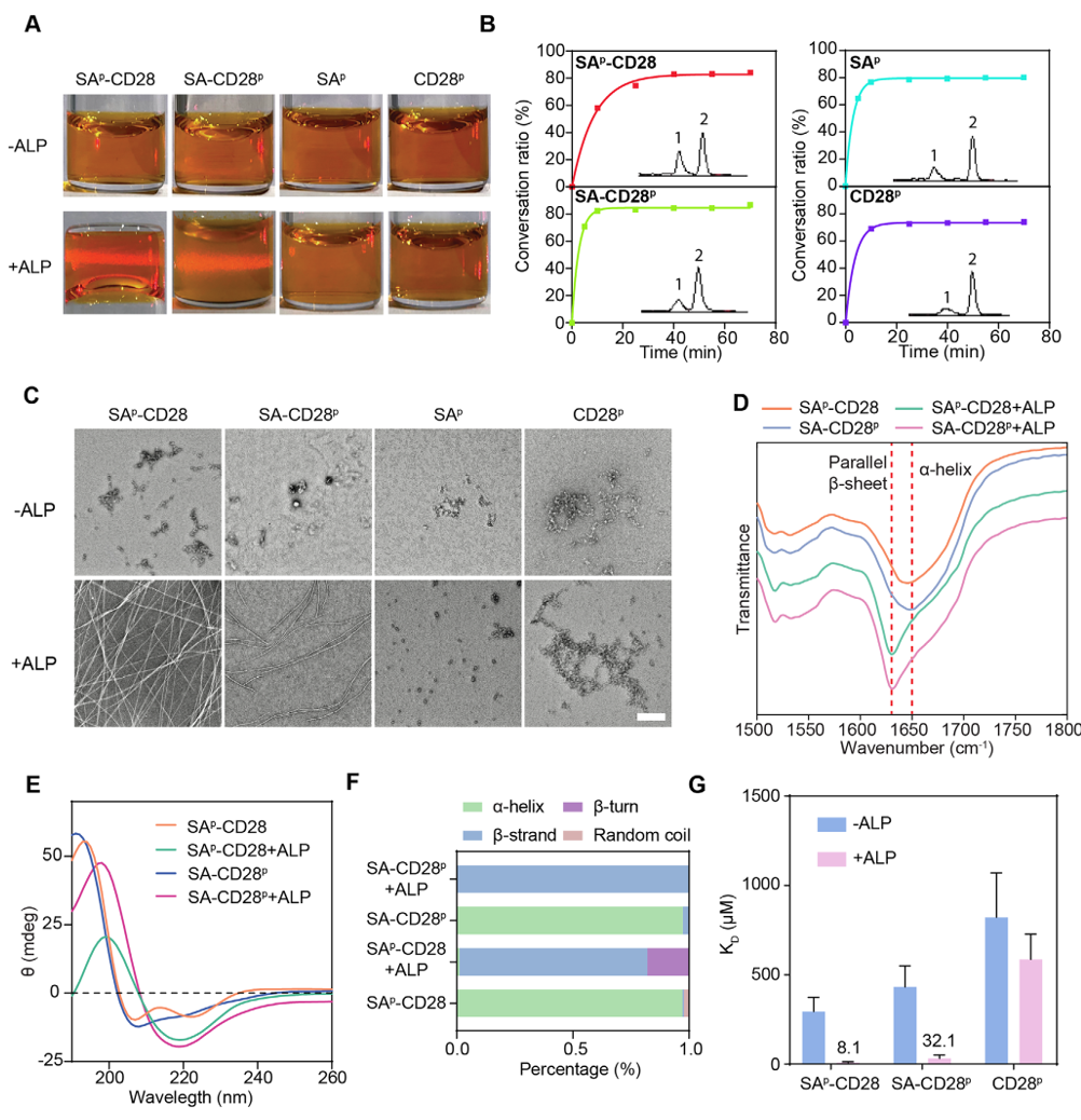 FIGURE 2
Characterization of the four peptide molecules before and after ALP dephosphorylation reactions. (A) Tyndall effects of the four peptides after incubation without ALP or with ALP for 12 h. (B) Conversion rates of the four peptides incubated with ALP (10 U/mL) over time. The HPLC chromatogram (inset images) shows two peaks, corresponding to the peptides before (peak 1) and after ALP dephosphorylation products (peak 2). (C) Transmission electron microscopy (TEM) images of the indicated peptides incubated without ALP or with ALP for 12 h. Scale bar: 200 nm. (D) FTIR spectra of SAp-CD28 and SA-CD28p in the presence or absence of ALP (10 U/mL). The dashed lines represent parallel β-sheets and α-helices, respectively. (E) CD spectrum of SAp-CD28 and SA-CD28p in the presence or absence of ALP (10 U/mL). (F) Secondary structure calculation of SApCD28 and SA-CD28p in the presence or absence of ALP (10 U/mL). (G) Microscale thermophoresis (MST) analysis of SAp-CD28, SA-CD28p and CD28p binding to the recombinant human CD28 proteins before and after ALP dephosphorylation reactions. KD values represent apparent binding affinities calculated from a 16-point titration. Data represent mean ± SD from three independent biological replicates (n = 3).
1.实验思路 以 Jurkat T-ALL 细胞为模型,正常 CD3+T、LO2 肝细胞做安全对照;共聚焦观察细胞摄取、核碎裂;多种内吞抑制剂明确摄取通路;MTT 定量细胞活力;流式区分凋亡 / 坏死比例;Bio 电镜观察核膜超微结构损伤。
2.实验结果
3.对应原图:图 3 共聚焦核染色、内吞抑制剂流式、细胞超微电镜、MTT 活力、Annexin V 流式坏死定量。
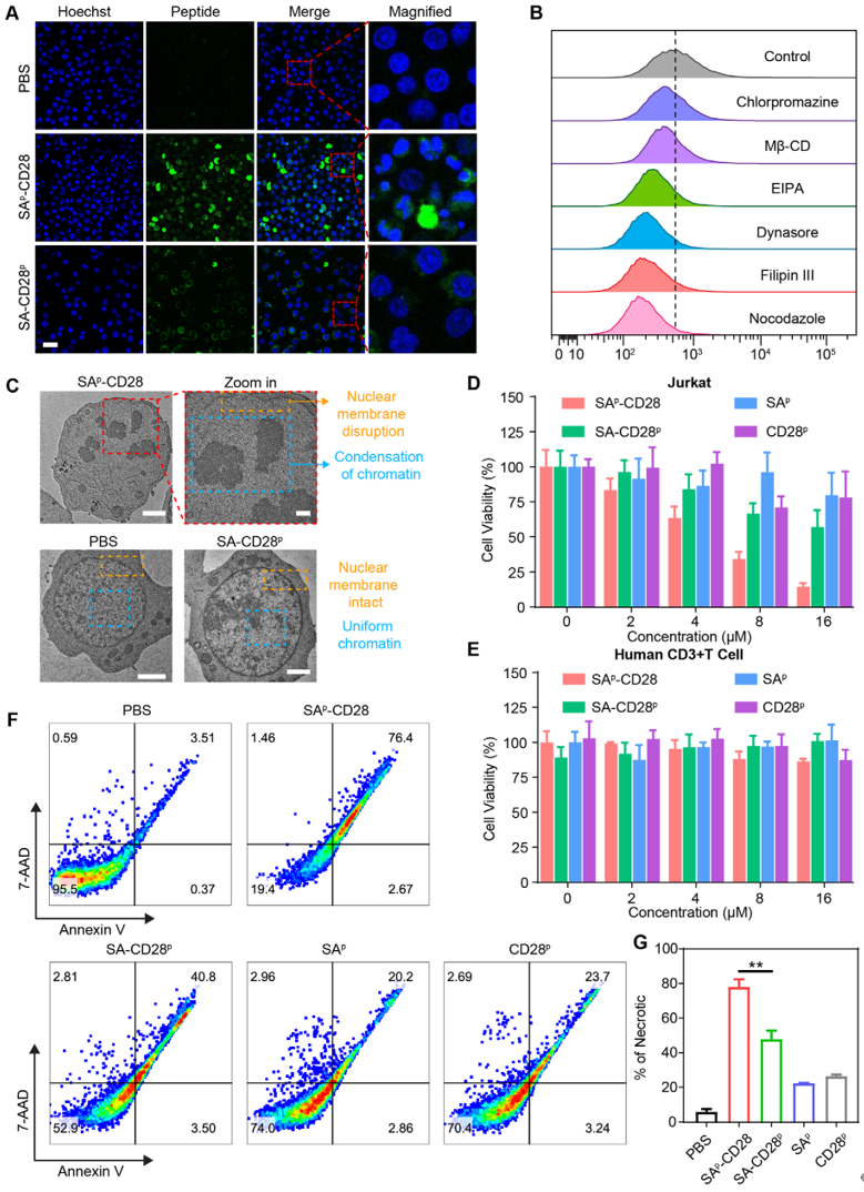 FIGURE 3
Nuclear damage and apoptosis triggered by peptide self-assembly at the cellular level. (A) Confocal laser scanning microscopy (CLSM) images of Jurkat cells treated with SAp-CD28 or SA-CD28p. Scale bar: 25 μm. (B) Flow chart of fluorescence intensity of Jurkat cells pretreated with different internalization inhibitors, then incubated with SAp-CD28 for 1 h. (C) Biological electron microscopic (Bio-EM) imaging of Jurkat cells after the indicated treatments for 8 h. Scale bar: 500 nm. (D, E) Relative cell viabilities of (D) Jurkat cells and (E) human CD3+ T cells incubated with different concentrations of the four peptides for 48 h (n = 4). (F) Annexin V-PE/7-AAD apoptosis detection of Jurkat cells after various treatments for 12 h. (G) Quantitative analysis of apoptosis flow cytometry results. Data are presented as mean ± SD (n = 3 independent experiments). Statistical analysis was performed using one-way ANOVA. **p < 0.01.
1.实验思路 PBS、SA-CD28p、SAp-CD28 三组细胞 RNA 测序,差异基因筛选、KEGG 通路富集,定位钙信号、PI3K-Akt、细胞骨架相关失调通路,为 WB 验证提供候选靶点。
2.实验结果 SAp-CD28 处理产生 278 个差异基因,显著富集 PI3K-Akt、钙信号、局灶黏附(细胞骨架)通路,提示存活通路抑制与钙稳态失衡。
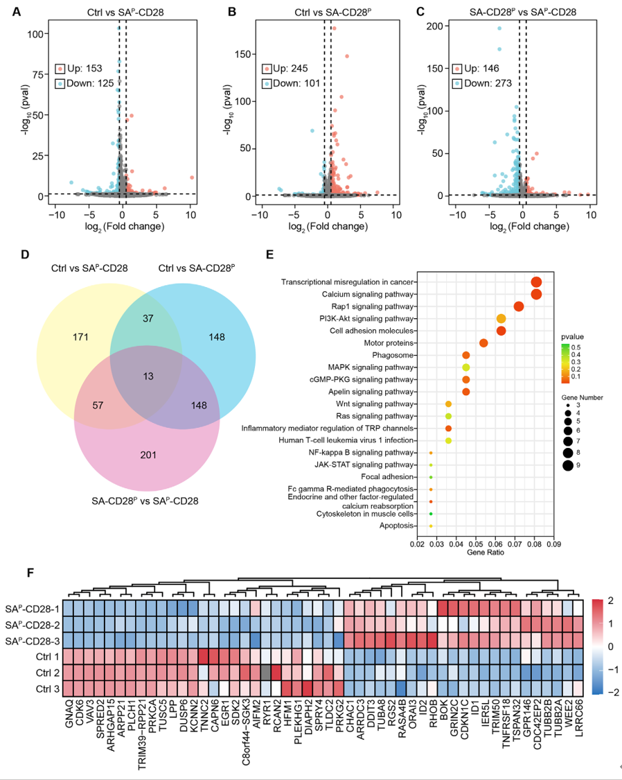 FIGURE 4
Transcriptomic profiling of cell apoptosis induced by peptides. (A–C) Volcano plot reveals gene regulation in Jurkat cells: Control vs SAp-CD28 (A), Control vs SAp-CD28 (B), and SA-CD28p vs SAp-CD28 (C). (D) Venn diagram illustrating common and unique deregulated genes in Jurkat cells treated by SAp-CD28 and SA-CD28p compared with PBS control. (E) KEGG pathway enrichment analysis of the differential pathways in Jurkat cells treated with SAp-CD28 versus PBS Control. (F) The heatmap of typical genes associated with apoptosis and nuclear damage in Jurkat cells after SAp-CD28 treatment in comparison with PBS control.
1.实验思路 针对转录组富集的 Akt、PLCγ 核心通路,使用特异性一抗检测总蛋白与磷酸化活化蛋白;采用 Absin HRP 二抗孵育显影,灰度定量,明确 SAp-CD28 对两条促存活通路的抑制作用;配套钙荧光、细胞骨架染色验证下游钙超载与骨架崩解。
2.实验操作含 Absin 产品关键步骤 蛋白电泳转膜后,封闭过夜;孵育兔源一抗(Akt、p-Akt、PLCγ1、p-PLCγ1);洗膜后稀释 Absin abs20039、abs20040 HRP 标记羊抗兔二抗室温孵育 1h;TBST 充分洗涤,HRP 化学发光成像采集条带,ImageJ 定量灰度值。
3.实验结果
Absin 产品关键价值:本模块唯一免疫学检测试剂,直接实现通路蛋白定量可视化,完成转录组数据蛋白水平验证,支撑全文核心杀伤机制推导。
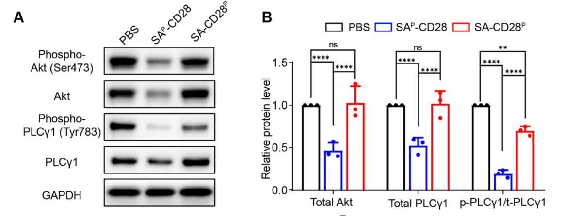 FIGURE 5
SAp-CD28 disrupts CD28 downstream signaling and induces calcium overload and cytoskeletal collapse. (A, B) Representative Western Blot bands (A) and quantitative analysis (B) of phospho-Akt, Akt, phospho-PLCγ, and PLCγ in Jurkat cells treated with SAp-CD28 or SA-CD28p versus PBS controls.
1.实验思路 Jurkat 细胞皮下构建裸鼠移植瘤,分为 PBS、单药阿糖胞苷、SA-CD28p、SAp-CD28 单药、SAp-CD28 + 阿糖胞苷联用 5 组;给药周期 28 天,监测肿瘤体积、小鼠体重;终点检测血清氧化损伤标志物 8-OHdG、肿瘤 H&E 坏死染色、Ki67 增殖染色、心肝肾脾肺脏器病理切片。
2.实验结果
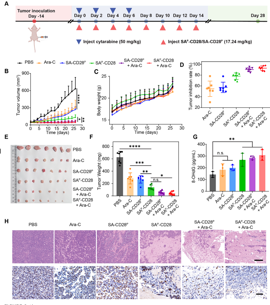 FIGURE 6
Antitumor effects of the self-assembling peptides in Jurkat tumor-bearing nude mice. (A) Schematic illustration of tumor inoculation and treatment in mice. (B) The growth curves of the tumors during the treatments. (C) The variations in mouse weights during the indicated treatments. (D) Tumor inhibition rates are determined by measuring tumor volumes at the end of treatment. (E) The digital images of the dissected tumors after the indicated treatments. (F) The average tumor weight at the end of the treatment. (G) Quantification of serum 8-OHdG as an oxidative stress biomarker in mice at the therapeutic endpoint. (H) Representative hematoxylin and eosin (H&E, top panel; scale bar: 200 μm) and Ki-67 immunohistochemistry (Ki-67, bottom panel; scale bar: 50 μm) staining of tumor tissues at the study endpoint after the indicated treatments. Data are presented as mean ± SD (n = 7 mice per group for B-D, F; n = 3 mice per group for (G)). Statistical significance was determined using two-way ANOVA (B) or one-way ANOVA (F, G). *p < 0.05, **p < 0.01, ***p < 0.001, ****p < 0.0001.
在本 CD28 靶向多肽治疗 T-ALL 的完整研究体系中,Absin HRP 标记二抗 abs20039、abs20040 是信号通路机制验证的核心支撑试剂,不可替代:

- 手机:13761418683