




- 手机:13761418683
联系人:爱必信
网址:http://www.absin.cn/
地 址:上海市浦东新区新浩路58号18号楼

- 文章标题:Asiaticoside enhances the antitumor efficacy of MSLN-targeted CAR-T cells in ovarian cancer
-
期刊:Journal of Translational Medicine;2026 最新 IF=9.7,JCR Q1 转化医学权威期刊;DOI:10.1186/s12967-026-08053-w,卷期页码:2026, 24:631。
- 单细胞分泌组验证基础表型:共培养证实 MSLN CAR-T 接触卵巢肿瘤后大量分泌免疫抑制因子 TGF-β1,诱发细胞耗竭;
- 体外细胞功能验证 AC 增效作用:RTCA、LDH 杀伤实验证实 AC 仅特异性增强 MSLN 阳性肿瘤的 CAR-T 杀伤,不影响抗原阴性细胞;转录组、流式验证 AC 下调耗竭标志物、上调 IFN-γ/ 穿孔素 / 颗粒酶 B 等效应分子;
- 分子机制解析:AC 通过抑制 TGF-β/SMAD 通路,阻断 SMAD2/3 磷酸化,抑制 Treg 分化;对比 TGFβ 受体抑制剂 SB431542,证实 AC 功能依赖 TGF-β 通路阻断;
- 腹腔转移卵巢癌体内药效:SKOV-3-luc 腹腔转移 NCG 小鼠,活体生物发光监测肿瘤负荷,证实 AC 联合 CAR-T 显著清除腹腔转移灶,无体重下降、脏器损伤;
- 皮下荷瘤模型 + 组织原位验证:皮下卵巢癌模型验证抑瘤效果;采用 Absin abs50012 多重荧光试剂盒染色肿瘤组织,证明 AC 提升瘤内 CD3?T 浸润、降低 Ki67 增殖与 CD31 血管生成,完成体内机制可视化验证。
- 体内肿瘤微环境原位可视化核心工具:本研究体内机制论证的关键实验依托 abs50012 四色多重荧光试剂盒实现,单张切片同步标记 T 细胞、肿瘤增殖、血管内皮三类关键标志物,直观建立 AC 给药、CAR-T 浸润、肿瘤生长抑制三者的空间关联,弥补单标 IHC 无法多指标共定位的缺陷。
- TSA 信号放大适配低丰度免疫标志物:试剂盒自带 TSA 酪酰胺放大系统,可清晰检出肿瘤组织中少量浸润 CD3?CAR-T 细胞,避免普通免疫荧光信号弱、假阴性问题,保证定量统计可靠。
- 标准化实验体系匹配高分转化医学发文要求:试剂盒全套试剂预装、洗脱流程稳定,染色重复性强,mIHC 成像数据满足Journal of Translational Medicine等转化医学 SCI 期刊组织学数据质控标准,支撑本文体内转化价值结论。
- 临床转化研究拓展价值:该试剂盒可批量处理人卵巢癌临床石蜡样本,同步检测肿瘤浸润 T 细胞、增殖、血管标志物,后续可用于 AC 联合 CAR-T 临床样本分层、疗效预测相关研究。
文献深度解析:Asiaticoside enhances the antitumor efficacy of MSLN-targeted CAR-T cells in ovarian cancer
2026-08-04
一、文献综述
(一)期刊基础信息
(二)全文核心结论
积雪草苷(AC)作为天然小分子药物,可靶向抑制TGF-β/SMAD信号轴,缓解 MSLN 靶向 CAR-T 细胞耗竭、阻断 Treg 分化,逆转卵巢癌肿瘤微环境免疫抑制;体外细胞共培养、腹腔转移瘤 / 皮下荷瘤小鼠模型均证实 AC 联合 CAR-T 具备更强抗肿瘤活性,无明显脏器毒性,是具备临床转化潜力的 CAR-T 实体瘤免疫佐剂。
(三)使用爱必信 abs50012-20T 产品作用
本研究采用 Absin 四色多重免疫荧光试剂盒 abs50012-20T 完成肿瘤组织多靶点原位共染。试剂盒依托 TSA 信号放大技术,单张石蜡切片同步标记 CD3、Ki67、CD31、DAPI 四指标,直观可视化肿瘤内 T 细胞浸润、肿瘤增殖、血管新生的空间关联。为 AC 提升 CAR-T 肿瘤浸润、抑制增殖与血管生成的体内结论提供原位组织学证据,数据符合高分转化医学期刊质控标准。
二、研究领域背景介绍
CAR-T 细胞疗法在血液肿瘤取得突破性疗效,但在卵巢癌等实体瘤中疗效受限,两大核心瓶颈:①肿瘤微环境高分泌 TGF-β1,持续刺激 CAR-T 细胞高表达 PD-1/TIM-3/LAG-3 耗竭标志物,杀伤功能丧失;②TGF-β1 诱导 CAR-T 向免疫抑制型 Treg 分化,形成负反馈免疫抑制环路。
现有改造 CAR-T 基因(显性负性 TGFβ 受体、SMAD7 过表达)策略存在基因编辑复杂、体内安全风险高等缺陷;天然小分子药物安全性高、给药便捷,是理想替代佐剂。积雪草苷(AC)是积雪草提取三萜皂苷,既往报道可调控 NK 细胞免疫,但 AC 能否调控 TGF-β 通路改善 CAR-T 实体瘤疗效尚无系统研究。
基于此,本文构建 MSLN 靶向 CAR-T 细胞,以卵巢癌 SKOV-3/OVCAR-3 模型,探究 AC 改善 CAR-T 抗肿瘤功能的分子机制与体内治疗潜力。
三、作者整体研究思路总结(递进式 5 大模块)
四、分模块详细研究思路
模块 1:单细胞分泌组证实 CAR-T 共培养后大量分泌 TGF-β1,易发生耗竭
1. 研究逻辑
单细胞细胞因子谱对比普通 T 细胞与 MSLN CAR-T 接触 SKOV3 肿瘤后的分泌特征,明确 TGF-β1 是 CAR-T 功能受损关键抑制因子。
2. 核心实验
IsoLight 单细胞多因子检测、t-SNE 单细胞分群可视化。
3. 关键结果
CAR-T 细胞共培养后 GM-CSF、颗粒酶 B 等效应因子升高,但免疫抑制 TGF-β1、IL-6 分泌显著高于普通 T 细胞;持续 TGF-β1 刺激驱动 CAR-T 耗竭。
 Fig. 1
Characterization of the single-cell secretome in CAR-T cells co-cultured with SKOV-3 cells. (A) PSI values comparing CAR-T cells against control T cells after stimulation with MSLN-positive targets. (B) Proportion of cytokine-secreting cells detected in CAR-T versus control T cell populations. (C) Signal intensities of secreted cytokines from CAR-T and T cells. (D) 2D t-SNE projection visualizing the landscape of single-cell cytokine secretion.
模块 2:体外实验证明 AC 特异性增强 MSLN-CAR-T 肿瘤杀伤活性
1. 研究逻辑
选取 MSLN 阳性(SKOV3、OVCAR3)、MSLN 阴性(ES-2)卵巢癌细胞,RTCA 实时细胞阻抗、LDH 释放杀伤实验验证 AC 增效的抗原依赖性;筛选安全有效 AC 工作浓度 20μM。
2. 核心实验
CCK8 细胞活力、RTCA 实时杀伤、LDH 细胞毒实验。
3. 关键结果
20μM AC 不影响 CAR-T 存活;仅在 MSLN 阳性肿瘤组,CAR-T+AC 组杀伤效率显著提升;ES-2 无抗原组各组杀伤无差异,排除 AC 直接细胞毒性。
 Fig. 2
AC enhances the antitumor function of MSLN-CAR-T cells in vitro. (A) Flow cytometric analysis of MSLN surface expression on OVCAR-3, SKOV-3, and ES-2 cells. (B) Western blot detection of MSLN protein levels in the indicated cell lines. (C, E, G) RTCA of CAR-T cells co-cultured with OVCAR-3 (C), SKOV-3 (E), and ES-2 (G) cells at an E: T ratio of 2:1. Specifically, CAR-T cells were pre-treated with TGF-β1 (10 ng/mL) for 24 h where indicated. Subsequently, AC (20 μM) was added to the co-culture system in the relevant groups (CAR-T+AC and CAR-T+TGF-β1+AC). (D, F, H) Quantification of specific lysis of OVCAR-3 (D), SKOV-3 (F), and ES-2 (H) cells at different E: T ratios (mean±SEM; n=3)
模块 3:AC 逆转 TGF-β1 诱导 CAR-T 耗竭,恢复效应分子分泌
1. 研究逻辑
RNA-seq 转录组筛选耗竭 / 效应基因;流式检测 PD-1/TIM-3/LAG-3 耗竭标志物、胞内 IFN-γ/ 穿孔素 / 颗粒酶 B。
2. 核心实验
RNA 测序、流式细胞术(耗竭标志物、胞内细胞因子染色)。
3. 关键结果
TGF-β1 处理显著上调 CAR-T 耗竭基因、下调效应基因;联合 AC 可完全逆转该转录表型;流式定量证实 AC 降低抑制性受体比例,提升杀伤因子阳性细胞占比。
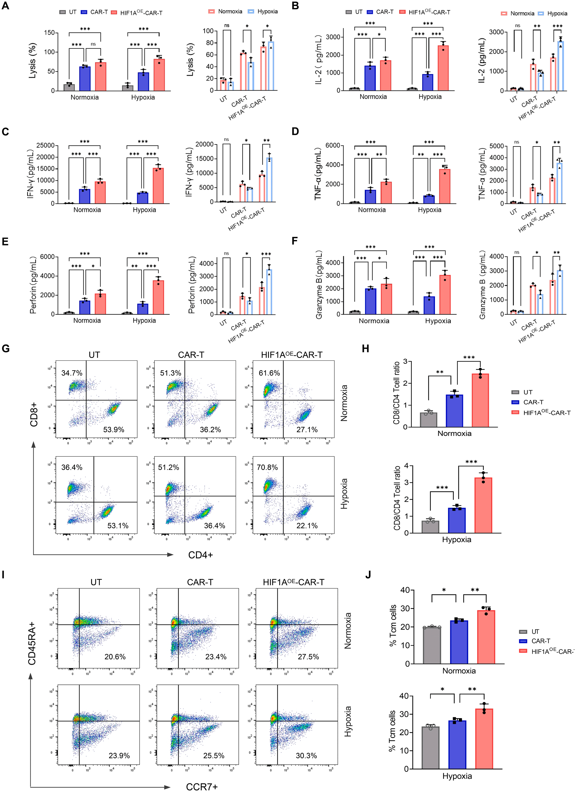 Fig. 3
AC mitigates CAR-T cell exhaustion and enhances the secretion of effector molecules. (A) Heatmap displaying the DEGs associated with T-cell exhaustion and effector functions across the indicated groups (n=3). (B-C) PD-1, TIM-3 and LAG-3 expression on CAR-T, CAR-T+TGF-β1, CAR-T+AC, and CAR-T+TGF-β1+AC groups co-cultured with SKOV-3 cells at an E: T ratio of 2:1 for 24 h by Flow Cytometry. Bar graphs (C) were pooled from 3 donors. (D-E) Intracellular staining of cytotoxic effector molecules (IFN-γ, Perforin, and Granzyme B) in CAR-T, CAR-T+TGF-β1, CAR-T+AC, and CAR-T+TGF-β1+AC groups co-cultured with SKOV-3 cells (E: T ratio=2:1, 24 h). Representative flow cytometry distributions (D) and statistical summary (E) from 3 donors. Data are expressed as mean±SEM (n=3). Statistical significance was determined by one-way ANOVA (*p<0.05; **p<0.01; ***p<0.001; ****p<0.0001)
模块 4:AC 抑制 TGF-β/SMAD 通路,阻断 CAR-T 向 Treg 分化
1. 研究逻辑
流式检测 CD4?FOXP3? Treg 比例;WB、磷酸化流式检测 SMAD2/3 磷酸化水平;转录组富集 TGF-β 通路基因;阳性对照 SB431542 验证通路依赖性。
2. 核心实验
FOXP3 Treg 流式、p-SMAD2/3 蛋白 WB、磷酸化流式、KEGG 通路富集、通路抑制剂对照实验。
3. 关键结果
TGF-β1 诱导 CAR-T 大量分化为 FOXP3? Treg,AC 可显著降低 Treg 比例;AC 显著抑制 SMAD2/3 磷酸化,下调 TGF-β 通路核心基因;AC 与 SB431542 通路抑制效果相当。
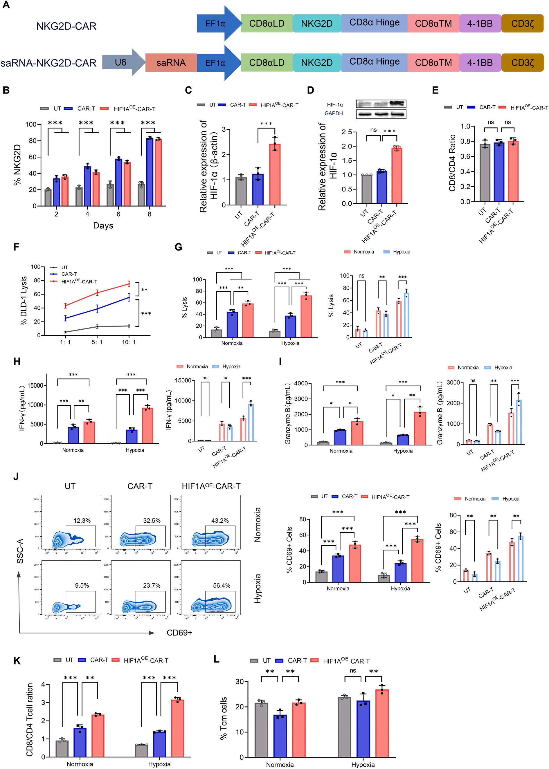 Fig. 4
AC inhibits Treg generation by reducing TGF-β1 release via the TGF-β/SMAD signaling pathway. (A-B) Flow cytometric analysis of CD4 and FOXP3 expression in CAR-T cells. Cells were co-cultured with SKOV-3 cells (E: T=2:1) for 24 h in the presence of TGF-β1, AC, or both. Representative plots (A) and pooled quantitative data (B) are pooled from 3 donors. (C-D) Assessment of TGF-β1 levels in the indicated groups by flow cytometry. Representative plots (C) and statistical summary (D). (E-F) Intracellular staining of p-SMAD2/3 levels. Cells were permeabilized and analyzed by flow cytometry. Representative plots (E) and pooled data (F). (G) Heatmap displaying the DEGs associated with the TGF-β signaling pathway across the indicated groups (n=3). (H) Western blot analysis of p-SMAD2/3 and total SMAD2/3 protein levels in CAR-T, CAR-T+TGF-β1, CAR-T+AC and CAR-T+TGF-β1+AC groups co-cultured with SKOV-3 cells at an E: T ratio of 2:1 for 24 h. One way ANOVA test (**p<0.01; ***p<0.001; ****p<0.0001); mean±SEM were shown (n=3)
模块 5:AC 联合 CAR-T 抑制卵巢癌腹腔转移,体内安全有效
1. 研究逻辑
构建 SKOV-3-luc 腹腔转移 NCG 小鼠,分组给药 CAR-T±AC,活体荧光成像动态监测肿瘤负荷;记录小鼠体重、终点腹腔转移结节、腹水、肝脏转移、脏器 HE 染色评估安全性。
2. 核心实验
活体生物发光 BLI、小鼠体重监测、腹腔大体拍照、脏器 HE 病理染色。
3. 关键结果
CAR-T+AC 组肿瘤荧光信号持续下降,4/5 小鼠无残留肿瘤;各组小鼠体重无明显差异;脏器 HE 未见坏死、炎性损伤,无全身毒性;对照组存在大量腹腔播散、肝转移。
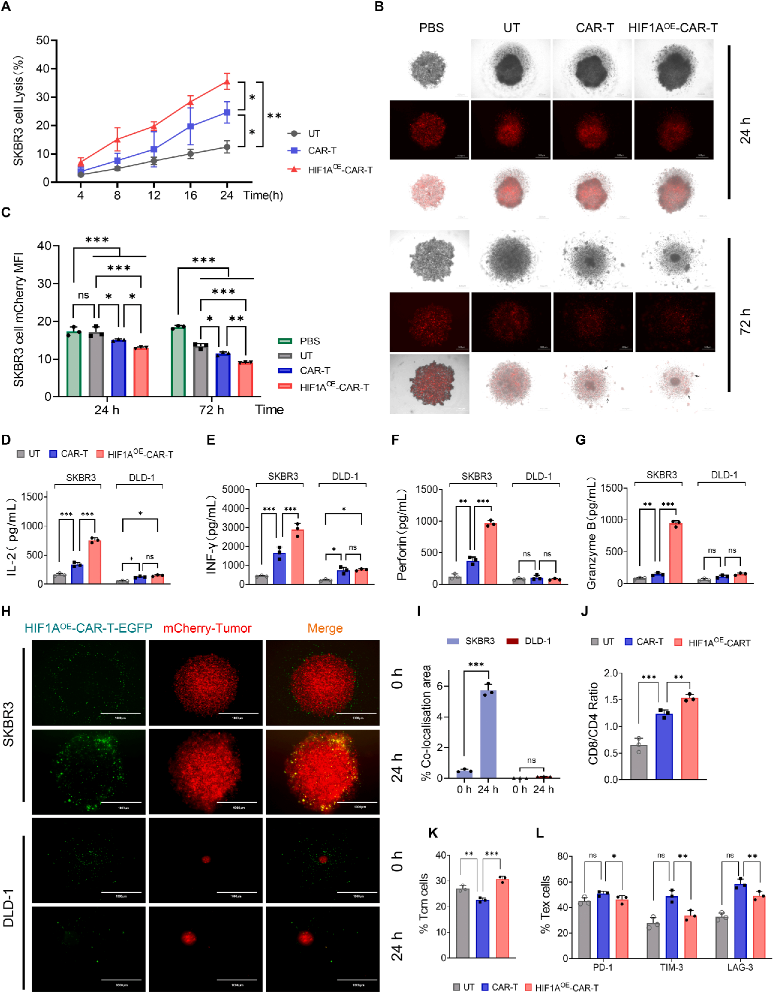 Fig. 5
AC potentiates CAR-T efficacy against SKOV-3-luc peritoneal metastasis of ovarian cancer. (A) Schematic diagram illustrating the experimental design for the SKOV-3-luc intraperitoneal metastasis mouse model. (B) BLI of tumor burden in mice bearing MSLN-positive SKOV-3 tumors during the treatment period. (C) Quantification of bioluminescence signals (Region of Interest, ROI) at indicated time points. (D) Monitoring of mouse body weights throughout the study. Data are expressed as mean±SEM (n=5 per group). ****p<0.0001
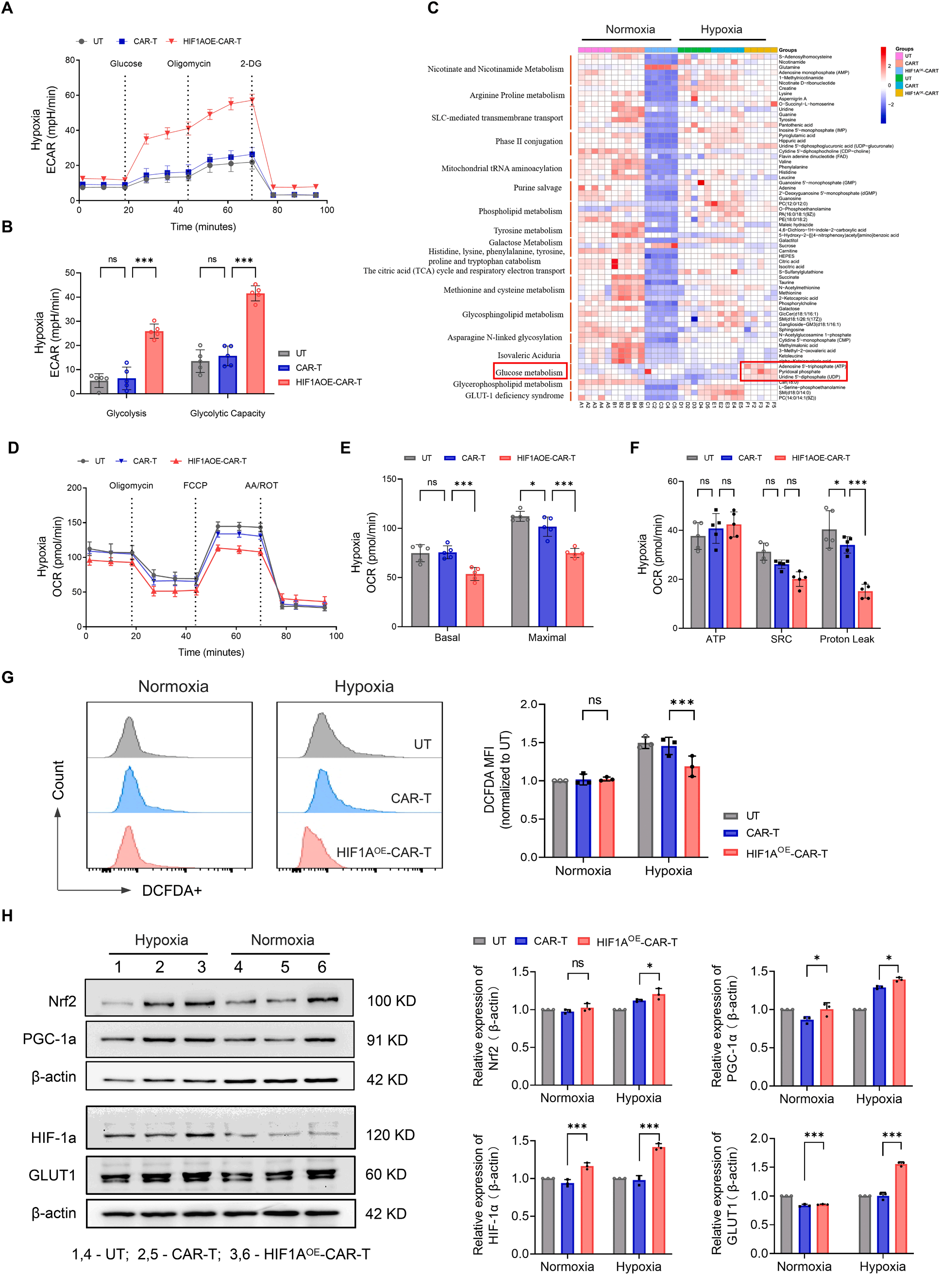 Fig. 6
AC restores CAR-T cell antitumor immunity suppressed by TGF-β1 in an ovarian cancer mouse model. (A) Macroscopic observation of metastatic nodules in the abdominal cavity at the study endpoint. Representative HE staining images are displayed in the right panels. Scale bars represent 100 μm and 20 μm. (B) Representative images showing the appearance of peritoneal lavage fluid from tumor-bearing mice. (C) Liver metastasis in NC and CAR-T+TGF-β1 groups demonstrated with photos of macroscopic and HE staining at the endpoint of the follow-up. The scale bars represented 100 μm and 20 μm, respectively
模块 6:皮下荷瘤模型验证 AC 体内抑瘤效果,Absin mIHC 完成肿瘤微环境原位检测
1. 研究逻辑
SKOV3 皮下异种移植瘤,测量肿瘤体积 / 重量;采用Absin abs50012-20T 四色多重荧光免疫组化试剂盒对肿瘤石蜡切片同步染色 CD3(T 细胞)、Ki67(增殖)、CD31(血管)、DAPI(核),原位解析 AC 重塑肿瘤微环境。
2. Absin 产品使用说明
实验使用 abs50012-20T 试剂盒处理 4μm 肿瘤石蜡切片,经抗原修复、HRP 封闭、多轮 TSA 荧光信号放大与抗体洗脱,同一张切片同时标记 CD3、Ki67、CD31 三个蛋白 + DAPI 核染;TSA 信号放大解决低丰度 CD3?T 细胞荧光弱问题,通过多通道成像直观对比各组肿瘤内 T 细胞浸润、肿瘤增殖、微血管密度差异。
3. 核心结果
CAR-T+AC 组肿瘤体积、重量最低;多重荧光切片显示该组 CD3?T 浸润显著增多,Ki67 肿瘤增殖、CD31 血管新生显著下调;心肝肾脾肺 HE 染色无病理损伤,证实体内安全性。
 Fig. 7
AC improves the therapeutic effects of CAR-T cells in SKOV-3 cells subcutaneous xenograft model. (A) The treatment scheme in subcutaneous tumor mouse models of SKOV-3. (B) Representative images of excised xenografts. (C) Tumor volumes are measured every three days. (D) Weights of xenograft tumors. (E) Representative mIHC images of xenograft tumors in (B) Yellow, CD3; Green, Ki67; Pink, CD31; Blue, DAPI. Scale bar represent 100 μm and 20 μm, respectively. Data are expressed as the mean±SEM; n=5 per group; *p<0.05; **p<0.01; ***p<0.001; ****p<0.0001
五、爱必信(Absin)abs50012-20T 产品整体作用总结

- 手机:13761418683