




- 手机:13761418683
联系人:爱必信
网址:http://www.absin.cn/
地 址:上海市浦东新区新浩路58号18号楼

- 论文标题:Overexpression of HIF-1α via small activating RNA enhances CAR-T cell function in hypoxic microenvironments
- 期刊:Pharmacological Research(2026,第 229 卷,最新影响因子 IF=12.2,中科院 1 区 Top 药理学权威期刊,SCI 收录开放获取期刊。)
-
DOI:10.1016/j.phrs.2026.108235 2026
- 临床生信验证:乳腺癌 TCGA 队列证实 HIF1A 高表达关联晚期肿瘤、不良预后、糖酵解重编程;
- saRNA 工具筛选:设计 5 条 HIF1A 启动子靶向 saRNA,qPCR/WB 筛选激活效率最优 saRNA-4;
- 非病毒 CAR-T 平台构建:piggyBac 电转搭建携带 saRNA 的 HER2 纳米抗体 CAR-T,优化转染效率并维持 T 细胞正常亚群;
- 体外缺氧功能验证:LDH 杀伤、细胞因子、流式记忆 / 耗竭表型证明 HIF1AOE 逆转缺氧损伤;
- 3D 肿瘤浸润模型:3D SKBR3/DLD-1 肿瘤球验证 HIF1AOE-CAR-T 深层浸润、更强抑瘤;
- 代谢分子机制:Seahorse 能量代谢 + WB 证明双重通路:GLUT1 促进糖酵解、Nrf2/PGC-1α 保护线粒体降低 ROS;
- 通用性与体内转化:NKG2D-CAR 重复全部表型验证策略广谱;NOD-SCID 乳腺癌异种移植证实 HIF1AOE-CAR-T 联合 CTLA4 纳米抗体协同抑瘤、延长生存期、无系统毒性。
- 临床:乳腺癌组织 HIF1A 显著上调,高表达对应高分期、短生存期、糖酵解通路富集;
- 细胞层面:5 条 saRNA 均可上调 HIF1A mRNA / 蛋白,saRNA-4 激活效果最强;
- 缺氧功能定量金标准:三款双抗体夹心试剂盒灵敏度高、批间差异小,可精准区分常氧 / 缺氧、不同 CAR-T 组间微量细胞因子差异,直观证明 HIF1A 逆转缺氧免疫抑制,为表型结论提供可重复量化数据;
- 多模型通用标准化试剂:适配细胞上清、肿瘤球共培养体系、小鼠人源 CAR-T 血清多种样本,统一检测标准,打通体外细胞实验到体内动物药效的数据连贯性,符合 Pharmacological Research 高分期刊数据质控要求;
- 支撑转化医学论证:体内血清细胞因子定量直接佐证 HIF1AOE-CAR-T 在实体瘤体内持续活化,联合 CTLA4 后效应因子进一步提升,有力支撑本方案临床转化潜力,是全文免疫功能论证不可或缺的实验工具。
文献深度解析:saRNA 上调 HIF-1α 增强缺氧微环境 CAR-T 功能
2026-08-04
一、文献基础综述
(一)期刊基础信息
(二)全文核心结论
本研究搭建piggyBac 非病毒转座子 CAR-T 制备平台,筛选最优 saRNA 特异性激活内源性 HIF1A 基因;HIF1A 过表达 CAR-T(HIF1AOE-CAR-T)通过GLUT1 糖酵解上调 + Nrf2/PGC-1α 线粒体保护双重代谢重塑,在 1% 缺氧条件下提升杀伤、细胞因子分泌、肿瘤球浸润,降低 T 细胞耗竭;HER2、NKG2D 双 CAR 模型验证策略普适;体内联合抗 CTLA4 纳米抗体可协同消退实体瘤,无明显脏器毒性,为实体瘤缺氧微环境 CAR-T 治疗提供安全非病毒新方案PubMed。
(三)爱必信 Absin 产品核心作用
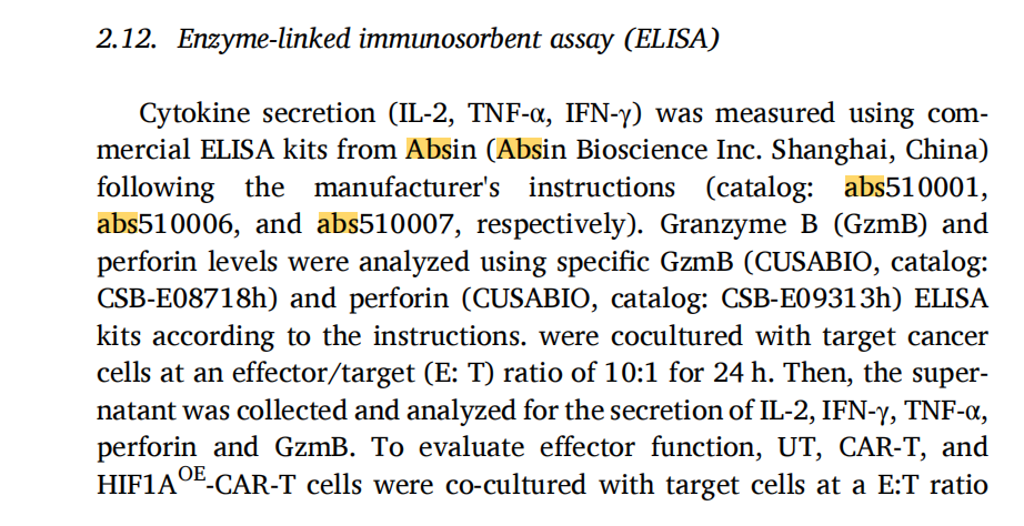
研究采用abs510001 人 IL-2、abs510006 人 TNF-α、abs510007 人 IFN-γ ELISA 试剂盒定量 CAR-T 上清关键效应细胞因子。三款夹心 ELISA 试剂盒精准检测常氧 / 缺氧不同处理组细胞因子浓度,直观证明 HIF1AOE 可逆转缺氧介导的细胞因子分泌抑制。试剂盒重复性与灵敏度满足多组体外、3D 肿瘤球、体内血清样本定量需求,为 CAR-T 功能表型提供关键定量数据支撑。
二、研究领域背景介绍
CAR-T 细胞疗法在血液肿瘤实现突破性疗效,但对实体瘤疗效极差,缺氧免疫抑制肿瘤微环境(TME) 是核心瓶颈:实体瘤血管紊乱、增殖过快造成局部 1% 低氧,直接抑制 CAR-T 浸润、促进 T 细胞耗竭、削弱杀伤因子释放。缺氧诱导因子 HIF-1α 是细胞低氧适应核心转录因子,调控 T 细胞糖代谢、线粒体稳态、趋化受体表达;持续 HIF-1α 过表达易诱发 T 终末耗竭,如何生理性精准上调 HIF-1α 是领域难题。
传统 CAR-T 病毒载体存在插入突变、装载量受限、成本高缺陷;saRNA 小激活 RNA 靶向基因启动子激活内源转录,无需外源转基因、安全性更高,但尚未与非病毒 CAR-T 系统结合用于实体瘤免疫治疗。既往研究仅单独探索 HIF-1α 或 saRNA,无研究整合 saRNA、piggyBac 非病毒递送、CAR-T 代谢重编程同步解决实体瘤缺氧耐受难题,本文填补该领域空白。
三、作者整体递进研究思路总览
全文遵循「临床线索→工具开发→体外 2D 功能→3D 肿瘤球验证→代谢机制解析→多 CAR 普适验证→体内联合治疗」7 层递进逻辑:
四、分模块详细研究思路
模块 1:临床生信 + saRNA 工具开发
1. 研究逻辑
临床样本锁定 HIF1A 作为靶点,构建并筛选高效 HIF1A 激活 saRNA;
2. 核心实验
TCGA 乳腺癌转录组生信、saRNA 序列设计、Jurkat / 原代 T 细胞转染、Cy3 荧光转染效率、qPCR(2^ΔΔCt)、Western blot;
3. 关键结果
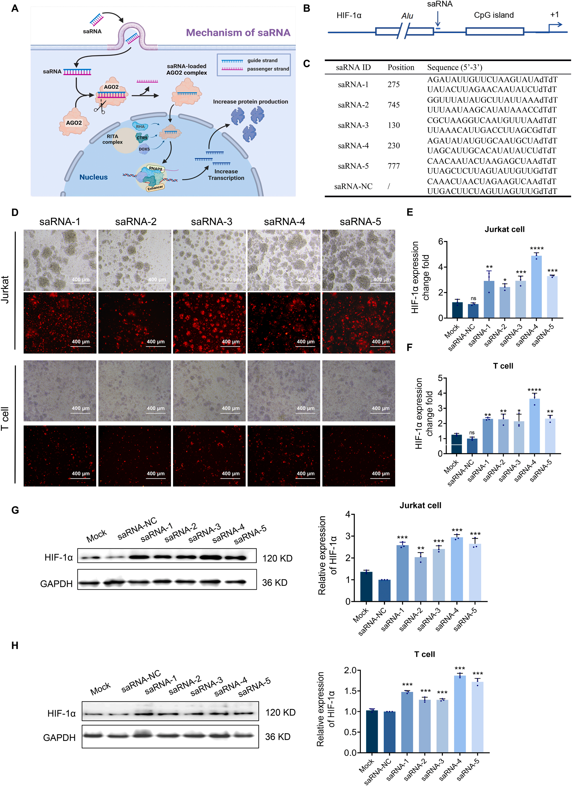 Fig. 1
Design and characterization of HIF-1α-targeting saRNAs in Jurkat and primary T cells. (A). Conceptual illustration of the mechanism of saRNAs in transcriptional activation. (B) Schematic of the HIF-1α promoter region, including CpG island, transcription start site, and saRNA target sites of saRNA-4. (C) Selected nucleotide sequences of HIF-1α for saRNAs. (D) Fluorescence microscopy images of Jurkat cells and primary human T cells transfected with 50 nM control duplex (saRNA-NC) or saRNA constructs for 48 h. Transfection efficiency was monitored using a Cy3 reporter (red fluorescence). Images captured at 40 × magnification. (E-F) qPCR analysis of HIF-1α mRNA in Jurkat cells (E) and primary T cells (F). Total RNA was extracted, reverse-transcribed into cDNA, and amplified using SYBR Green qPCR mix. HIF-1α expression was normalized to GAPDH (2ΔΔCt). Triplicate assays were performed in three independent experiments. (G-H) Western blot analysis of HIF-1α protein in Jurkat cells (G) and primary T cells (H). SDS-PAGE was used to separate proteins (30 μg), which were then transferred to PVDF membranes and probed with human anti-HIF-1α and human anti-GAPDH antibodies. Band intensities were quantified and normalized to the GAPDH control. Data represent mean ± SEM (n = 3). Statistical significance was assessed by one-way ANOVA with Bonferroni’s post hoc test (*p < 0.05, **p < 0.01).
模块 2:piggyBac 非病毒 saRNA-HER2-CAR-T 构建与表征
1. 研究逻辑
构建携带 saRNA 的纳米抗体 CAR 质粒,电转优化获得高转染 CAR-T,验证 HIF1A 上调且 T 细胞亚群稳定;
2. 核心实验
HER2 纳米抗体筛选、piggyBac 载体构建、电转工艺梯度优化、EGFP 荧光示踪、流式 CAR 阳性率、CD4/CD8、记忆 T 分群、HIF1A qPCR;
3. 关键结果
优化后第 9 天 CAR 转染效率 84%±12.9%,CD4/CD8 比值、中央记忆 T(Tcm)与未转染 T 无差异;saRNA 组 HIF1A 表达显著高于普通 CAR-T;
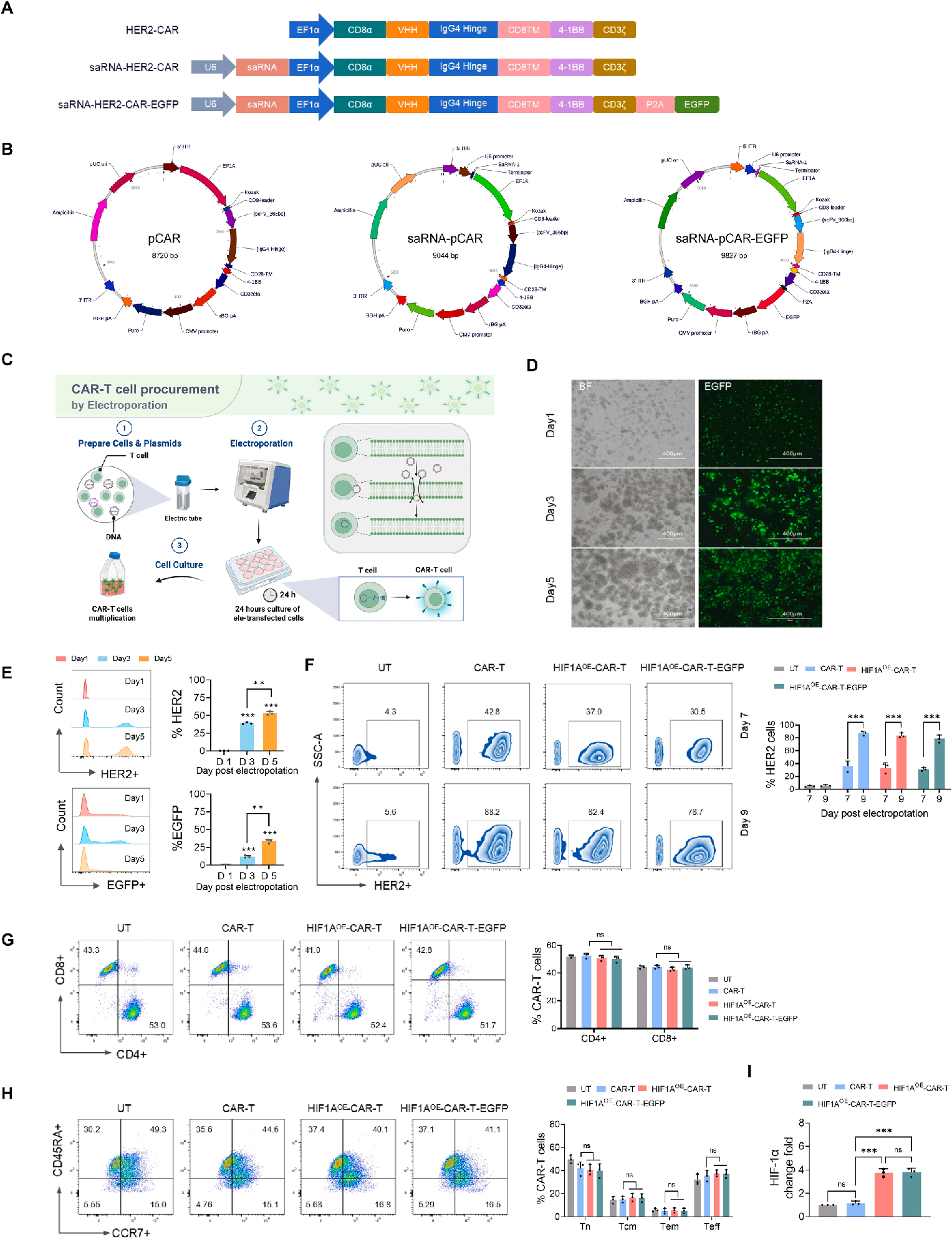 Fig. 2
Electroporation-based generation and cell characterization of HER2-CAR-T cells. (A) Schematics of three nanobody-based HER2-CAR constructs incorporating CD8α leader, HER2 nanobody, IgG4 hinge, CD28 transmembrane domain, 4–1BB, and CD3ζ domains. The U6 promoter was used to integrate the saRNA sequence. (B) CAR expression cassettes cloned into piggyBac transposon vectors, and their recombinant plasmid maps. (C) Workflow of T cell electroporation with piggyBac transposon and transposase plasmids. Cells after electroporation are cultured in preheated medium to promote recovery and proliferation. (D) Fluorescence microscopy showing EGFP expression after electroporation of saRNA-pCAR-EGFP. Bright field and corresponding fluorescence images were acquired at Days 1, 3, and 5 post-electroporation. (E) Flow cytometry analysis of CAR expression at days 1, 3, and 5 post-electroporation. (F) Representative flow cytometry and statistical analysis of the CAR-positive rate in CAR-T, HIF1AOE-CAR-T, and HIF1AOE-CAR-T-EGFP cells on days 7 and 9 post-electroporation. (G) Flow cytometric analysis of the CD4+ and CD8+ CAR-T cell subpopulations. Cells were collected on day 9 post-electroporation and stained with anti-CD3, anti-CD4, and anti-CD8 antibodies, followed by flow cytometric analysis with gating on CD3+ T cells. (H) Flow cytometric analysis of T cell subsets, including na?ve T cells (Tn, CD45RA+CCR7+), central memory T cells (Tcm, CD45RA-CCR7+), effector memory T cells (Tem, CD45RA-CCR7- ), and terminal effector T cells (Teff, CD45RA+CCR7- ). Representative flow cytometry dot plots, and Statistical analysis of T cell proportions. (I) Fold change analysis of HIF-1α in CAR-T cell constructs; GAPDH is the internal control. All data are presented as mean ± SEM (n = 3). Statistical analysis was performed using two-way ANOVA with a Bonferroni post hoc test. (*p < 0.05, **p < 0.01, ***p < 0.001).
模块 3:常氧 / 缺氧下 HIF1AOE-CAR-T 体外杀伤与细胞因子功能
1. 研究逻辑
2D 共培养对比 UT、普通 CAR-T、HIF1AOE-CAR-T 在 1% 缺氧下杀伤、细胞因子、T 细胞亚群;
2. 核心实验
LDH 细胞毒性实验、细胞因子 ELISA、流式 CD4/CD8、记忆 T(CD45RA/CCR7);
3. Absin 产品关键应用
采用 abs510001(IL-2)、abs510006(TNF-α)、abs510007(IFN-γ)三款人源细胞因子 ELISA 试剂盒,检测 24h 共培养上清,定量缺氧条件下各组细胞因子分泌水平;
4. 关键结果
缺氧显著抑制普通 CAR-T 杀伤、IL-2/IFN-γ/TNF-α、穿孔素 / 颗粒酶 B;HIF1AOE-CAR-T 完全逆转该抑制,CD8 比例、Tcm 占比显著升高,耗竭标志物降低;
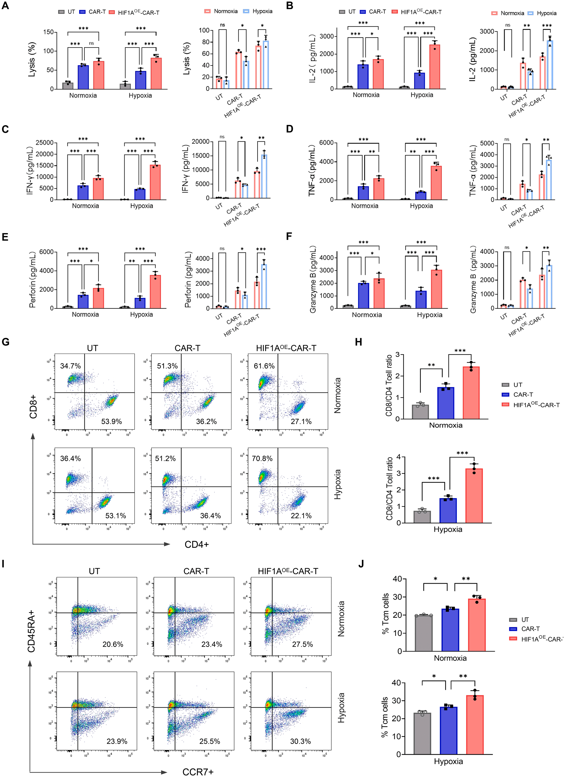 Fig. 3
Anti-tumor activity of HIF-1α-overexpressing CAR-T cells under normoxic and hypoxic conditions. (A). Cytotoxicity of UT, CAR-T, and HIF1AOE-CART cells against SKBR3 cells was assessed by LDH release assay at an E:T ratio of 5:1 for 24 h under normoxia (21% O2) and hypoxia (1% O2). (B-F) ELISA quantification of cytokine release following 24 h of co-culture with SKBR3 cells under normoxia and hypoxia. (B) IL-2 (C) IFN-γ (D) TNF-α (E) perforin, and (F) GzmB. (G-H) Flow cytometry analysis of CD4+ and CD8+ T cell subsets in CD3+ gated cells after 72 h of co-culture. Cells were collected and stained with anti-CD4, anti-CD8, and anti-CD3 antibodies for 30 min in the dark and subjected to flow cytometry analysis. (G) Representative double fluorescence dot plot and (H) statistical analysis of CD8+/CD4+ ratio. (I-J) Flow cytometry analysis of memory T cell subsets using CD45RA and CCR7 expression in normoxic and hypoxic conditions. (I) Representative double fluorescence dot plot and (J) Quantification of Tcm population. All data are presented as mean ± SEM (n =3). Statistical comparisons were made using two-way ANOVA with the Bonferroni multiple comparison test. *p < 0.05, **p < 0.01, ***p < 0.001.
模块 4:NKG2D-CAR 模型验证策略普适性
1. 研究逻辑
更换实体瘤广谱 NKG2D 靶向 CAR,重复全部体外实验,证明 saRNA-HIF1A 策略不依赖单一抗原;
2. 核心实验
NKG2D-CAR 载体构建、电转、缺氧 LDH 杀伤、Absin ELISA 细胞因子、CD69 活化、T 亚群流式;
3. Absin 产品
同模块 3,使用 abs510001/abs510006/abs510007 检测 IFN-γ、TNF-α;
4. 关键结果
NKG2D-HIF1AOE-CAR-T 同样具备缺氧抵抗、高杀伤、高细胞因子、富集 CD8 与 T 细胞记忆,证实方案广谱通用;
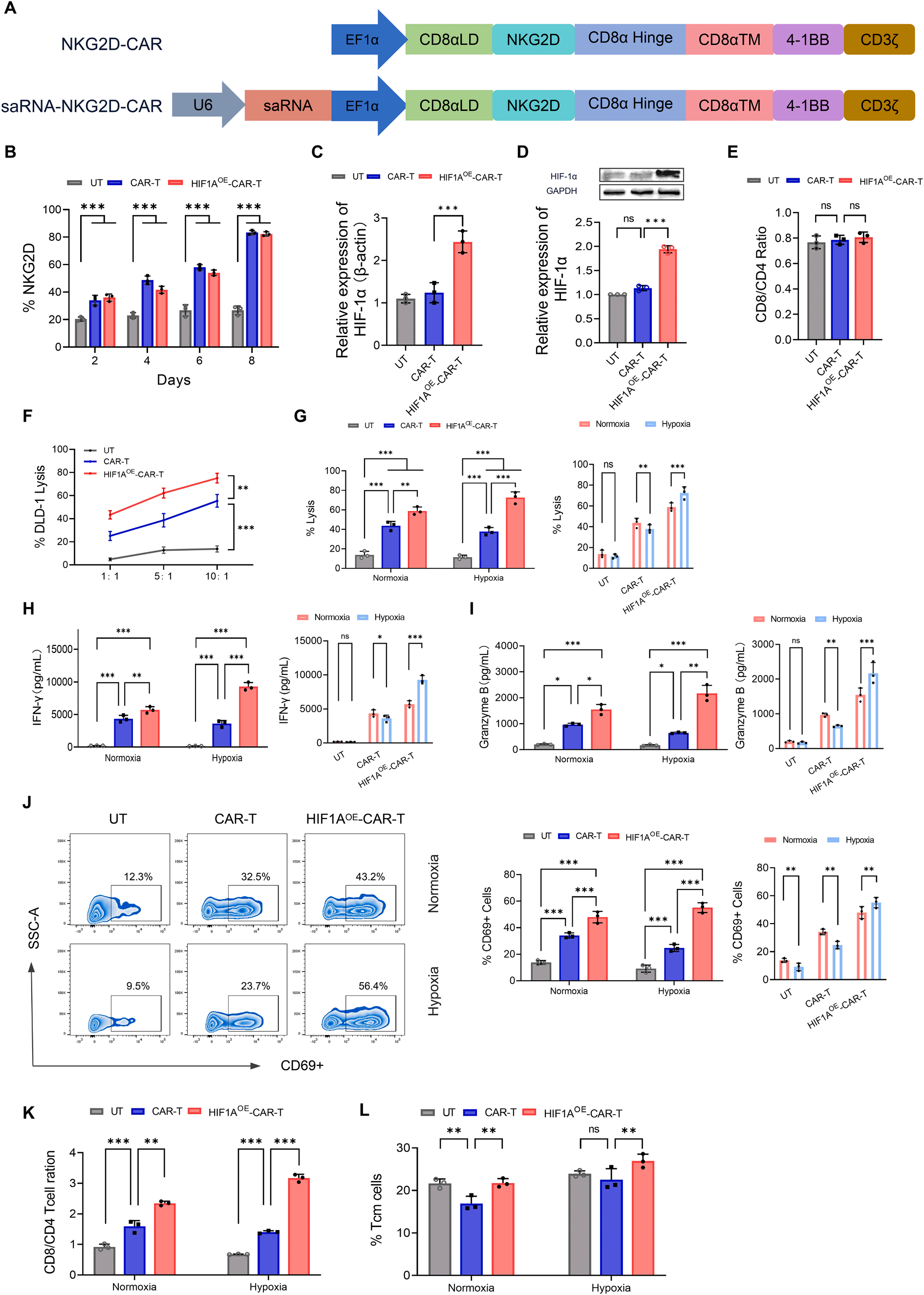 Fig. 4
HIF-1α overexpressing NKG2D-CAR-T cells demonstrate enhanced anti-tumor activity, supporting the broader applicability of the strategy. (A) Schematic illustration of NKG2D-CAR and HIF1AOE-CAR constructs. The U6 promoter was used to drive the integration of the saRNA sequence for HIF-1α activation. (B) Flow cytometry analysis of NKG2D-CAR expression on days 2, 4, 6, and 8 post-electroporation. (C-D) Validation of HIF-1α upregulation at the mRNA level via RT-qPCR (C) and at the protein level via Western blot analysis with corresponding densitometry quantification (D) in untransduced (UT), conventional CAR-T, and HIF1AOE-CART cells. (E) Quantification of the Activation ratio in different T cell groups. (F) Dose-dependent cytotoxicity of UT, CAR-T, and HIF1AOE-CAR-T cells against DLD-1 cells measured by LDH release assay after 24 h co-culture at different effector-to-target (E:T) ratios (1:1, 5:1, and 10:1). (G) Cytotoxic activity of UT, CAR-T, and HIF1AOE-CAR-T cells against DLD-1 cells assessed by LDH release assay at an E:T ratio of 5:1 for 24 h under normoxia (21% O?) and hypoxia (1% O?). (H–I) ELISA quantification of cytokine secretion following 24 h co-culture with DLD-1 cells under normoxic and hypoxic conditions: (H) IFN-γ and (I) granzyme B (GzmB). (J) Representative flow cytometry plots and statistical analysis of CD69 expression in CAR-T and HIF1AOE-CAR-T cells after 24 h of co-culture. (K–L) Flow cytometry analysis of T cell subsets after 72 h of co-culture. Cells were stained with anti-CD3, anti-CD4, and anti-CD8 antibodies and analyzed by flow cytometry. (K) Quantification of the CD8?/CD4? ratio. (L) Analysis of memory T cell subsets defined by CD45RA and CCR7 expression under normoxic and hypoxic conditions. Data are presented as mean ± SEM (n = 3). Statistical comparisons were performed using two-way ANOVA with Bonferroni multiple comparison test. *p < 0.05, **p < 0.01, ***p < 0.001.
模块 5:3D 肿瘤球浸润、抑瘤实验
1. 研究逻辑
3D 类器官模拟实体瘤空间缺氧微环境,评估 CAR-T 肿瘤穿透能力;
2. 核心实验
SKBR3/DLD-1 mCherry 肿瘤球构建、CAR-T 共培养、LDH、共聚焦荧光浸润成像、Absin ELISA 细胞因子、流式耗竭(PD1/TIM3/LAG3);
3. Absin 产品
abs510001/abs510006/abs510007 定量肿瘤球共培养上清 IL-2、IFN-γ、TNF-α;
4. 关键结果
HIF1AOE-CAR-T 可穿透肿瘤球核心,mCherry 肿瘤荧光显著降低,细胞因子分泌远高于普通 CAR-T,耗竭标志物表达下调;
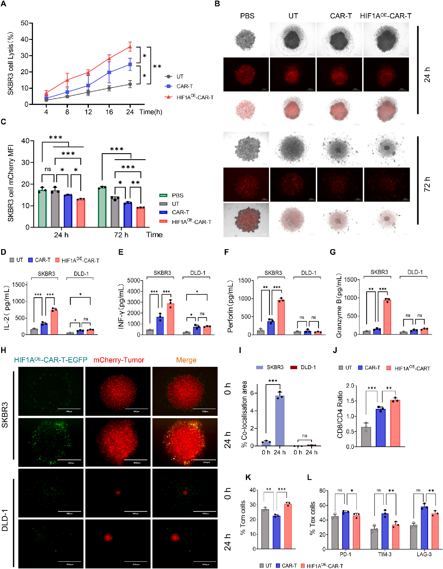 Fig. 5
Anti-tumor effects and infiltration capabilities of HIF1AOE-CAR-T cells in a 3D tumor spheroid model. (A). Cytotoxicity assessment of UT, CAR-T, and HIF1AOE-CAR-T cells against SKBR3 tumor spheroids after 24 h of co-culture at an effector to target ratio (E: T) of 10:1, assessed by LDH release assay. (B) Representative microscopic images of SKBR3-mCherry 3D tumor spheroids co-cultured with PBS (control), UT, CAR-T, or HIF1AOE-CAR-T at a 10:1 effector-to-target ratio for 24 and 72 h. Top: bright-field images, middle: mCherry fluorescence (red) indicating tumor cell viability, and bottom: merged images. Scale bar = 500 μm. (C) Quantitative analysis of mCherry fluorescence (red) intensity measured after 24 and 72 h of co-culture. (D-G) Quantification of effector molecule secretions. Levels of (D) IL-2, (E) IFN-γ, (F) perforin, and (G) granzyme B secreted by UT, CAR-T, and HIF1AOE-CAR-T cells after 24 h of co-culture with SKBR3-tumor spheroids were measured using ELISA. (H) Representative confocal microscopy images showing infiltration of HIF1AOE-CAR-T cells (green) into SKBR3-mCherry or DLD-1- mCherry tumor spheroids (red) after 24 h of co-culture. Scale bar = 1000 μm (left panel). Quantification of T cell infiltration by co-localization area analysis (right panel). (I) CD8+/CD4+ T cell ratio after 48 h of co-culture with SKBR3-mCherry tumor spheroids. After tumor dissociation, T cell subsets were analyzed using flow cytometry. (J) Memory T cell distribution following 48 h of co-culture with SKBR3 spheres. The proportion of central memory T cell subsets was determined by flow cytometry using anti-CD45RA and anti-CCR7 flow cytometry antibodies. (K) Expression of T cell exhaustion markers (PD-1, TIM-3, and LAG-3) after 48 h of coculture, analyzed by flow cytometry. Data represent the mean ± SEM from three independent experiments. Statistical significance was determined using one-way or two-way ANOVA with Bonferroni multiple comparison tests. *p < 0.05, **p < 0.01, ***p < 0.001.
模块 6:HIF1A 调控 CAR-T 代谢双重分子机制
1. 研究逻辑
Seahorse 能量代谢 + WB 解析 HIF1A 重塑糖酵解与线粒体稳态分子通路;
2. 核心实验
糖酵解压力测试 ECAR、线粒体压力测试 OCR、代谢组、ROS 流式、WB(GLUT1、Nrf2、PGC-1α、HIF1A);
3. 关键结果
缺氧下 HIF1AOE-CAR-T 糖酵解能力、线粒体最大呼吸显著提升;GLUT1 上调促进糖代谢,Nrf2/PGC-1α 通路激活减少 ROS、维持线粒体完整;
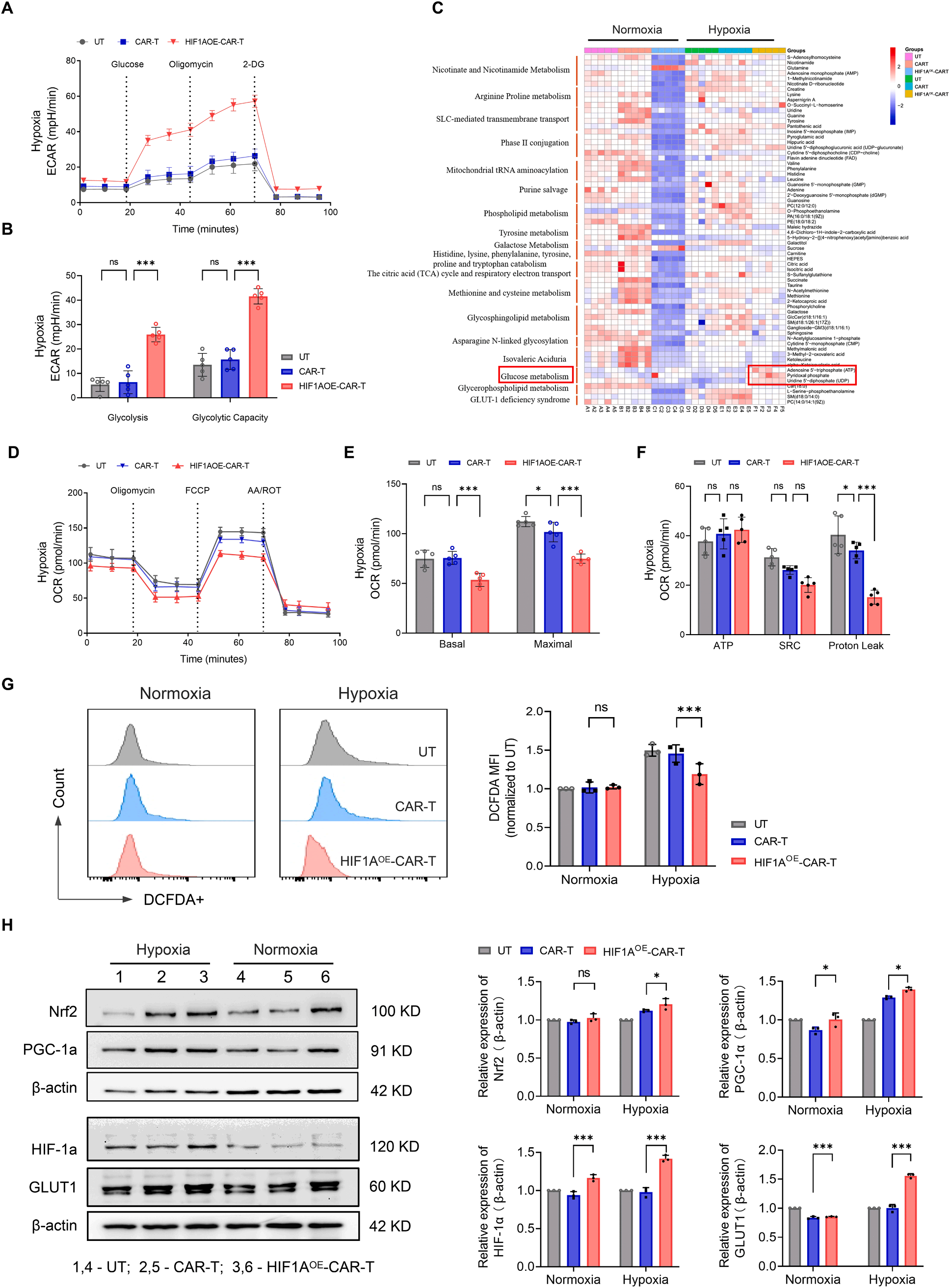 Fig. 6
HIF1AOE-CAR-T cells reprogram metabolic pathways under hypoxia. (A) Seahorse glycolysis stress test showing extracellular acidification rate (ECAR) profiles of UT, CAR-T, and HIF1AOE-CAR-T cells cultured under hypoxia for 48 h. (B) Quantification of glycolysis and glycolytic capacity derived from ECAR measurements. Data represent mean values from five replicate wells (n = 5). (C) Heatmap of hierarchical clustering of differential metabolites (VIP > 1.0, P < 0.05) and their associated KEGG pathways of metabolomic profiles in UT, CAR-T, and HIF1AOE-CAR-T cells under normoxia and hypoxia conditions. Z-score–normalized metabolite levels are shown (red, upregulated; blue, downregulated). (D) Oxygen consumption rate (OCR) profiles obtained using the Seahorse Cell Mito Stress Test in UT, CAR-T, and HIF1AOE-CAR-T cells cultured under hypoxia for 48 h. (E-F) Quantification of mitochondrial respiration parameters, including basal respiration, maximal respiration (E), ATP-linked respiration, spare respiratory capacity (SRC), and proton leak (F). Data represent mean values from five replicate wells (n = 5). (G) Flow cytometry analysis of intracellular ROS levels (MFI) in UT, CAR-T, and HIF1AOE-CAR-T cells following 24 h exposure to normoxia or hypoxia. Data represent the mean ± SEM from three independent experiments. (H) Western blot analysis of glycolysis and mitochondrial function-related proteins (Nrf2, PGC-1α, HIF-1α, GLUT1) in UT, CAR-T, and HIF1AOE-CAR-T cells under normoxic and hypoxic conditions. β-actin served as a loading control. Statistical comparisons were made using one-way or two-way ANOVA with Bonferroni multiple comparison tests. *p < 0.05, **p < 0.01, ***p < 0.001 were considered statistically significant.
模块 7:体内乳腺癌异种移植联合 CTLA4 纳米抗体治疗
1. 研究逻辑
NOD-SCID 荷瘤模型验证单药、联合 CTLA4 纳米抗体体内抑瘤、生存期、脏器毒性;
2. 核心实验
SKBR3-Luc 皮下荷瘤、尾静脉回输 CAR-T、腹腔 CTLA4 纳米抗体、活体生物发光、肿瘤体积 / 重量、生存曲线、肿瘤 IHC Ki67/CD3、小鼠血清 Absin 细胞因子 ELISA、脏器 H&E 病理;
3. Absin 产品
采用 abs510001/abs510006/abs510007 检测小鼠外周血 IL-2、IFN-γ、TNF-α,评估体内 CAR-T 活化水平;
4. 关键结果
HIF1AOE-CAR-T 单药抑瘤优于普通 CAR-T;联合 CTLA4 纳米抗体实现最强肿瘤消退、最长生存期;肿瘤内 CD3+T 浸润增多、增殖 Ki67 降低;心肝肾脾肺无损伤,无系统毒性;
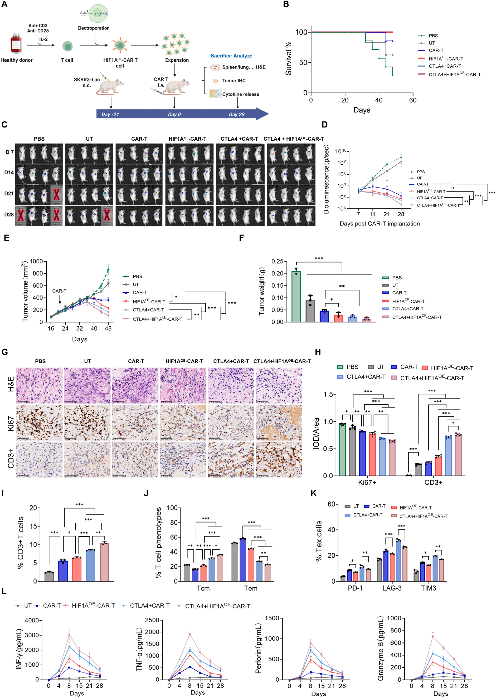 Fig. 7
CTLA4 nanobody synergizes with HIF-1α overexpressing CAR-T cells to achieve enhanced tumor regression and survival in SKBR3 xenografts. (A). Schematic diagram of animal experiment design. Female NSG mice were subcutaneously inoculated with Luc-SKBR3 cells (5 × 10?/mouse) to establish xenograft tumors. All animals were included following successful tumor establishment. Predefined exclusion criteria included failure of tumor engraftment, tumor volume not reaching 100 mm3 before randomization, or signs of unrelated illness; no animals or data points met these criteria and were excluded. Mice were treated with PBS, UT, CAR-T, HIF1AOE-CAR-T, or HIF1AOE-CAR-T + CTLA4. The combination group additionally received intraperitoneal injections of Nb CTLA4 (3 mg/kg). All experiments were performed under identical SPF conditions using standardized procedures. (B) Kaplan-Meier survival analysis of treated groups (n = 7 mice/group). (C) Representative IVIS images showing tumor progression at various time points. (D) Quantification of tumor burden by total flux (photons/sec) from IVIS imaging (n = 4 mice/ group). (E) Tumor volume was measured every 3 days using callipers and plotted as growth curves (n = 7 mice/group). (F) Tumors were excised at the study endpoint for analysis of tumor weight (n = 4 mice/group). (G-H) Histological analysis of tumor tissues by H&E and IHC staining showed reduced Ki67 expression and enhanced CD3+ T cell infiltration in treated groups (n = 4 mice/group). (I-K) Flow cytometry analysis of peripheral blood at endpoint for (I) circulating CD3+ T cell, (J) Tcm cells and (K) frequency of Tex cells. (L) Serum cytokines and effector molecule levels (TNF-α, IFN-γ, perforin, and GzmB) quantified by ELISA (n = 4 mice/ group). Data represent mean ± SEM. Statistical comparisons were made using one-way or two-way ANOVA with Bonferroni multiple comparison tests. *p < 0.05, **p < 0.01,***p < 0.001.
五、Absin(爱必信)试剂盒整体作用总结
本研究体外 2D 细胞、3D 肿瘤球、体内动物血清三大功能验证环节全程依赖 abs510001(IL-2)、abs510006(TNF-α)、abs510007(IFN-γ)三款人源细胞因子 ELISA 试剂盒,是论证 HIF1AOE 提升 CAR-T 效应功能的核心定量工具,整体科研价值分为三层:

- 手机:13761418683