




- 手机:13761418683
联系人:爱必信
网址:http://www.absin.cn/
地 址:上海市浦东新区新浩路58号18号楼

- 文章标题:Sustained‐Release Ulcer‐Protective Microarray Patch With Dihydrocapsaicin for Diabetic Wound Regeneration
- 期刊:Exploration(Wiley 综合顶级期刊,2026 最新 IF=30.4,CiteScore=32.8,JCR Q1 综合 / 纳米材料 / 生物工程三区,OA 期刊)
-
DOI:10.1002/EXP.20250129,发表年份 2026
- Ce-MOF 兼具 SOD/CAT 双重纳米酶活性,可长效清除创面 ROS;多孔载体实现 DHC 长达 28 天平稳缓释,解决游离 DHC 水溶性差、体内快速清除短板;
- 体外细胞层面:显著降低内皮氧化损伤、抑制细胞凋亡,调控巨噬由促炎 M1 向修复 M2 型转换,提升内皮迁移与管状结构形成能力;
- 体内双动物模型(大鼠背部全层创面 + 糖尿病足溃疡)验证:MN-MOF@DHC 组 16 天创面基本完全愈合,胶原沉积、新生 / 成熟血管密度显著优于其余对照组;
- 材料优势:机械强度充足适配增厚糖尿病皮肤、可控降解、生物安全性高,为糖尿病足全程修复提供新型多功能透皮给药方案。
- 病理恶性循环:高糖环境大量生成 AGE,创面 ROS 持续蓄积、线粒体损伤、内皮凋亡;巨噬长期停滞 M1 促炎表型,VEGF 表达抑制,血管新生受阻,炎症 / 增殖 / 重塑三期紊乱,愈合周期大幅延长;
- 传统疗法短板:抗生素耐药加剧,普通敷料无生物活性;生长因子半衰期极短无法全程起效;游离 DHC 水溶性差、生物利用度低,给药后快速代谢;
- 常规微针缺陷:载药量有限、机械强度不足难以穿透增厚糖尿病皮肤,无内源 ROS 清除能力,仅单纯递送药物,缺乏协同修复功能。
- 材料合成与理化表征:制备 Ce-MOF 并负载 DHC,光交联制备 GelMA 复合微针;通过 SEM/TEM/XRD/FTIR、纳米酶活性、体外释药、力学 / 溶胀 / 降解实验验证材料结构、长效释药、抗氧化、皮肤适配性能(对应 Figure 1);
-
体外细胞分层机制验证
- 抗氧化、抗凋亡:TBHP 诱导 HUVEC 氧化损伤,ROS、线粒体膜电位、凋亡通路检测;
- 免疫调控:LPS 诱导巨噬 M1 极化,验证 M1→M2 重编程;
- 促血管生成:内皮迁移、管形成、血管标志物免疫荧光;
- 体内动物疗效双层验证:构建 2 型糖尿病大鼠背部创面、糖尿病足溃疡两大临床模拟模型,分组干预,通过创面愈合速率、H&E/Masson 病理、组织 ROS、巨噬分型、血管密度定量评价体内修复效果;
- 机制总结:阐明 Ce-MOF 纳米酶与 DHC 协同修复通路,指出临床转化仍需临床试验与工艺优化。
- 水热法合成 Ce-UiO-66 MOF,吸附负载 DHC 制备 MOF@DHC 纳米颗粒;
- 10% 取代度 GelMA 水凝胶混合纳米颗粒,模具紫外光交联制备空白微针、MN-MOF、MN-MOF@DHC;
- 形貌、晶体、化学键表征;Ce-MOF CAT/SOD/ 羟基自由基清除活性测试;DHC 缓释、拉伸 / 压缩力学、溶胀、降解、皮肤穿透实验。
- DCFH-DA/MitoSOX 检测胞内、线粒体 ROS;JC-1 检测线粒体膜电位;TUNEL 标记凋亡细胞;WB 检测 Cleaved-Caspase3/Bax/Bcl2 凋亡蛋白;
- abs9149 核心操作步骤(原文 2.18 免疫荧光):细胞 4% 多聚甲醛固定 PBS 洗涤后,配制0.1% abs9149(Triton X-100)PBS 通透液,室温孵育 30 min,温和溶解细胞膜磷脂双分子层,让荧光探针、凋亡抗体顺利进入胞内结合靶标;通透完成后封闭、一抗孵育、成像。
- abs9149曲拉通X-100全文免疫荧光、免疫组化唯一通用通透试剂,适配贴壁细胞、20 μm 动物冰冻切片,温和透化不破坏抗原蛋白,保障 M1/M2 分型、内皮血管、氧化应激、凋亡全部荧光实验可顺利成像,是所有可视化实验基础必备试剂;
- 氧化应激检测体系(DCFH-DA/MitoSOX/JC-1/DHE):实现细胞、线粒体、组织多尺度 ROS 精准定量,直观证明 Ce-MOF 与 DHC 协同抗氧化效应;
- 凋亡检测工具(TUNEL 试剂盒、Cleaved-Caspase3/Bax/Bcl2 一抗):完整阐释材料保护内皮、阻断氧化诱导凋亡分子通路;
- 免疫分型 & 血管特异性一抗(iNOS/CD206/CD31/VEGF/α-SMA):体内外同步验证抗炎重编程、促血管再生两大核心疗效; 整套 Abs 试剂兼容性强,无交叉干扰,操作标准化,实验结果重复性高,统计学差异稳定,为本篇糖尿病创面再生高分论文提供细胞通透→氧化应激→凋亡→免疫调控→血管再生完整闭环检测解决方案。
文献解析:缓释二氢辣椒素微针贴片用于糖尿病创面修复
2026-08-04一、文献综述
(一)文献基础信息
(二) 全文核心结论
本研究构建MN-MOF@DHC 复合微针贴片,一站式解决糖尿病创面氧化应激过载、慢性炎症持续、血管再生缺陷三大临床难题:
(三) 使用 Absin产品作用
abs9149曲拉通X-100整体作用:覆盖材料体外细胞、动物体内组织全流程机制验证,从细胞通透、ROS 定量、凋亡检测、免疫分型到血管标记形成完整标准化检测体系,保障数据重复性与统计学可靠性。
二、领域背景与科学问题引出
(一)糖尿病足溃疡(DFU)临床痛点
全球糖尿病患者达 5.29 亿,糖尿病足溃疡是高致残、高致死严重并发症,现有临床方案存在多重缺陷:
(二)现有研究局限
DHC 天然具备抗氧化、抗炎、促血管活性,但游离给药稳定性差;单一微针仅物理递送,现有 MOF 微针无法实现近 30 天长效释药,无法匹配糖尿病漫长修复周期。
(三)本文科学切入点
是否可将 Ce 基 ROS 清除纳米酶 MOF 与 DHC 整合至 GelMA 微针,构建兼具透皮穿刺、28 天持续释药、内源抗氧化、免疫重编程、促血管再生一体化贴片,同步打破 DFU 氧化、炎症、血管三重病理屏障?本文围绕该问题完成材料制备→体外细胞机制→体内双创面疗效完整验证。
三、作者整体研究思路总结
全文三段式闭环研究逻辑:
四、分模块详细研究思路
模块 1:MN-MOF@DHC 材料合成与理化表征
1. 实验流程
2. 关键结果
Ce-MOF 粒径约 86 nm,晶体结构匹配文献;具备浓度依赖三重自由基清除能力;MN-MOF 力学强度优于纯 GelMA,可穿刺糖尿病增厚皮肤;MN-MOF@DHC 实现 28 天持续释药,微针 16–20 天可控降解适配创面周期。
3. Absin 试剂应用
纳米酶自由基比色检测采用 Absin 微量比色试剂盒定量 405/450/550 nm 吸光度,量化抗氧化催化效率。
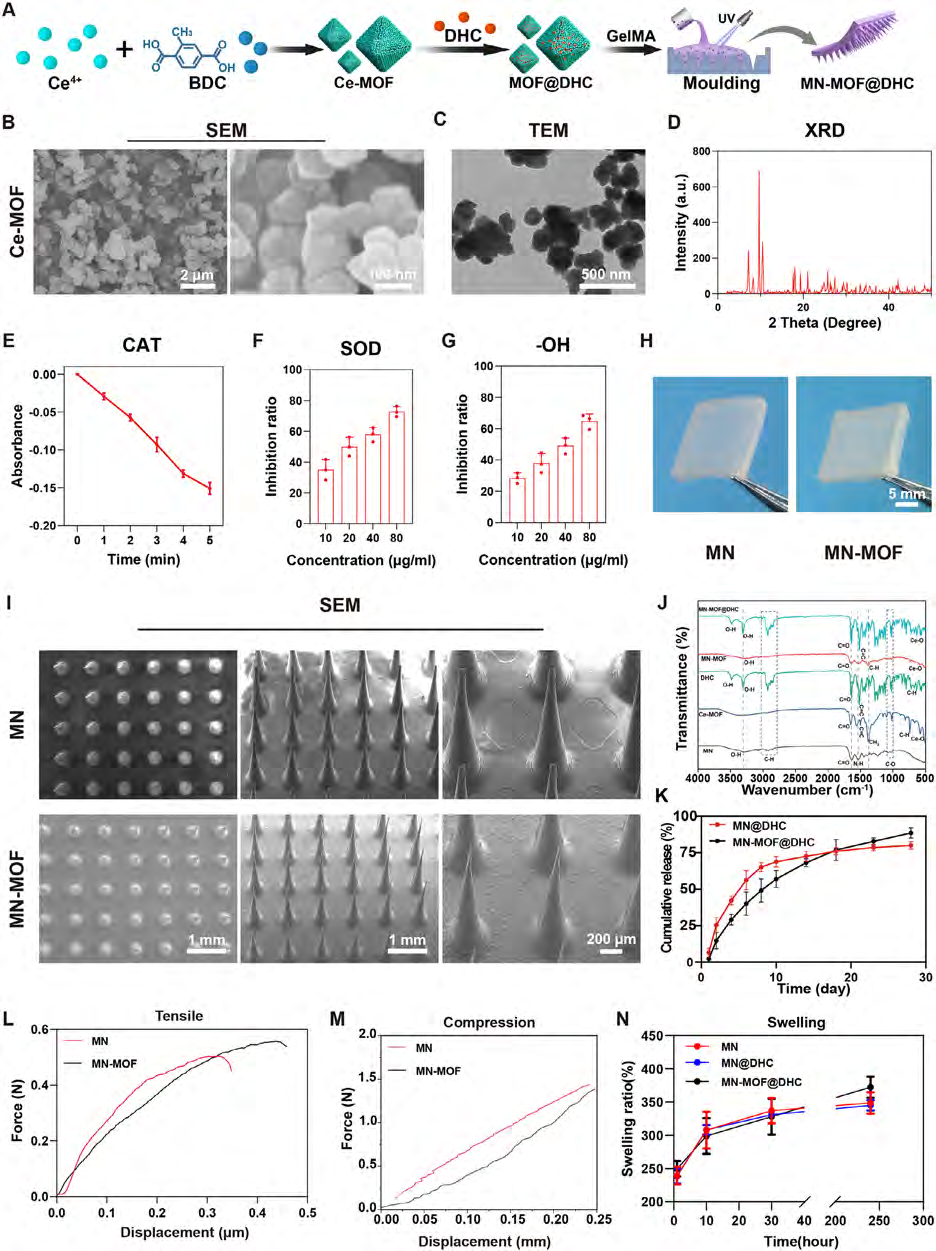 FIGURE 1
Synthesis and characterization of MN-MOF@DHC. (A) Schematic diagram of preparation of MN-MOF@DHC. (B) SEM images of Ce-MOF. (C) TEM images of Ce-MOF. (D) XRD of Ce-MOF. (E–G) CAT-mimicking, SOD-mimicking, and OH-scavenging ability of Ce-MOF. (H) Gross morphology of MN and MN-MOF. (I) SEM images of MN and MN-MOF. (J) FTIR of MN, Ce-MOF, DHC, MN-MOF, and MN-MOF@DHC. (K) Dihydrocapsaicin (DHC) release curve of microneedles following incubation in a saline solution. (L–N) Tensile curve, compressive curve, and swelling ratio of the microneedles. (n = 3, error bars, means ± SD; all analyses were done using one-way ANOVA with Tukey’s post hoc test *p < 0.05, **p < 0.01, ***p < 0.001, and **** P < 0.0001).
模块 2:体外抗氧化、抑制内皮凋亡
1. 实验流程
TBHP 诱导 HUVEC 氧化损伤,设置 Control/TBHP/DHC/MN-MOF/MN-MOF@DHC 分组;
2. 关键结果
TBHP 组 ROS 大量蓄积、线粒体膜电位崩解、凋亡细胞显著增多;MN-MOF@DHC 协同作用下 ROS 大幅下降,线粒体损伤、细胞凋亡显著缓解,促凋亡蛋白下调、抗凋亡蛋白上调,效果优于单一处理组。
3. abs9149 产品优势
温和非离子去垢,不会破坏线粒体、凋亡蛋白抗原构象,荧光成像信号稳定,组间统计学差异清晰。
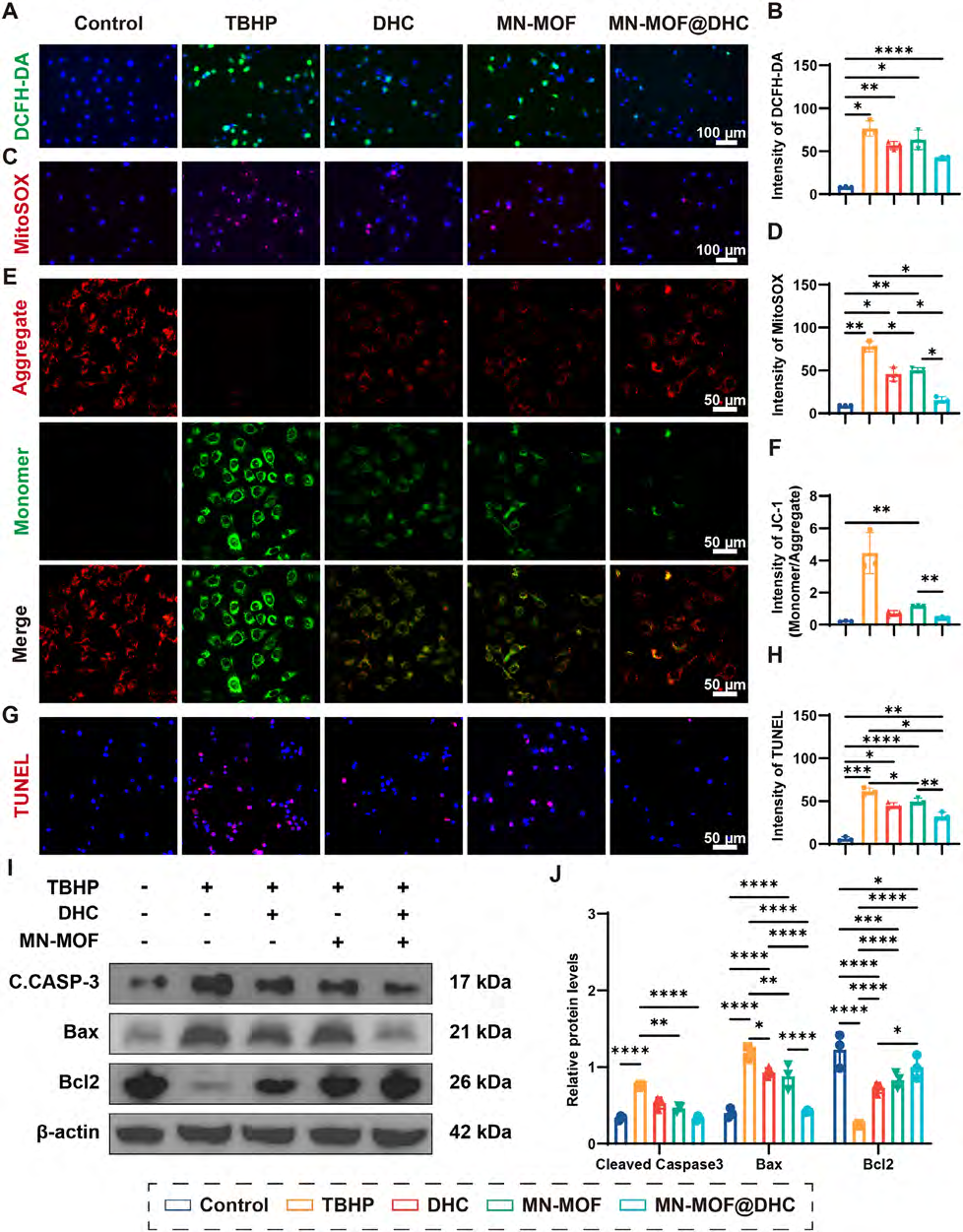 FIGURE 2
Assessment of the anti-oxidant, anti-apoptosis properties of MN-MOF@DHC in vitro. (A, C) Images of HUVECs were obtained after staining with DCFH-DA and MitoSOX, captured with diverse experimental conditions. (B, D) Semiquantitative analysis of fluorescence intensity. (E) Mitochondrial membrane potential was assessed by JC-1 assay in HUVECs under diverse experimental conditions. (F) Semiquantitative analysis of JC-1 fluorescence intensity. (G) TUNEL staining of HUVECs under different conditions. (H) Semiquantitative analysis of TUNEL staining. (I–J) The protein levels of cleaved caspase-3 (C.CASP-3), Bax, and Bcl-2 were measured by western blotting and semiquantitative evaluation. (n = 3, error bars, means ± SD; all analyses were done using one-way ANOVA with Tukey’s post hoc test *p < 0.05, **p < 0.01, ***p < 0.001, and **** P < 0.0001).
-100实物图
模块 3:调控巨噬 M1/M2 极化重塑抗炎微环境
1. 实验流程
LPS 诱导 RAW264.7 巨噬 M1 极化,给予各组微针浸提液干预;iNOS (M1)、CD206 (M2) 免疫荧光染色,配套 ELISA、qPCR 检测抗炎因子; abs9149 操作:巨噬爬片固定后,0.1% abs9149 PBS 室温通透 30 min,暴露胞内 iNOS、CD206 抗原,后续一抗孵育共聚焦成像定量荧光强度。
2. 关键结果
LPS 组 iNOS 高表达、CD206 极低;MN-MOF@DHC 显著下调 M1 标志物,上调修复型 M2 标志物,IL-4/IL-10/IL-13 抗炎因子、Arg1/Mrc1 M2 基因显著升高,证实可解除创面慢性炎症阻滞。
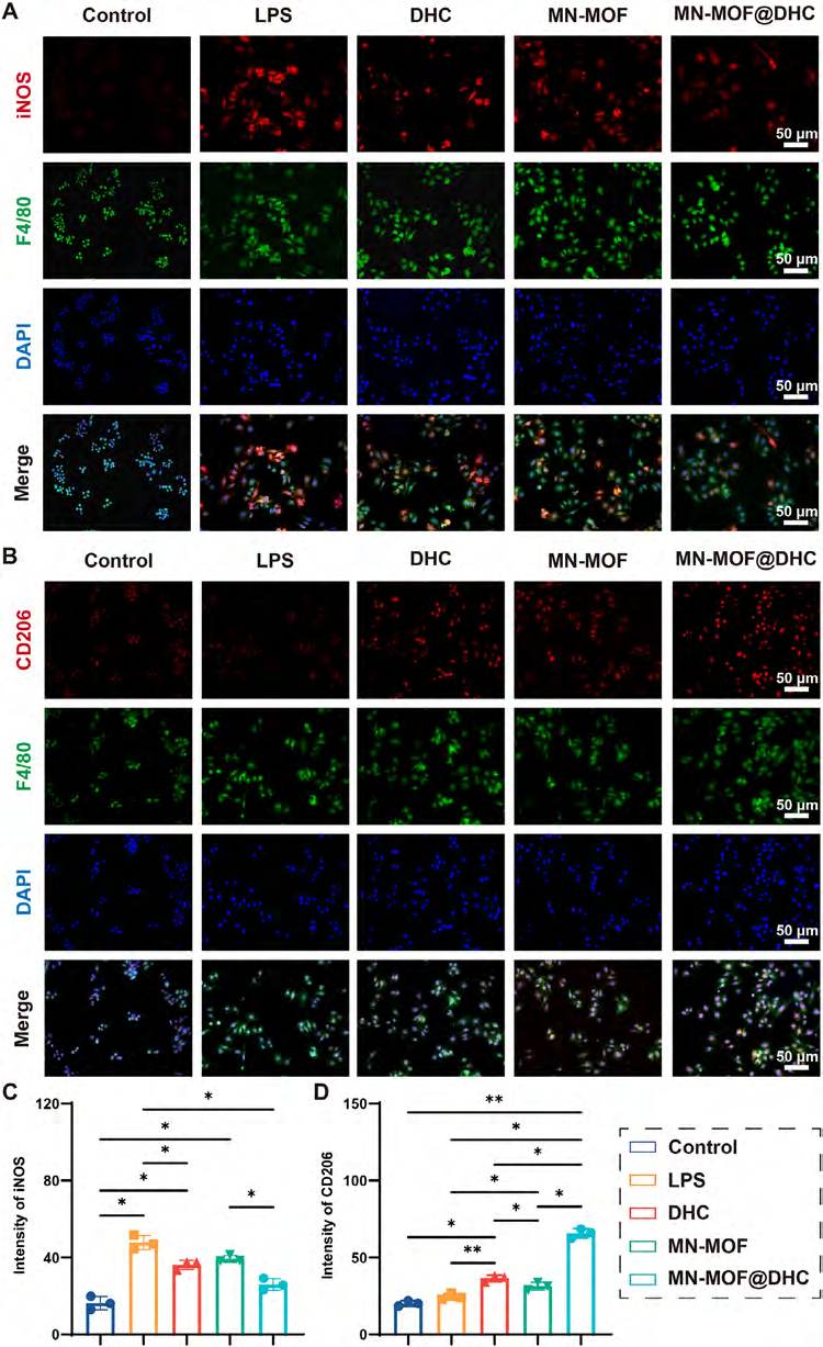 FIGURE 3
Regulation of macrophage phenotypes with MN-MOF@DHC. (A) Immunofluorescence images of iNOS expression in macrophages cocultured with DHC, MN-MOF, and MN-MOF@DHC. (B) Immunofluorescent images of CD206 expression in macrophages cocultured with DHC, MN-MOF, and MN-MOF@DHC. (C) semiquantitative assessment of iNOS in macrophages. (D) semiquantitative assessment of CD206 in macrophages. (n = 3, error bars, means ± SD; all analyses were done using one-way ANOVA with Tukey’s post hoc test *p < 0.05, **p < 0.01, ***p < 0.001, and **** P < 0.0001).
模块 4:促进内皮迁移与血管新生
1. 实验流程
TBHP 损伤 HUVEC,Transwell 迁移、Matrigel 管形成实验;CD31、VEGF 免疫荧光标记血管内皮; abs9149 操作:细胞爬片经 0.1% abs9149 通透,充分暴露胞内 CD31、VEGF 促血管蛋白,荧光半定量分析表达水平。
2. 关键结果
TBHP 损伤后内皮迁移、成管能力严重受损;MN-MOF@DHC 组迁移率达 64.7%,管状网络数量最多,CD31、VEGF 表达显著上调,证实缓释 DHC 协同 Ce-MOF 激活 VEGF 通路促进血管再生。
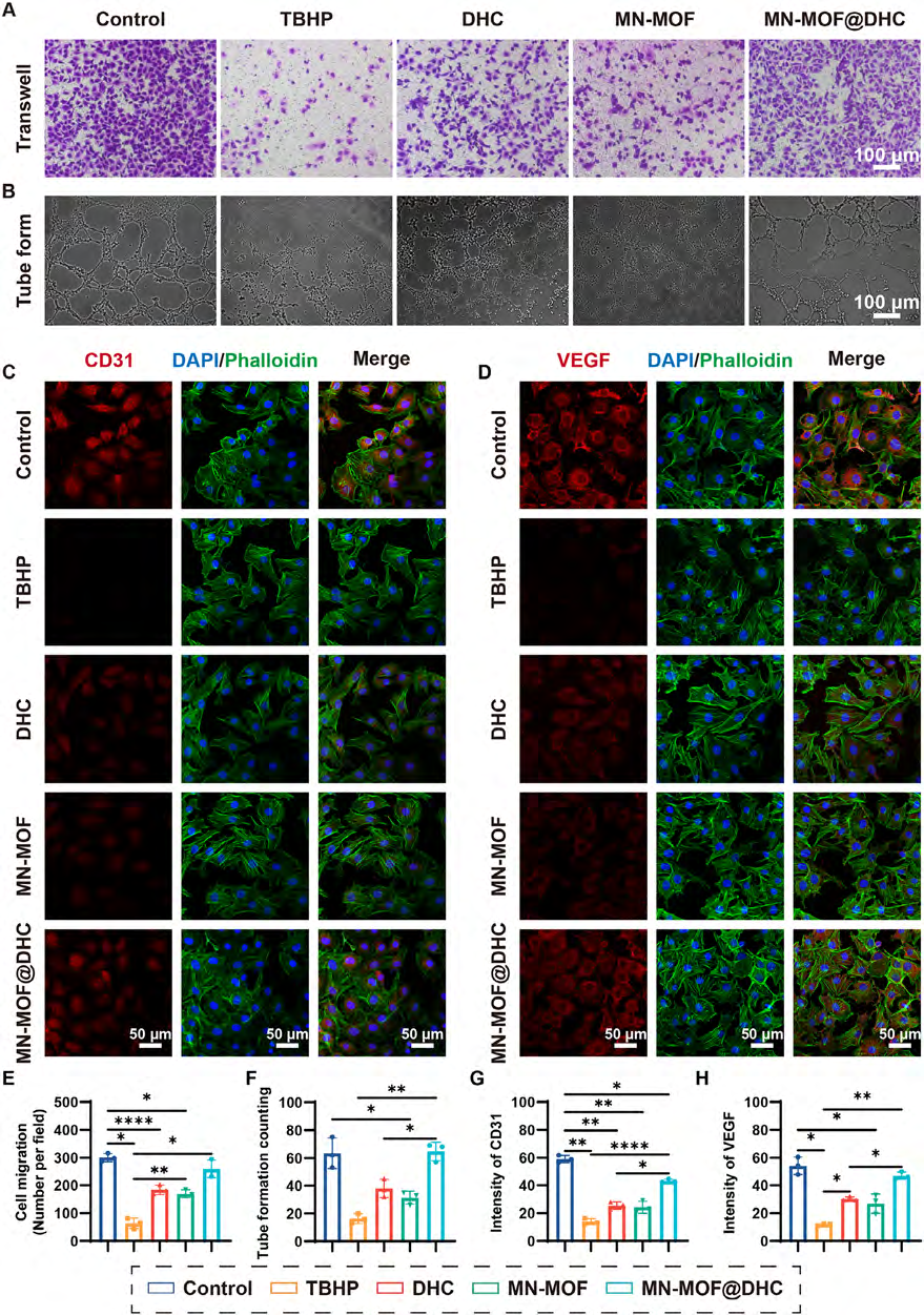 FIGURE 4
In vitro assessment of the angiogenesis properties of MN-MOF@DHC. (A) Transwell migration of HUVECs under various conditions. (B) Tube formation of HUVECs under various conditions. (C,D) Immunofluorescent images of CD31 and VEGF in HUVECs cocultured with various conditions. (E,F) Statistical evaluation of transwell migration and tube formation. (G-H) Semiquantitative analysis of CD31 and VEGF fluorescence intensity. (n = 3, error bars, means ± SD; all analyses were done using one-way ANOVA with Tukey’s post hoc test *p < 0.05, **p < 0.01, ***p < 0.001, and **** P < 0.0001).
模块 5:体内大鼠背部全层糖尿病创面愈合
1. 实验流程
高脂饮食 + STZ 诱导 2 型糖尿病 SD 大鼠,背部构建 15 mm 全层皮肤缺损,四组贴片干预;第 0/4/8/12/16 天拍照统计愈合率;第 8、16 天取材做 H&E 病理切片。
2. 关键结果
MN-MOF@DHC 愈合速率最优,第 12 天创面闭合率近 90%,16 天基本愈合;H&E 切片显示上皮化完整、肉芽组织更厚,创面缺损长度显著低于其余三组。
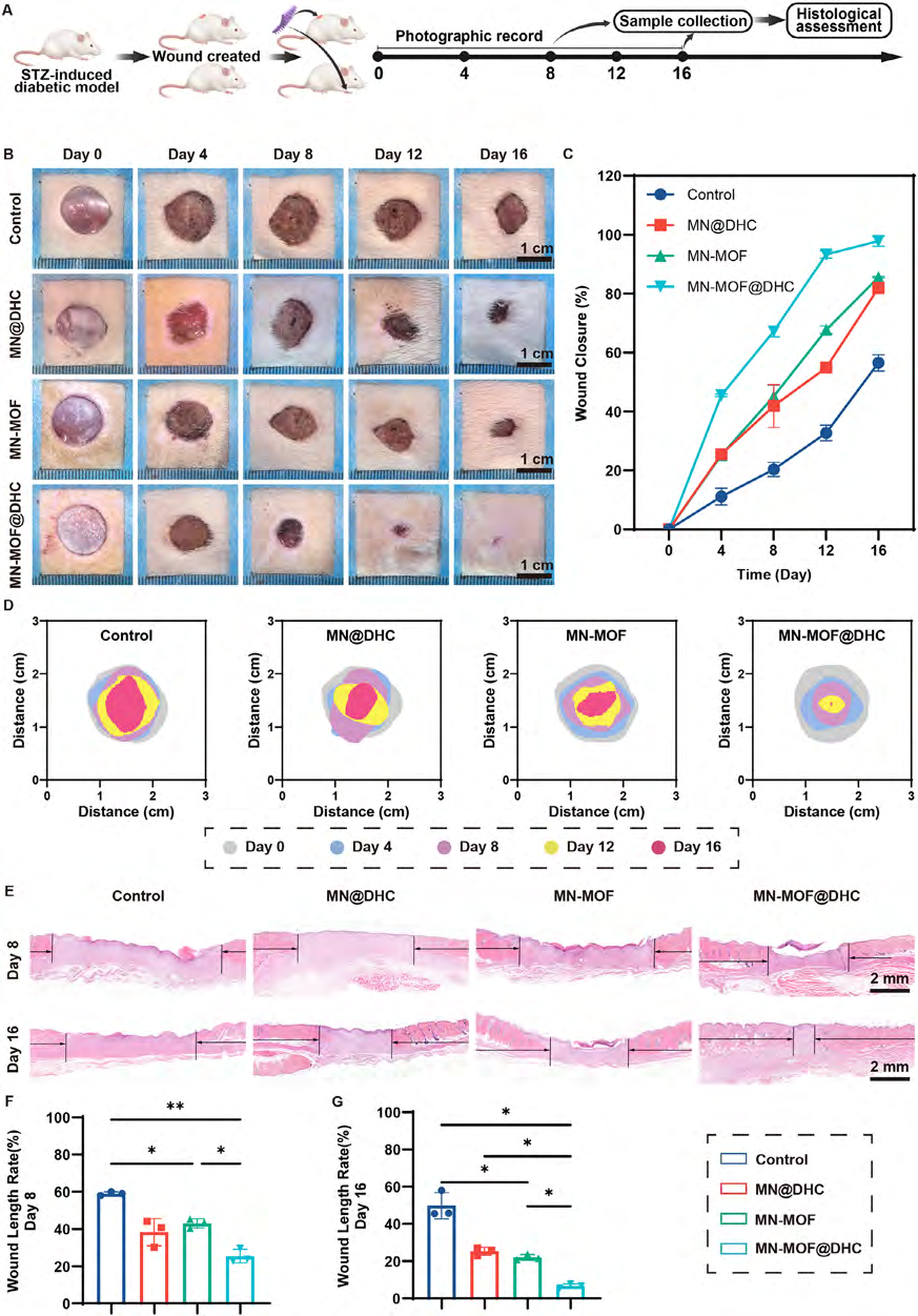 FIGURE 5
MN-MOF@DHC enhanced wound closure in diabetic rats. (A) Schematic diagram of the application process of therapeutic microneedles in animal experiments. (B) Illustrative images showed the diabetic wounds across various treatment groups. (C) Measurement and quantitative analysis of the residual wound areas were conducted on day 4, 8, 12, and 16 post-treatment for the various groups. (D) Traces of the healing process of diabetic wounds under various treatment groups. (E) Histological examination through H&E staining of the wound tissue in various groups on days 8 and 16. (F-G) Quantitative analysis of wound length rate on days 8 and 16. (n = 3, error bars, means ± SD; all analyses were done using one-way ANOVA with Tukey’s post hoc test *p < 0.05, **p < 0.01, ***p < 0.001, and **** P < 0.0001).
模块 6:体内组织抗氧化、抗炎、抗凋亡验证
1. 实验流程
创面制备 20 μm 冰冻切片,DHE 组织 ROS 染色、iNOS/CD206/Bax 免疫荧光; abs9149 操作:冰冻切片采用 0.1% abs9149 PBS 通透 15–30 min,打通组织脂质屏障,深层创面抗原充分暴露,保障 ROS、炎症、凋亡荧光均匀显色。
2. 关键结果
MN-MOF@DHC 组 DHE(ROS)荧光最弱,M1 标志物 iNOS 下调、M2 标志物 CD206 上调,促凋亡 Bax 蛋白表达最低,体外机制在体内组织层面完全复现。
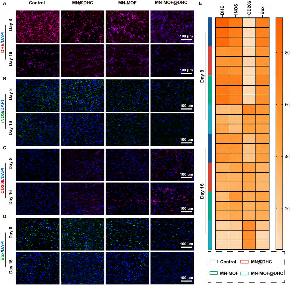 FIGURE 6
MN-MOF@DHC reduced oxidative stress, inflammation, and apoptosis in diabetic wound tissues. (A) Immunofluorescence images of wound tissue sections from a diverse group following DHE staining on days 8 and 16. (B-C) Immunofluorescence staining images of iNOS and CD206 captured on days 8 and 16. (D) Immunofluorescence images of Bax on days 8 and 16. (E) Semiquantitative analysis of DHE, iNOS, CD206, and Bax fluorescence intensity on days 8 and 16. (n = 3, error bars, means ± SD; all analyses were done using one-way ANOVA with Tukey’s post hoc test *p < 0.05, **p < 0.01, ***p < 0.001, and **** P < 0.0001)
模块 7:体内胶原沉积与成熟血管生成
1. 实验流程
创面切片 Masson 三色胶原染色;CD31 免疫组化、CD31/α-SMA 双荧光标记新生 / 成熟血管,统计血管密度;切片预处理均使用 abs9149 通透。
2. 关键结果
MN-MOF@DHC 组胶原沉积量最高、纤维排列规整;新生血管、成熟血管密度显著高于对照组,VEGF/Ang-1 通路协同激活促进血管成熟。
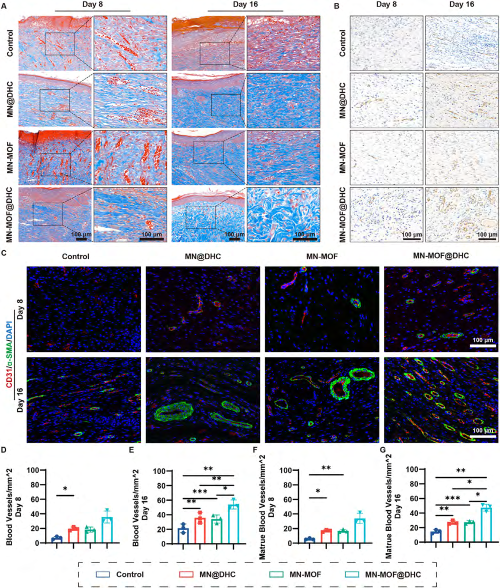 FIGURE 7
In vivo assessment of the angiogenic properties of MN-MOF@DHC in a diabetic wound model. (A) Visual representation of Massonstained wounds on days 8 and 16 post-treatment with MN@DHC, MN-MOF, and MN-MOF@DHC. (B) Immunohistochemicalstaining images depicting CD31 expression in wound tissues on days 8 and 16. (C) Immunofluorescence staining images for CD31 and α-SMA in diabetic wound tissues on days 8 and 16. (D-E) Quantitative analysis of newly formed blood vessels. (F-G) Quantitative assessment of mature blood vessels. (n = 3, error bars, means ± SD; all analyses were done using one-way ANOVA with Tukey’s post hoc test *p < 0.05, **p < 0.01, ***p < 0.001, and **** P < 0.0001).
模块 8:糖尿病足溃疡 DFU 临床模拟模型验证
1. 实验流程
糖尿病大鼠足部构建 6 mm 溃疡创面,分组干预;定期拍照、H&E 病理、CD31/α-SMA 血管荧光定量,切片透化使用 abs9149。
2. 关键结果
足部创面愈合规律与背部创面一致,MN-MOF@DHC 组 16 天基本愈合,肉芽组织有序、上皮再生完整,成熟血管密度显著提升,证实该微针适配临床糖尿病足病灶。
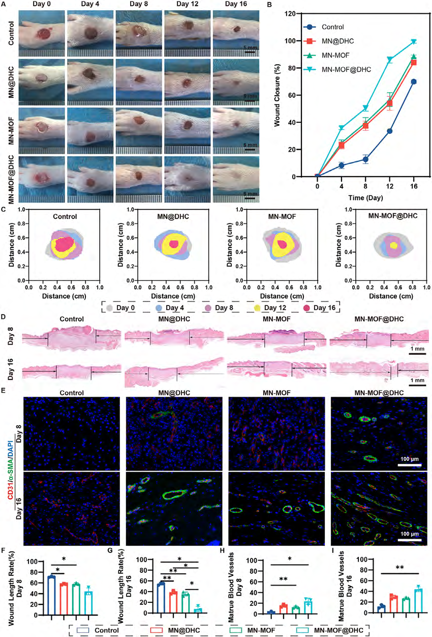 FIGURE 8
In vivo evaluation of MN-MOF@DHC on a diabetic feet wound model. (A) Representative photographs of the diabetic feet wounds under various treatment groups. (B) Measurement of the residual areas of foot wounds following treatment administered by various groups. (C) Traces of diabetic feet wound healing processes under diverse treatment groups. (D) Histological examination through H&E staining of the diabetic feet wound areas in various groups on days 8 and 16. (E) Immunofluorescence images of CD31 and α-SMA in feet diabetic wound tissues on days 8 and 16. (F-G) Quantitative analysis of feet wound length rate on days 8 and 16. (H-J) Quantitative measurement of mature blood vessels in feet wound tissues. (n = 3, error bars, means ± SD; all analyses were done using one-way ANOVA with Tukey’s post hoc test *p < 0.05, **p < 0.01, ***p < 0.001, and **** P < 0.0001).
五、Absin全系列产品整体作用总结
Absin 全套试剂贯穿本文材料表征、体外细胞、动物组织全部机制验证环节,是高分论文数据严谨性的核心支撑:

- 手机:13761418683