




- 手机:13761418683
联系人:爱必信
网址:http://www.absin.cn/
地 址:上海市浦东新区新浩路58号18号楼

- 材料制备与多维度理化表征:合成 CuO?纳米花、Nb?C/CuO?异质结,叶酸 PEG 修饰并负载 R848、MTX,TEM、XPS、光热、ESR 等验证光学与催化性能(对应 Fig.1);
- 体外靶向与细胞毒性:4T1 乳腺癌细胞验证叶酸摄取、ROS、线粒体损伤、凋亡杀伤能力(对应 Fig.2);
- 铜死亡与免疫原性死亡分子机制:WB 检测铜死亡核心蛋白,CLSM 检测 CRT/HMGB1 等 ICD 标志物,体外 DC 成熟实验验证免疫激活(对应 Fig.3);
- 体内生物分布与原位 4T1 抑瘤:ICP-MS、活体荧光验证肿瘤富集,分组评估肿瘤生长、脏器安全性、转移抑制(对应 Fig.4);
- 体内多维度免疫重塑:流式检测肿瘤、淋巴结内 DC、巨噬细胞、CD4/CD8 T 细胞浸润,血清炎症因子定量(对应 Fig.5);
- 双侧荷瘤模型验证远端抑瘤与免疫记忆:原发 + 远端双肿瘤评估全身免疫抑制转移,脾脏记忆 T 细胞检测(对应 Fig.6);
- RNA 转录组测序:差异基因 GO/KEGG/GSEA 富集,从分子层面证实铜死亡、氧化应激、免疫通路协同激活(对应 Fig.7);
- 综合总结材料优势、临床转化前景与后续长期毒性研究展望。
- TEM 证实 CuO?纳米花、Nb?C 片层成功复合,元素 mapping 均匀分布 Nb、Cu、O;
- XPS 验证 Cu2?、过氧键 O-O 存在,证明 CuO?稳定复合;
- NbCuF 光热转换效率 41.48%,5 次升降温无衰减,体内激光照射肿瘤局部升温至 46℃;
- ESR 证明异质结可高效产生羟基自由基、单线氧,酸性 + 高 GSH TME 下 ROS 生成显著增强;
- FA 修饰 NbCuF 细胞摄取量远高于无靶向 NbCu,具备叶酸受体靶向特异性;
- NbCuF@MR + 激光组 ROS 水平最高,线粒体膜电位严重丢失,凋亡率高达 96%;
- MDA-MB-231、BT-474 双乳腺癌细胞重复验证广谱杀伤效果;
- NbCuF@MR+L 组 DLAT 寡聚化显著上调,FDX1、LIAS 表达下调,铜死亡通路完全激活;铜螯合剂 TTM 可逆转细胞死亡,证实死亡依赖铜离子;
- 激光联合组大量 CRT 外翻、HMGB1 释放,胞外 ATP 升高,强效 ICD;
- 处理后肿瘤上清可显著促进 CD80?CD86?成熟 DC 比例上升;克隆形成能力几乎完全抑制;
- NbCuF 组肿瘤荧光信号最强,24 h 肿瘤 Nb、Cu 蓄积最高,主要脏器可逐步代谢清除;
- NbCuF@MR + 激光组肿瘤抑制率近 90%,小鼠体重无下降,心肝脾肺肾无病理损伤,生物安全;
- 仅联合治疗组肝脏无肿瘤转移灶,Ki67 增殖蛋白显著下调,TUNEL 凋亡信号大幅升高,肿瘤内 CD8?T 大量浸润;
- NbCuF@MR+L 组成熟 DC(CD80?CD86?)提升 4.6 倍,肿瘤 CD8?T、CD4?T 分别提升 13.7、5.4 倍;
- TAMs 完成 M2→M1 极化,免疫抑制微环境被逆转;血清促炎细胞因子 TNF-α、IL-6、IFN-γ 显著升高,全身抗肿瘤免疫激活;
- NbCuF@MR + 激光组原发、远端肿瘤生长均被强烈抑制,远端肿瘤 CD8?T 大量浸润;
- 脾脏效应记忆 T 细胞比例显著升高,证明治疗诱导长效免疫记忆,可长期抵抗肿瘤复发与远处转移;
- 差异基因富集 TCA 循环(铜死亡核心通路)、谷胱甘肽代谢、凋亡、NF-κB 氧化应激通路;
- Toll 样受体、细胞因子互作、T 细胞分化、TNF 免疫通路显著上调,完整印证表型实验中免疫激活结论;
- 温和高效组织解离,保护免疫抗原:该胶原酶低胰蛋白酶活性,消化肿瘤、脾脏、淋巴结结缔组织时不会破坏 CD3、CD4、CD8、CD80、CD86、CD206 等细胞膜表面标志物,最大程度保留免疫细胞天然表型,避免消化造成流式假阴性结果;
- 多实体组织通用标准化试剂:适配乳腺肿瘤、引流淋巴结、脾脏多种致密结缔组织样本,统一消化体系,保证原位瘤、双侧转移瘤多批次实验数据横向对比稳定,符合 CEJ 顶刊实验重复性质控标准;
- 支撑核心免疫创新结论落地:全文 “M2 巨噬细胞极化、DC 成熟、CD8 效应 T 浸润、免疫记忆” 所有关键流式数据均依赖该试剂制备单细胞悬液,是论证纳米平台重塑肿瘤免疫微环境不可或缺的基础工具,为材料 - 铜死亡 - 免疫协同创新机制提供可靠实验支撑。
高分 SCI 文献深度解析:TME 响应 MXene 纳米异质结协同光疗、代谢干扰强化铜死亡与肿瘤免疫治疗
2026-08-06
一、文献基础综述
(一)期刊基础信息
论文标题:TME-activated MXene nanoheterojunctions engineer cancer cells through phototherapy and metabolic interference for enhanced cuproptosis and immunotherapy
期刊:Chemical Engineering Journal(CEJ)
2026 最新 IF:12.5,JCR 一区 Top 期刊,化工、生物材料领域顶刊
DOI:10.1016/j.cej.2026.178527
(二) 全文核心结论
本研究构建叶酸靶向 Nb?C/CuO?纳米异质结纳米载体 NbCuF@MR,共载免疫佐剂 R848 与代谢抑制剂甲氨蝶呤(MTX)。该纳米平台可特异性响应肿瘤微环境(酸性、高 GSH)靶向释放铜离子,结合 808 nm 近红外激光光热 / 光动力双重作用放大氧化应激,通过干扰线粒体 TCA 循环诱导强效铜死亡;铜死亡同步触发免疫原性细胞死亡(ICD),重塑免疫抑制型冷肿瘤微环境,促进树突状细胞成熟、M2 型肿瘤相关巨噬细胞向 M1 极化、CD4?/CD8?效应 T 细胞大量浸润。体内原位、双侧 4T1 乳腺癌模型证实单轮给药即可显著抑制原发瘤生长、远端转移并建立长效抗肿瘤免疫记忆,转录组完整验证铜死亡、氧化应激、免疫相关通路协同激活,为铜死亡联合光免疫治疗提供可转化纳米新策略。
(三)爱必信 Absin 产品应用简述
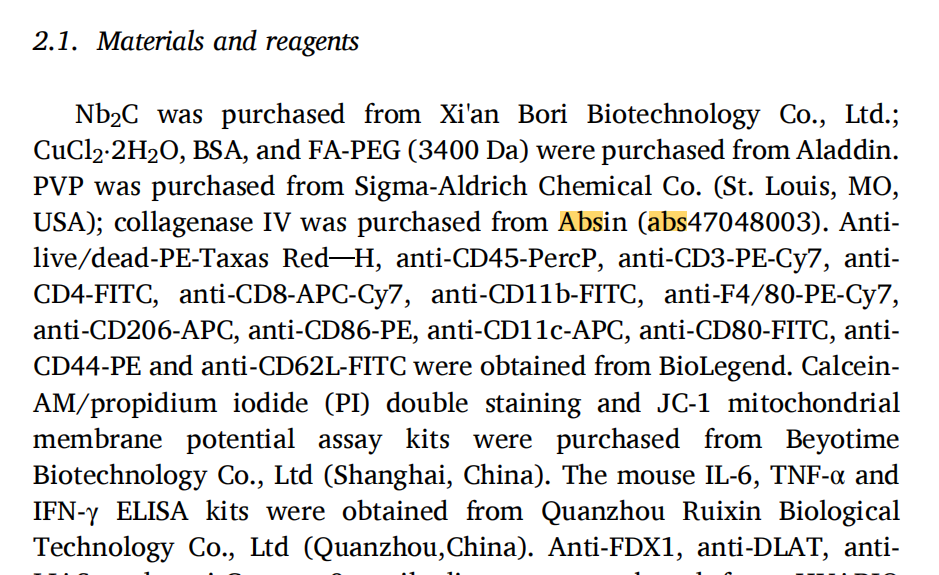
实验采用胶原酶 IV(货号 abs47048003) 完成小鼠肿瘤、脾脏、淋巴结实体组织的高效解离,完整保留免疫细胞膜表面抗原活性,为后续流式免疫细胞分群、肿瘤单细胞功能检测提供高质量单细胞悬液,保障体内免疫表型数据精准可靠。
二、研究领域背景介绍
1.铜死亡(Cuproptosis)领域瓶颈:铜死亡是全新铜依赖线粒体程序性死亡,靶向肿瘤线粒体脂酰化蛋白 DLAT、FDX1、LIAS 实现肿瘤杀伤,但游离铜离子全身递送会造成正常器官毒性;肿瘤高浓度谷胱甘肽(GSH)会螯合铜离子大幅削弱铜死亡效果,CuO?纳米材料自身稳定性差、易提前降解,限制体内应用转化。
2.肿瘤免疫治疗核心痛点:实体瘤属于 “冷肿瘤”,缺氧、高 GSH、大量 M2 型巨噬细胞、成熟 DC 匮乏、效应 T 细胞浸润不足形成强免疫抑制微环境;单纯免疫激动剂难以有效浸润肿瘤,无法启动持久全身抗肿瘤免疫。
3.MXene 纳米材料优势与空白:Nb?C MXene 具备优异近红外光热、产氧、耗竭 GSH 性能,但单一 MXene 无铜死亡诱导能力;现有铜基纳米体系无法同步实现靶向铜递送、光疗增效、代谢重编程、免疫激活四重功能,缺少集铜死亡放大、光疗、免疫调节一体化靶向纳米平台。
4.本文创新切入点:针对以上三重领域瓶颈,首次将 Nb?C MXene 与 CuO?构建 TME 响应纳米异质结,叶酸靶向实现肿瘤精准铜递送,同步搭载 R848/MTX 完成代谢重塑,808 nm 激光放大铜死亡与 ICD,打通铜死亡 - 光疗 - 免疫正反馈环路,填补多功能铜死亡纳米免疫治疗材料研究空白。
三、作者整体递进研究思路总览
全文遵循材料合成表征→体外细胞功能验证→铜死亡 / ICD 机制解析→体内生物分布与单药抑瘤→肿瘤微环境免疫重塑验证→双侧肿瘤远端转移 + 免疫记忆验证→转录组分子通路佐证8 层完整递进逻辑:
四、分模块详细研究思路
模块 1:NbCuF@MR 纳米异质结合成与理化表征
1、研究逻辑:分步构建 CuO?、Nb?C 异质结,表面 FA-PEG 靶向修饰,共载 R848+MTX,系统验证形貌、元素、光热、产 ROS、TME 响应催化性能;
2、核心实验:纳米合成、TEM/HAADF-STEM 元素 mapping、XPS 全谱 / 高分辨谱、808 nm 光热升温循环稳定性、ESR 检测?OH/1O?、模拟肿瘤微环境催化实验;
3、关键结果
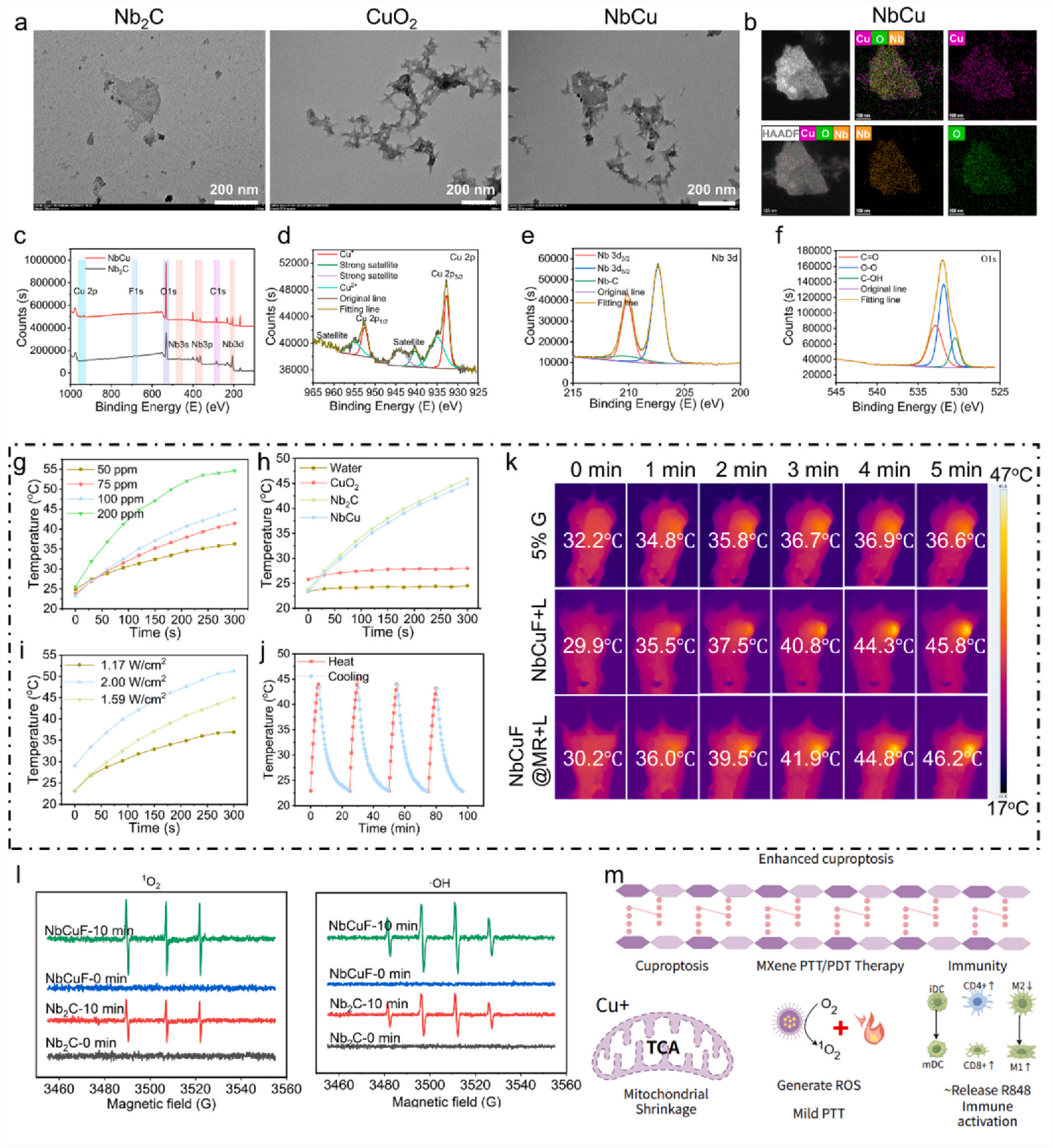
Fig. 1. (a) TEM images of Nb2C, CuO2 and NbCu (scale bar: 200 nm); (b) HAADF-STEM image and corresponding elemental mapping images (Cu, O, Nb) (scale bar: 100 nm); (c) wide-scan XPS spectra of NbCu and high-resolution XPS spectra of (d) Cu 2p, (e) Nb 3d and (f) O 1s of NbCu; (g) photothermal heating curves of NbCuF under 808 nm laser irradiation for 5 min at different concentrations (1.59 W/cm2 ); (h) photothermal heating curves of NbCuF, Nb?C, and CuO? under identical concentration and power density (100 ppm, 1.59 W/cm2 ) during 808 nm laser irradiation for 5 min; (i) photothermal heating curves of NbCuF under 808 nm laser irradiation for 5 min at different power densities (100 ppm); (j) thermal stability of NbCuF. (k) Representative infrared thermal images of tumors in mice. (l) ESR spectra of Nb2C and NbCuF; (m) multiple mechanisms of action produced by NbCuF@MR after entering tumor cells.
模块 2:体外细胞靶向摄取、光疗联合细胞毒性、线粒体损伤
1、研究逻辑:验证叶酸介导肿瘤细胞特异性摄取,检测纳米体系在 808 nm 激光下 ROS 爆发、线粒体膜电位崩塌、细胞凋亡;
2、核心实验:Cy5.5 荧光共聚焦摄取、DCFH-DA ROS 染色、JC-1 线粒体膜电位、Calcein-AM/PI 活死染色、Annexin V/7-AAD 流式凋亡;
3、关键结果
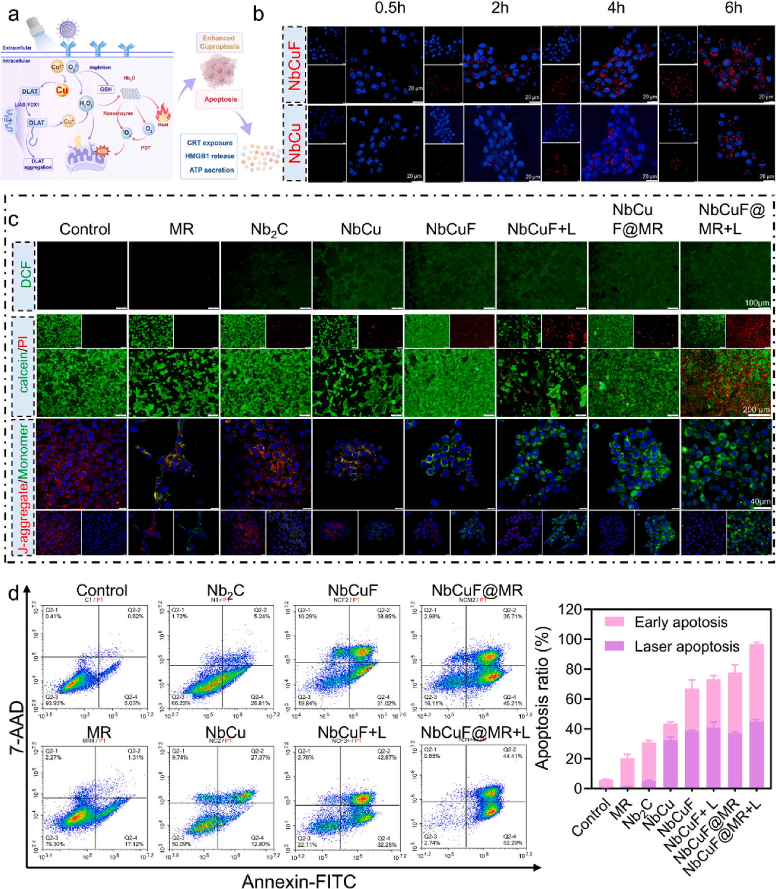
Fig. 2. In vitro evaluation of cell-targeting of NbCuF@MR nanoplatforms. (a) Cuproptosis and ICD effects induced by NbCuF@MR upon uptake by tumor cells. (b) CLSM images showing the intracellular uptake of Cy5.5-NbCu and Cy5.5-NbCuF in 4T1 cells at different times. (c) Measurements of antitumor efficacy and mechanism of NbCuF@MR against 4T1 cells through live/dead cell staining (scale bar: 100 μm), ROS staining (scale bar: 200 μm) and JC-1 staining (scale bar: 40 μm) after different treatments (1.59 W/cm2 , 5 min). (d) Apoptosis of 4T1 cells following different treatments was estimated using the annexin V-FITC/7-AAD assay and the corresponding quantitative results (1.59 W/cm2 , 5 min).
模块 3:体外铜死亡通路激活与免疫原性死亡(ICD)诱导
1、研究逻辑:分子层面证明纳米体系诱导经典铜死亡,同时释放 DAMPs 激活树突状细胞;
2、核心实验:WB 检测 FDX1、LIAS、DLAT、Cleaved Caspase-9;CLSM 观测细胞膜 CRT 外翻、胞外 HMGB1 释放;骨髓来源 DC(BMDC)共培养流式成熟度检测;细胞克隆形成抑制实验;
3、关键结果
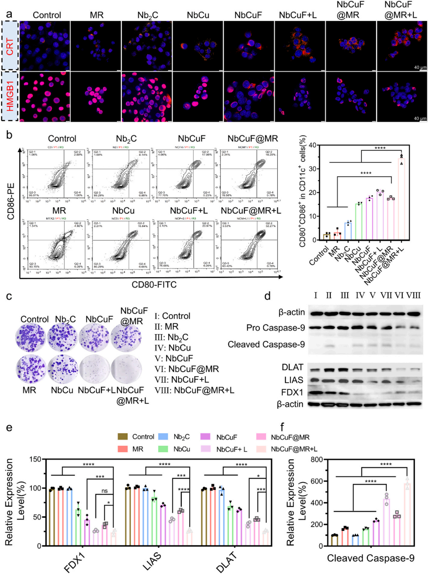
Fig. 3. Mechanistic investigations of the antitumor effects of NbCuF@MR. (a) CLSM images of CRT expression and HMGB1 release in 4T1 cells after different treatments (1.59 W/cm2 , 5 min). (b) Flow cytometry showing the stimulatory effects of different treatments on BMDC maturation (1.59 W/cm2 , 5 min). (c) Colonyformation ability of 4T1 cells treated with the different nanoparticles (1.59 W/cm2 , 5 min). (d) Western blotting analysis of DLAT (*p = 0.0153, ***p = 0.0002, ****p < 0.0001), LIAS (***p = 0.0002, ****p < 0.0001), FDX1 (ns = 0.9726, *p = 0.0466, ***p = 0.0009, ****p < 0.0001) and Caspase-9 expression (n = 3, independent experiments). (e) (f) Quantification of the corresponding protein levels.
模块 4:体内生物分布、原位 4T1 肿瘤抑瘤、脏器安全性
1、研究逻辑:尾静脉给药验证纳米肿瘤富集,8 组对照评估体内抑瘤、抗转移、生物安全性;
2、核心实验:Cy5.5 活体荧光、ICP-MS 脏器 Nb/Cu 定量;小鼠分组给药、激光处理,隔日记录肿瘤体积 / 体重;肿瘤 H&E、Ki67、TUNEL、CD8 免疫荧光;肝脏转移灶病理检测;
3、关键结果
4、Absin 产品使用标注:本阶段肿瘤、脾脏、引流淋巴结单细胞制备环节使用 Absin 胶原酶 IV abs47048003 消化实体瘤组织,温和降解胶原基质,完整保留免疫细胞表面标志物,避免消化过度损伤 CD4/CD8/CD86 等抗原,保证后续流式检测数据准确。
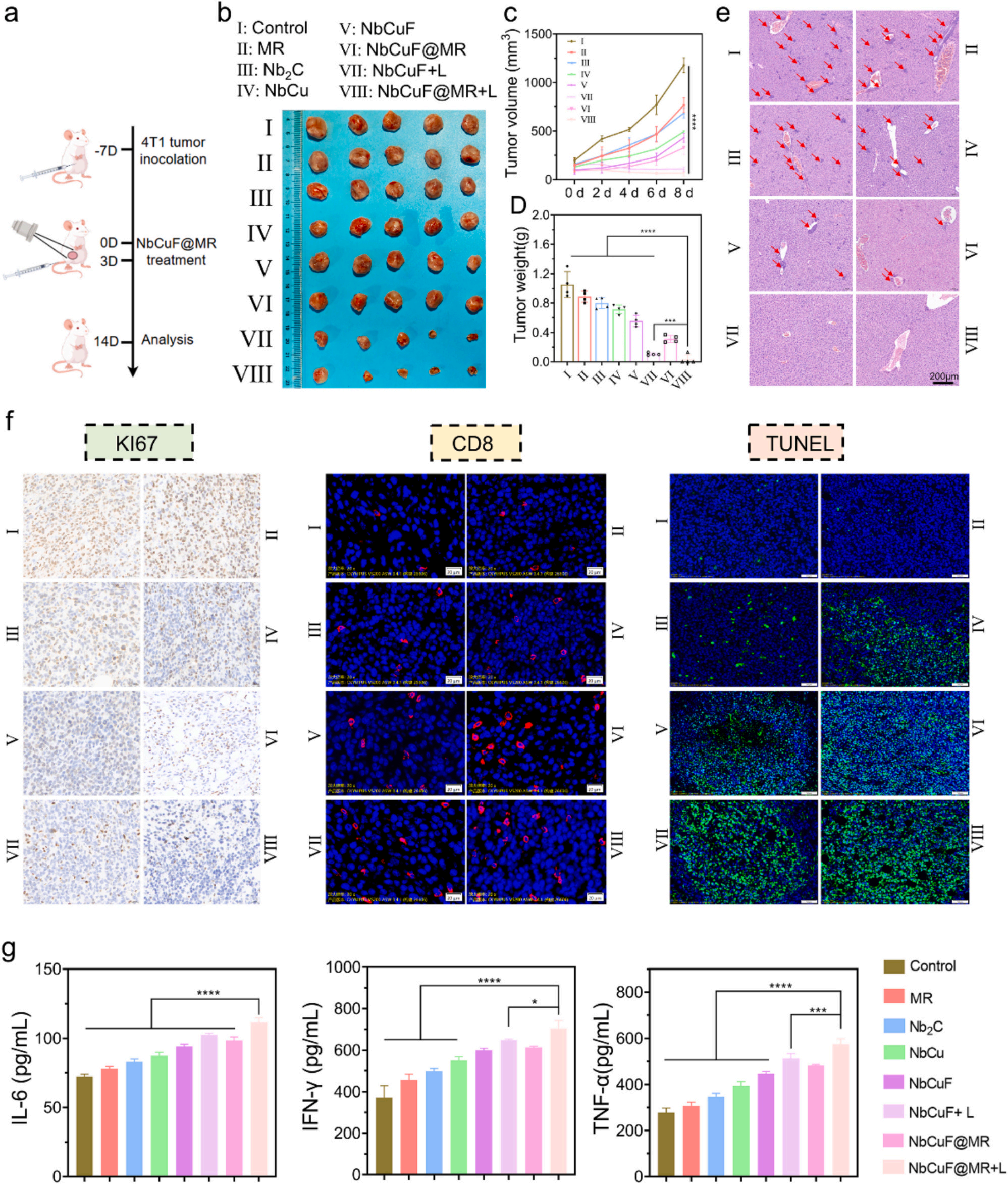
Fig. 4. In vivo antitumor effect. (a) Timeline of orthotopic 4T1 breast cancer mice treated with NbCuF@MR (2.0 W/cm2 , 5 min). (b) Tumor photographs. (c) Tumor growth curves of mice during the observation period after different treatments. (d) Average weights of tumors harvested after different treatments (ns = 0.7397, ***p = 0.0007, ****p < 0.0001; n = 5 independent experiments). (e) H&E staining of mouse liver tissues after different treatments, the red arrow indicates the metastatic tumor site. (f) KI67, H&E, CD8, TUNEL, CRT and FDX1 staining images of tumors after different treatments. (g) Secretion levels of TNF-α (***p = 0.0001, ****p < 0.0001), IL-6 and IFN-γ (*p = 0.0479, NbCuF@MR + L vs. NbCuF@MR ***p = 0.0006, NbCuF@MR + L vs. NbCuF ***p = 0.0001, ****p < 0.0001) in mouse serum after treatments (n = 4 independent experiments).
模块 5:体内肿瘤微环境免疫重塑(巨噬细胞、DC、T 细胞)
1、研究逻辑:多组织流式系统解析治疗后免疫微环境变化,验证冷肿瘤向热肿瘤转化;
2、核心实验:脾脏、肿瘤、引流淋巴结组织经胶原酶 IV 消化制备单细胞悬液,多色流式分群 CD4?/CD8?T、M1/M2 巨噬细胞、成熟 DC;血清 TNF-α、IL-6、IFN-γ ELISA 定量;免疫荧光 CD8 染色;
3、关键结果
4、Absin 产品使用标注:全程依赖 abs47048003 胶原酶 IV 完成实体瘤、淋巴结、脾脏组织解离,获得高活性单细胞悬液,是流式免疫分群实验前置关键试剂。
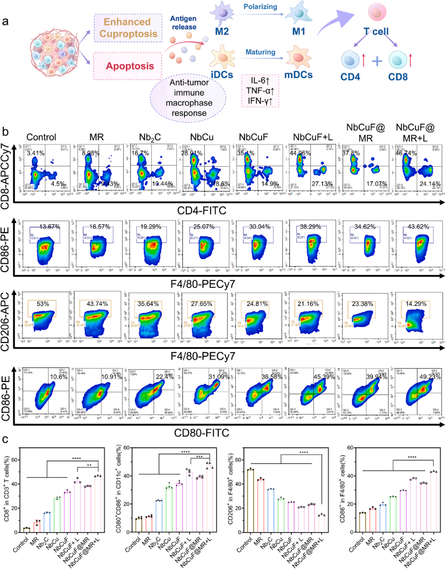
Fig. 5. In vivo evidence supporting the antitumor effects of NbCuF@MR. (a) Schematic illustration of an antitumor immune response associated with cuproptosisrelated stress. (b) Representative flow cytometric analysis following the administration of the various treatments. (c) Quantification of immune cells in 4T1 tumorbearing mice (CD8+: **p = 0.0034, ****p < 0.0001; CD80+CD86+: ***p = 0.0009, ****p < 0.0001; n = 3 independent experiments).
模块 6:双侧 4T1 荷瘤模型验证远端转移抑制与长效免疫记忆
1、研究逻辑:构建原发 + 远端双肿瘤模拟肿瘤转移模型,评估全身系统性免疫应答与免疫记忆形成;
2、核心实验:双侧皮下荷瘤造模,分组治疗测量双侧肿瘤体积重量;肿瘤组织 CD8、TUNEL 免疫荧光;脾脏记忆 T 细胞(CD44?CD62L?)流式检测;
3、关键结果
4、Absin 产品使用标注:双侧肿瘤、脾脏组织解离统一使用 Absin abs47048003 胶原酶 IV,标准化消化流程保证多组样本平行可比,减少实验误差。
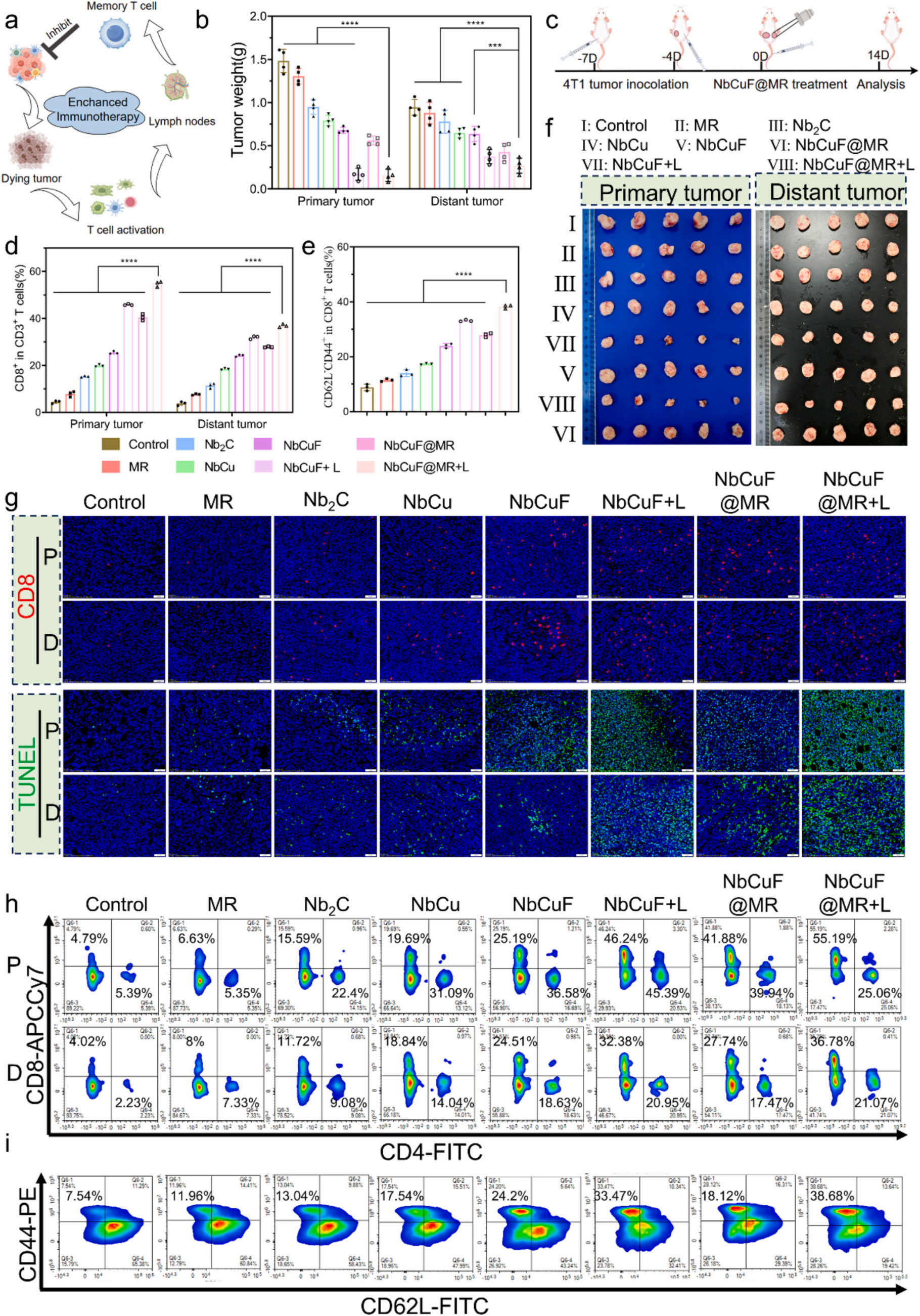
Fig. 6. In vivo antitumor efficacy of NbCuF@MR in a bilateral tumor model. (a) Mechanism of NbCuF@MR-induced long-term immune memory effects. (b) Average weights of tumors harvested after different treatments (c) Timeline of bilateral 4T1 breast cancer mice treated with NbCuF@MR (2.0 W/cm2 , 5 min). (d) Quantitative analysis of CD8+ T cells (CD45+CD3+CD8+) in tumors. (e) Quantitative analysis of splenic effector memory T cell (CD45+CD44+CD62L?) in spleens. (f) Tumor photographs. (g) CD8 and TUNEL staining images of tumors after different treatments. Representative flow cytometric analysis of splenic effector memory T cells in tumors (h) and CD8+ T cells in spleens (i).
模块 7:RNA 转录组测序解析全局分子通路变化
1、研究逻辑:转录组全景解析治疗后肿瘤基因差异,从分子通路层面佐证铜死亡、氧化应激、免疫通路协同激活;
2、核心实验:Control 与 NbCuF@MR+L 组肿瘤 RNA 提取,测序后韦恩图、火山图、热图、KEGG/GO/GSEA 富集分析;
3、关键结果
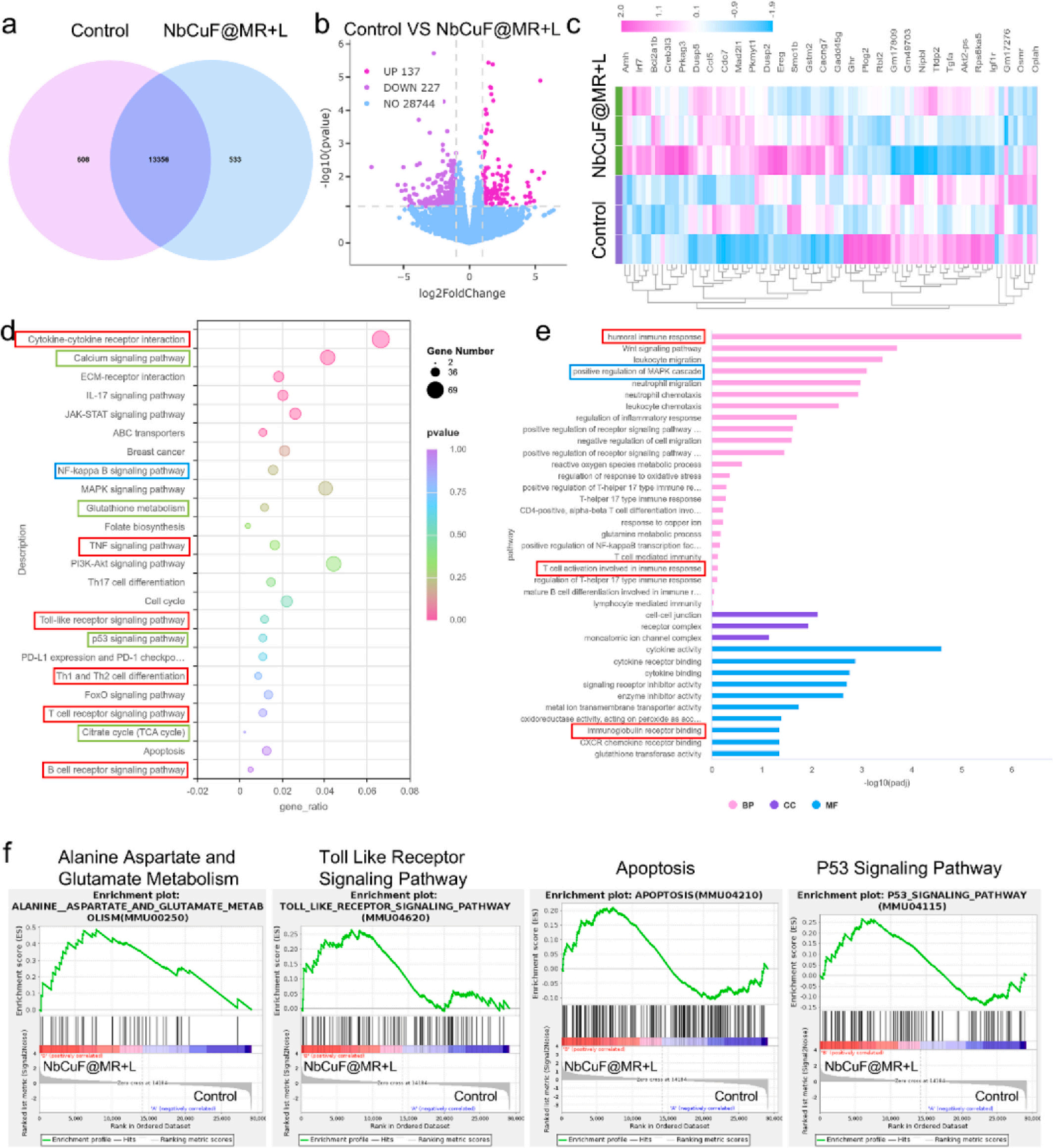
Fig. 7. Transcription analysis of 4T1 cells by RNA transcriptome analysis. (a) A Venn diagram revealed the number of genes transcribed in each treatment group. (b) Heat map of gene alterations between the control or NbCuF@MR + L group. (c) Heat map of gene alterations between the control and NbCuF@MR + L group. (d) Volcano plots displayed the differentially expressed genes by KEGG enrichment analysis. (e) GO enrichment analysis of differentially expressed pathways. (f) GSEA showed the gene sets of alanine aspartate and glutamate metabolism, toll-like receptor signaling pathway, apoptsis, and P53 signaling pathway
五、Absin产品整体作用总结
Absin 货号 abs47048003 胶原酶 IV 是本研究体内免疫表型全套实验的基础前置核心试剂,支撑全文免疫重塑核心结论的数据可靠性,价值分为三层:

- 手机:13761418683