




- 手机:13761418683
联系人:爱必信
网址:http://www.absin.cn/
地 址:上海市浦东新区新浩路58号18号楼

- PCA 与 OPLS?DA 显示高低产奶牛氨基酸代谢谱完全分离,置换检验证明模型无过拟合;
- 共筛选 13 个差异氨基酸,L?Pro 在高产组显著上调;
- KEGG 富集精氨酸 / 脯氨酸代谢通路显著富集,提示 L?Pro 与泌乳性能强相关;
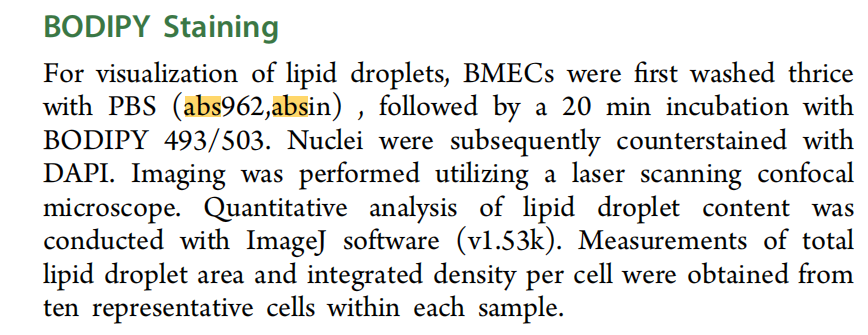
- Absin abs962 PBS 应用:奶牛乳腺活检组织离体后,预冷 abs962 PBS 反复冲洗去除血液杂质,保证代谢组样本纯度。
- 0.5 mM L?Pro 为最优浓度,显著上调 CDK1、PCNA、Cyclin D1,EdU 阳性细胞比例上升,显著促进细胞增殖;
- α/β? 酪蛋白 mRNA 与蛋白表达显著升高,β? 酪蛋白荧光强度提升;
- 脂合成基因 FASN/SREBP1/DGAT1/2 上调,胞内与分泌 TAG 含量、脂滴面积显著增加;
- L?Pro 组 516 个差异基因,显著富集 mTOR、PI3K?Akt 通路;GSEA 证实 mTOR 通路显著激活;
- L?Pro 剂量依赖性提升 p?mTOR、p?S6K、p?4EBP1;RAPA 完全逆转 L?Pro 促增殖、促乳脂 / 酪蛋白合成效应;
- SNAT2(SLC38A2)基础表达最高,L?Pro 呈剂量依赖性上调;
- SNAT2 敲除完全消除 L?Pro 对 mTOR 通路激活、细胞增殖、乳脂 / 蛋白合成的促进作用;证明 SNAT2 是 L?Pro 进入乳腺细胞必需载体;
- 高产奶牛乳腺 P4HA2 mRNA、蛋白显著高于低产组;SNAT2 敲低后 L?Pro 无法上调 P4HA2;
- MD 模拟显示 L?Pro 与 P4HA2 结合构象稳定;DARTS/CETSA 证实 L?Pro 结合提升 P4HA 蛋白稳定性;
- P4HA2 敲除完全阻断 L?Pro 诱导 mTOR 磷酸化、细胞增殖、乳脂酪蛋白合成;
- 单独过表达 P4HA2 可在无外源 L?Pro 条件下轻度激活 mTOR,促进乳合成;证明 P4HA2 正向调控泌乳;
- Co?IP 直接证明细胞内 P4HA2 与 mTOR 物理结合;
- P4HA2 过表达提升 mTOR?LAMP2 共定位,敲低则降低;L?Pro 处理可提升共定位,敲除 P4HA2 后该效应消失;明确机制:L?Pro?P4HA2 复合物促进 mTOR 向溶酶体迁移激活 mTORC1;
- 中剂量组(每日 50g L?Pro)产奶量、FCM、ECM 提升最显著,乳脂率、乳蛋白、乳糖均显著高于对照组;处理 × 时间交互显著,饲喂时间越长提升效果越明显;高剂量提升幅度弱于中剂量;
SCI 文献深度解析:《The Functional Amino Acid L Proline Enhances Lactation Performance by Activating the SNAT2-P4HA2-mTORC1 Axis》
2026-08-20
一、文献基础综述
(一)期刊与基础信息
期刊:Journal of Agricultural and Food Chemistry(JAFC,ACS 老牌农林 1 区 TOP 期刊);2026 最新影响因子 6.7;
DOI:10.1021/acs.jafc.5c15936;发表卷期:2026, 74, 15921?15944;
论文标题:The Functional Amino Acid L?Proline Enhances Lactation Performance by Activating the SNAT2?P4HA2?mTORC1 Axis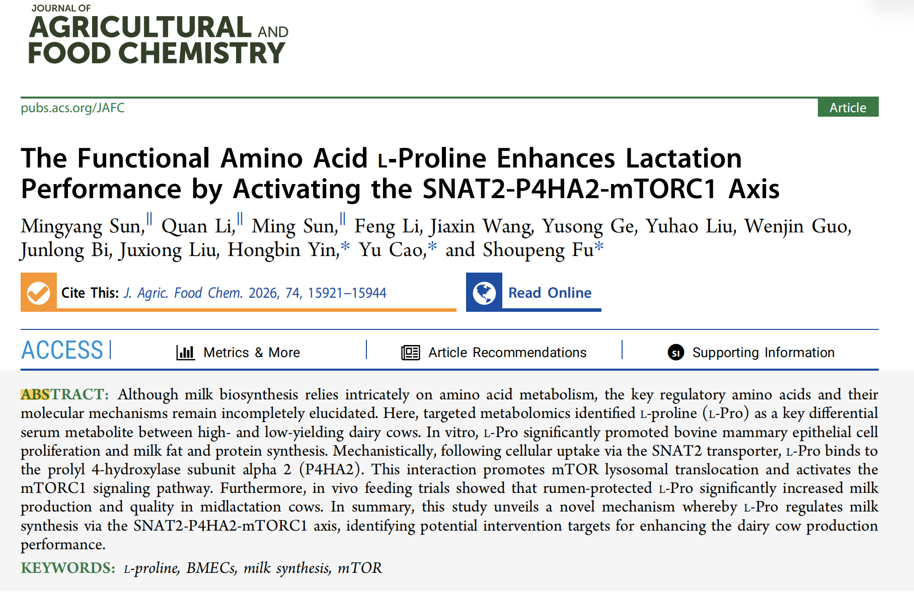
(二)全文核心结论
靶向代谢组学证实高产奶牛血清 L? 脯氨酸(L?Pro)显著富集;体外牛乳腺上皮细胞(BMEC)实验证明 L?Pro 可促进细胞增殖、提升乳蛋白与乳脂合成;机制层面:L?Pro 经 SNAT2 转运入胞后直接结合 P4HA2,促进 mTOR 向溶酶体转位并激活 mTORC1 通路;体内围保护 L?Pro 饲喂试验证实,中等添加剂量可显著提升泌乳中期奶牛产奶量、乳脂率、乳蛋白率,首次完整揭示SNAT2?P4HA2?mTORC1调控泌乳全新信号轴,为奶牛精准营养、功能性氨基酸添加剂开发提供靶点与理论依据。
(三)爱必信 abs962 PBS 缓冲液产品作用
本研究细胞免疫荧光、WB 蛋白提取、细胞传代清洗全程使用 abs962 无菌 PBS 缓冲液,完成 BMEC 细胞、组织样本的温和洗涤;标准化等渗缓冲环境保障细胞活性与抗原完整性,稳定免疫荧光、蛋白印迹实验结果重复性。
二、研究领域背景介绍
泌乳性能是奶牛养殖核心经济指标,乳蛋白、乳脂合成效率直接决定养殖收益。传统营养研究仅将赖氨酸、蛋氨酸等限制性氨基酸作为乳合成底物补充,近年功能性氨基酸理论提出:部分非必需 / 条件必需氨基酸除合成蛋白外,可作为信号分子调控乳腺细胞代谢与增殖。
乳腺上皮细胞乳合成高度依赖 mTORC1 通路,但目前仍存在两大科研瓶颈:①缺乏筛选调控泌乳关键氨基酸的系统代谢组体系;②氨基酸调控 mTORC1 的上游完整分子链条尚不清晰。脯氨酸是乳腺胶原基质核心组成,泌乳期乳腺持续重塑对脯氨酸需求极高,高产奶牛乳汁脯氨酸浓度远高于血浆,但 L?Pro 对泌乳的调控功能、分子通路无系统体内外联合验证。
本研究以高低产奶牛血清靶向氨基酸代谢组为切入点,锁定差异代谢物 L?Pro,结合细胞分子机制 + 奶牛饲喂体内试验,完整阐明 L?Pro 作为新型功能性氨基酸调控乳合成的分子轴,填补脯氨酸在奶牛泌乳营养调控领域的机制空白。
三、作者整体递进研究思路总结
全文遵循临床群体代谢组筛选→体外细胞功能验证→转录组挖掘通路→分子互作机制解析→体内奶牛饲喂功效验证完整 8 层转化畜牧营养研究逻辑链:
1.高低产荷斯坦奶牛血清靶向氨基酸代谢组,筛选 L?Pro 为关键差异代谢物;
2.分离鉴定原代 BMEC,梯度 L?Pro 处理,验证其促增殖、促乳脂 / 乳蛋白合成功能;
3.RNA?seq 转录组富集 mTOR 通路,使用雷帕霉素抑制剂反向验证 mTOR 为核心下游通路;
4.筛选脯氨酸转运蛋白,确认 SNAT2 为 L?Pro 主要胞内转运载体,siRNA 敲除回证转运功能;
5.结合组学内质网富集线索锁定靶蛋白 P4HA2,通过分子动力学、DARTS、CETSA 验证 L?Pro 与 P4HA2 直接结合;
6.P4HA2 敲低 / 过表达、Co?IP、共定位荧光证实 P4HA2 介导 mTOR 溶酶体转位激活 mTORC1;
7.完整搭建 SNAT2?P4HA2?mTORC1 调控轴,完成上下游全套回救实验;
8.40 头泌乳奶牛分组饲喂不同剂量过瘤胃 L?Pro,体内验证产奶量与乳品质提升效果。
四、分模块详细研究思路
模块 1:高低产奶牛血清靶向氨基酸代谢组筛选 L?Pro(对应原文 Figure 1,附图:ref 49a0?Figure1)
1.研究逻辑:分组高低产奶牛尾静脉采血,靶向氨基酸代谢组分析,筛选差异代谢物并富集通路,锁定脯氨酸代谢通路为核心差异通路;
2.核心实验:PCA、OPLS?DA 分组判别、置换检验、差异代谢物火山图、相关性热图、KEGG 通路富集;
3.关键实验结果
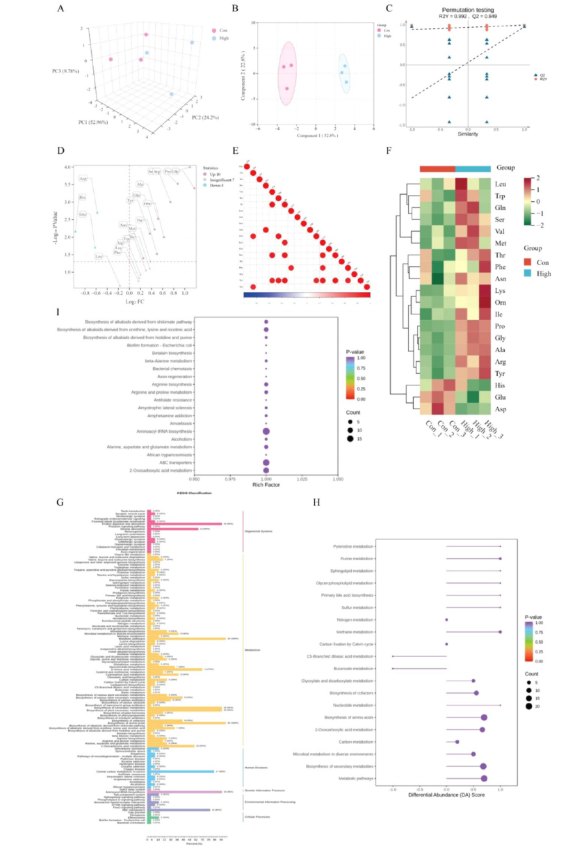
Figure 1. Targeted amino acid metabolomic analysis of serum from high? and low?yielding dairy cows. (A) Principal component analysis (PCA) score plot comparing high? and low?yielding dairy cows. (B) Orthogonal Partial Least Squares Discriminant Analysis (OPLS?DA) score plot comparing high? and low?yielding dairy cows. (C) Permutation test validation of the OPLS?DAmodel. (D) Volcano plot illustrating differential serum amino acids/metabolites between groups. (E) Correlation heatmap of differential serum amino acids/metabolites. (F) Clustering heatmap showing relative abundance patterns of differential serum amino acids/metabolites. (G) Barplot displaying results of KEGG pathway enrichment analysis. (H) Plot of KEGG pathway DA scores. (I) Visualization of KEGG pathway enrichment results.
模块 2:体外 L?Pro 促进 BMEC 增殖、乳蛋白与乳脂合成
1.研究逻辑:CK18 鉴定原代乳腺上皮细胞,设置 0/0.125/0.25/0.5/1 mM L?Pro 梯度,从细胞增殖、酪蛋白表达、甘油三酯合成三个维度验证功能;
2.核心实验:CK18 免疫荧光、CCK8、EdU 增殖、qPCR、WB、β? 酪蛋白免疫荧光、BODIPY 脂滴染色、胞内 / 上清 TAG 定量;
3.关键实验结果
4.Absin abs962 PBS 应用:免疫荧光、BODIPY 脂滴染色前,使用 abs962 PBS 洗涤细胞爬片 3 次,去除培养基残留杂质,降低非特异性荧光背景。
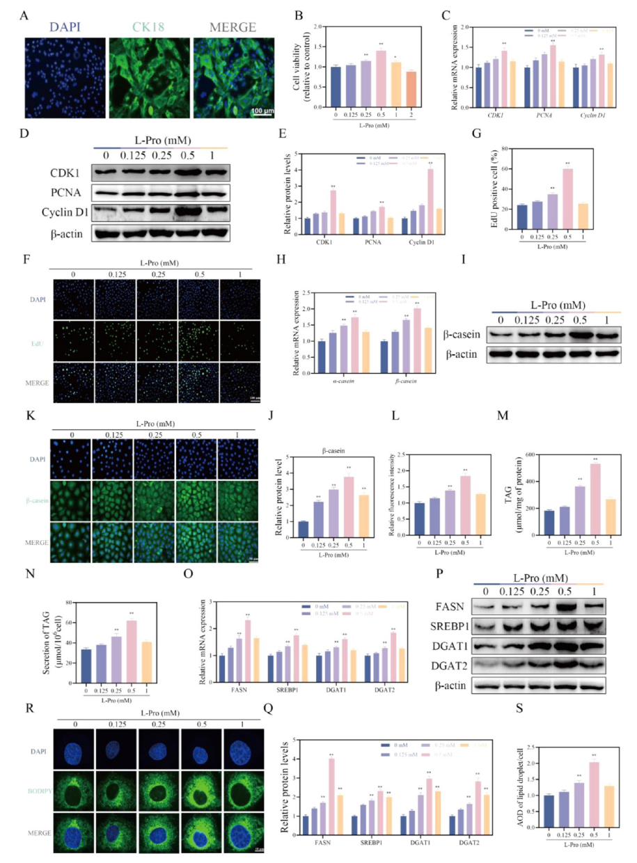
Figure 2.
L?Proline enhances milk synthesis in BMECs. (A) Identification of BMECs using Cytokeratin 18 (CK18) immunofluorescence staining. BMECs were stimulated with different concentrations of L?Pro (0, 0.125, 0.25, 0.5, and 1 mM) for 24 h. (B) Cell proliferation assessed by the CCK?8 assay. (C) Relative mRNA expression levels of CDK1, PCNA, and Cyclin D1 determined by qPCR. (D,E) Protein expressionlevels of CDK1, PCNA, and Cyclin D1 assessed by Western blotting and corresponding quantification. (F,G) Assessment of cell proliferation using an EdU incorporation assay. (H) Relative mRNA expression levels of α?casein and β?casein determined by qPCR. (I,J) Protein expression level of β?casein assessed by Western blotting and corresponding quantification. (K,L) Immunofluorescence detection of β?casein representative images and quantification of relative fluorescence intensity per cell. (M) Intracellular TAG content measured using a detection kit. (N) TAG content secreted into the culture medium measured using a detection kit. (O) Relative mRNA expression levels of FASN, SREBP1, DGAT1, and DGAT2 determined by qPCR. (P,Q) Protein expression levels of FASN, SREBP1, DGAT1, and DGAT2 assessed by Western blotting and corresponding quantification. (R) Representative confocal microscopy images showing lipid droplets stained with BODIPY 493/503 (green) and nuclei counterstained with DAPI (blue). (S) Quantification of lipid droplet area and integrated density (AOD) per cell using ImageJ software.
模块 3:转录组测序 + 抑制剂验证 L?Pro 依赖 mTORC1 通路发挥功能
1.研究逻辑:L?Pro 处理 BMEC 转录组筛选差异基因,GSEA 富集 mTOR 通路;使用 mTOR 抑制剂雷帕霉素(RAPA)阻断通路,反向验证 mTOR 为必需下游;
2.核心实验:RNA?seq、PCA、差异基因火山图、GO/KEGG 富集;RAPA 预处理 + L?Pro 共刺激,WB 检测 mTOR 磷酸化、增殖、乳合成指标;
3.关键实验结果
4.Absin abs962 PBS 应用:WB 细胞裂解前用冰 abs962 PBS 清洗细胞,去除血清与培养基蛋白污染,保证蛋白定量准确性。
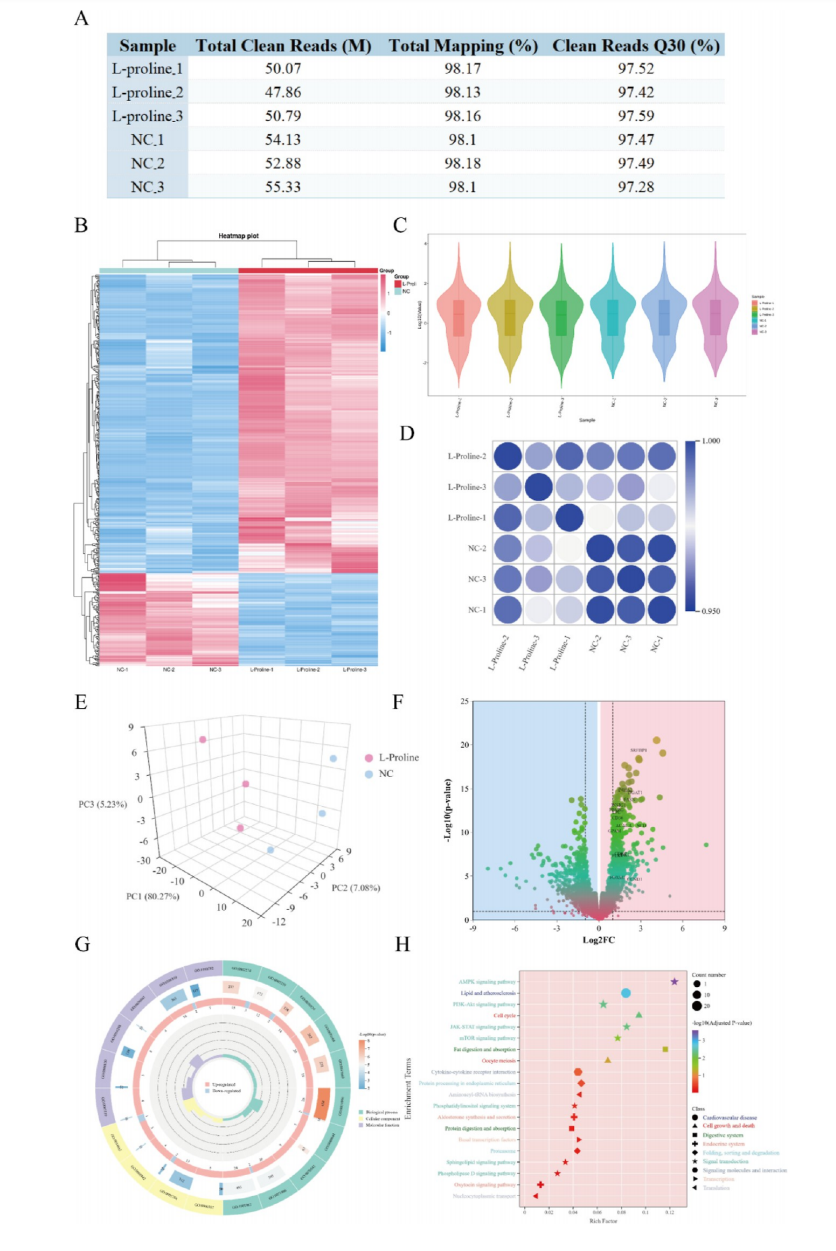
Figure 3. Transcriptomic analysis of BMECs following L?proline treatment. (A) Summary table of RNA?seq data quality metrics. (B) Clustering heatmap analysis. (C) Violin plot illustrating expression level distributions. (D) Sample correlation results from transcriptome sequencing data.(E) PCA results. (F) Volcano plot highlighting differentially expressed genes (DEGs). (G) Gene Ontology (GO) enrichment analysis of differentially expressed genes. (H) KEGG pathway enrichment analysis of differentially expressed genes.
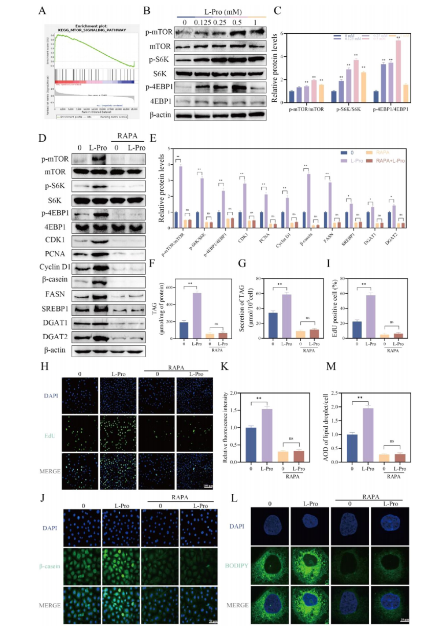
Figure 4. (A) Gene Set Enrichment Analysis (GSEA) results showing enrichment of the mTOR signaling pathway following L?Pro stimulation. BMECs were stimulated with indicated concentrations of L?Pro (0, 0.125, 0.25, 0.5, and 1 mM) for 24 h. (B,C) Western blot analysis and quantification of phosphorylated and total mTOR, S6K, and 4E?BP1 protein levels. BMECs were pretreated with rapamycin (RAPA; 200 nM)or vehicle for 1 h, followed by stimulation with 0.5 mM L?Pro or control for an additional 24 h. (D,E) Western blot analysis and quantification of phosphorylated/total mTOR pathway proteins (mTOR, S6K, 4E?BP1) and key milk synthesis?related proteins (CDK1, PCNA, Cyclin D1, β? casein, FASN, SREBP1, DGAT1, DGAT2). (F) TAG content measured using a detection kit. (G) TAG content secreted into the culture medium measured using a detection kit. (H,I) Assessment of cell proliferation using an EdU incorporation assay. (J,K) Immunofluorescence staining for β?casein and quantification of relative fluorescence intensity per cell. (L,M) Confocal microscopy images of lipid droplets stained with BODIPY 493/503 and quantification of lipid droplet area and integrated density (AOD) per cell.
模块 4:SNAT2 作为关键转运蛋白介导 L?Pro 入胞
1.研究逻辑:筛选 6 种脯氨酸转运体,qPCR 验证 SNAT2 响应 L?Pro 上调;siRNA 敲低 SNAT2,回证 L?Pro 无法激活下游 mTOR 与乳合成;
2.核心实验:转运体基因 qPCR、SNAT2 三株 siRNA 敲低效率验证;敲低后增殖、TAG、酪蛋白、mTOR 通路检测;
3.关键实验结果
4.Absin abs962 PBS 应用:siRNA 转染后细胞传代、EdU 染色、脂滴染色步骤均采用 abs962 PBS 清洗,维持细胞渗透压稳定。
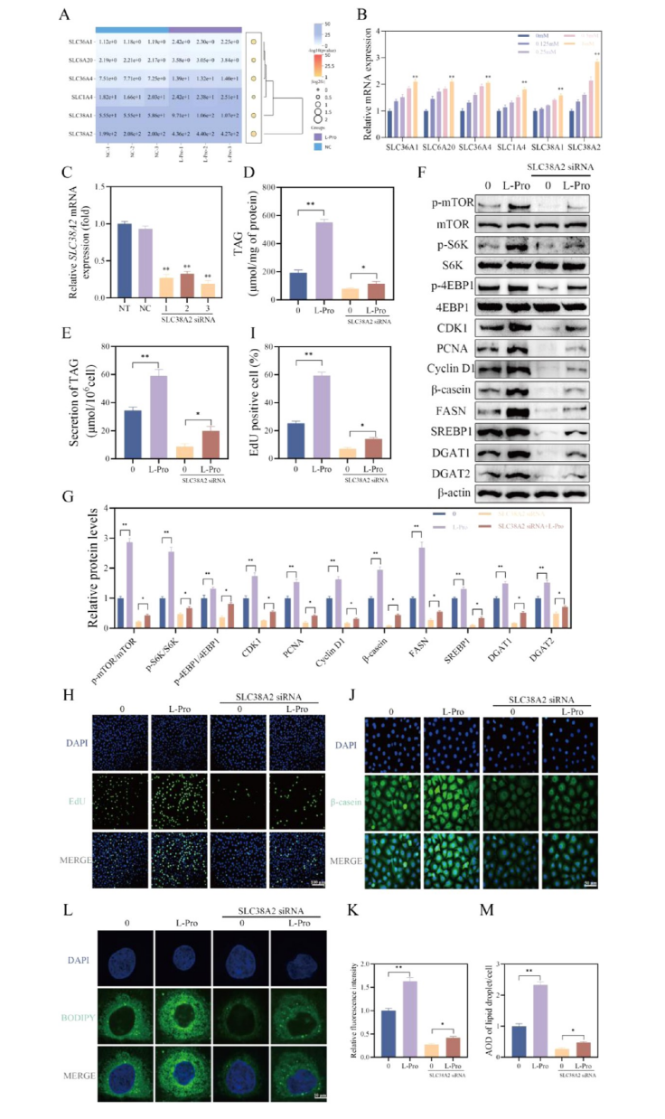 Figure 5. Amino acid transporter SNAT2 mediates L?proline transport and participates in regulating milk synthesis in BMECs. (A) Expression levels of genes mediating proline transport identified from transcriptome sequencing data. BMECs were stimulated with different concentrations of L?Pro (0, 0.125, 0.25, 0.5, and 1 mM) for 24 h. (B) Relative mRNA expression of transporter genes (SLC36A1, SLC6A20, SLC36A4,SLC1A4, SLC38A1, SNAT2) determined by qPCR. (C) Validation of SNAT2 knockdown efficiency by qPCR after transfecting BMECs with three different siRNAs targeting SNAT2. BMECs were transfected with control siRNA (NC) or SNAT2?siRNA?3, followed by stimulation with 0.5 mM L?Pro or control for 24 h. (D) Intracellular TAG content analyzed using a detection kit. (E) TAG content in the culture medium analyzed using a detection kit. (F,G) Western blot analysis and quantification of phosphorylated/total mTOR pathway proteins (mTOR, S6K, 4E?BP1) and key milk synthesis?related proteins (CDK1, PCNA, Cyclin D1, β?casein, FASN, SREBP1, DGAT1, DGAT2). (H,I) Assessment of cell proliferation using an EdU incorporation assay. (J,K) Immunofluorescence staining for β?casein and quantification of relative fluorescence intensity per cell. (L,M) Confocal microscopy images of lipid droplets stained with BODIPY 493/503 and quantification of lipid droplet area and integrated density (AOD) per cell.
模块 5:鉴定 P4HA2 为 L?Pro 直接结合靶蛋白
1.研究逻辑:高产奶牛乳腺 P4HA2 高表达,L?Pro 上调 P4HA;分子动力学、DARTS 蛋白酶保护、CETSA 热漂移实验证明 L?Pro 直接结合并稳定 P4HA2;
2.核心实验:高低产乳腺组织 qPCR/WB、梯度 L?Pro 处理 P4HA2 表达、分子动力学模拟、DARTS、CETSA;
3.关键实验结果
4.Absin abs962 PBS 应用:CETSA、DARTS 蛋白提取全程使用 abs962 PBS 配置裂解缓冲液,维持蛋白天然构象。
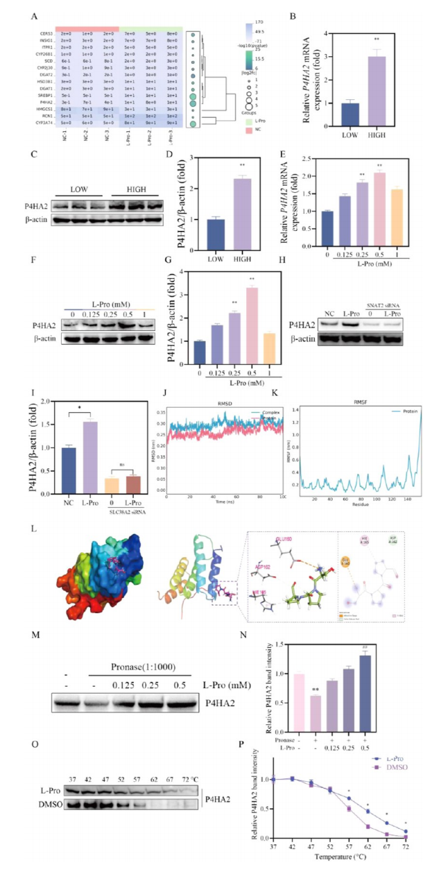 Figure 6. Identification of P4HA2 as a potential target for L?proline. (A). Expression information on major differentially expressed genes in transcriptome sequencing. (B) Relative P4HA2 mRNA expression levels in mammary tissues from high? versus low?yielding dairy cows, determined by qPCR. (C,D) Western blot analysis and quantification of P4HA2 protein levels in mammary tissues from high? versus low?yielding dairy cows. BMECs were treated with indicated concentrations of (L?Pro; 0, 0.125, 0.25, 0.5, and 1 mM) for 24 h. (E) Relative P4HA2 mRNA expression levels determined by qPCR. (F,G) Western blot analysis and quantification of P4HA2 protein levels. BMECs were transfected with control siRNA (NC) or SNAT2?siRNA?3, followed by stimulation with 0.5 mM L?Pro or control for 24 h. (H,I) Western blot analysis and quantification of P4HA2 protein levels following SNAT2 knockdown and L?Pro stimulation. (J) Root mean square deviation (RMSD) analysis of the L?Pro?P4HA2 complex during MD simulation. (K) Root mean square fluctuation (RMSF) analysis of the L?Pro?P4HA2 complexduring MD simulation. (L) Structural representation of the L?proline binding pose within P4HA2, illustrating key interactions. (M,N) Drug affinity responsive target stability (DARTS) assay results showing P4HA2 stability in BMEC lysates following incubation with indicated L?Pro concentrations (0.125, 0.25, and 0.5 mM) and subsequent protease treatment. (O,P) Cellular thermal shift assay (CETSA) results assessing P4HA2 thermal stability in BMEC lysates treated with 0.5 mM L?Pro versus control across a temperature gradient (37?72 °C).
模块 6:P4HA2 介导 L?Pro 激活 mTORC1 通路
1.研究逻辑:P4HA2 siRNA 敲低、质粒过表达双向操作,验证 P4HA2 是 L?Pro 调控乳合成必需中间分子;
2.核心实验:P4HA2 敲低 / 过表达、WB mTOR 通路、增殖、TAG、β? 酪蛋白、脂滴定量;
3.关键实验结果
4.Absin abs962 PBS 应用:β? 酪蛋白免疫荧光爬片使用 abs962 PBS 多次漂洗,去除一抗二抗非特异性结合。
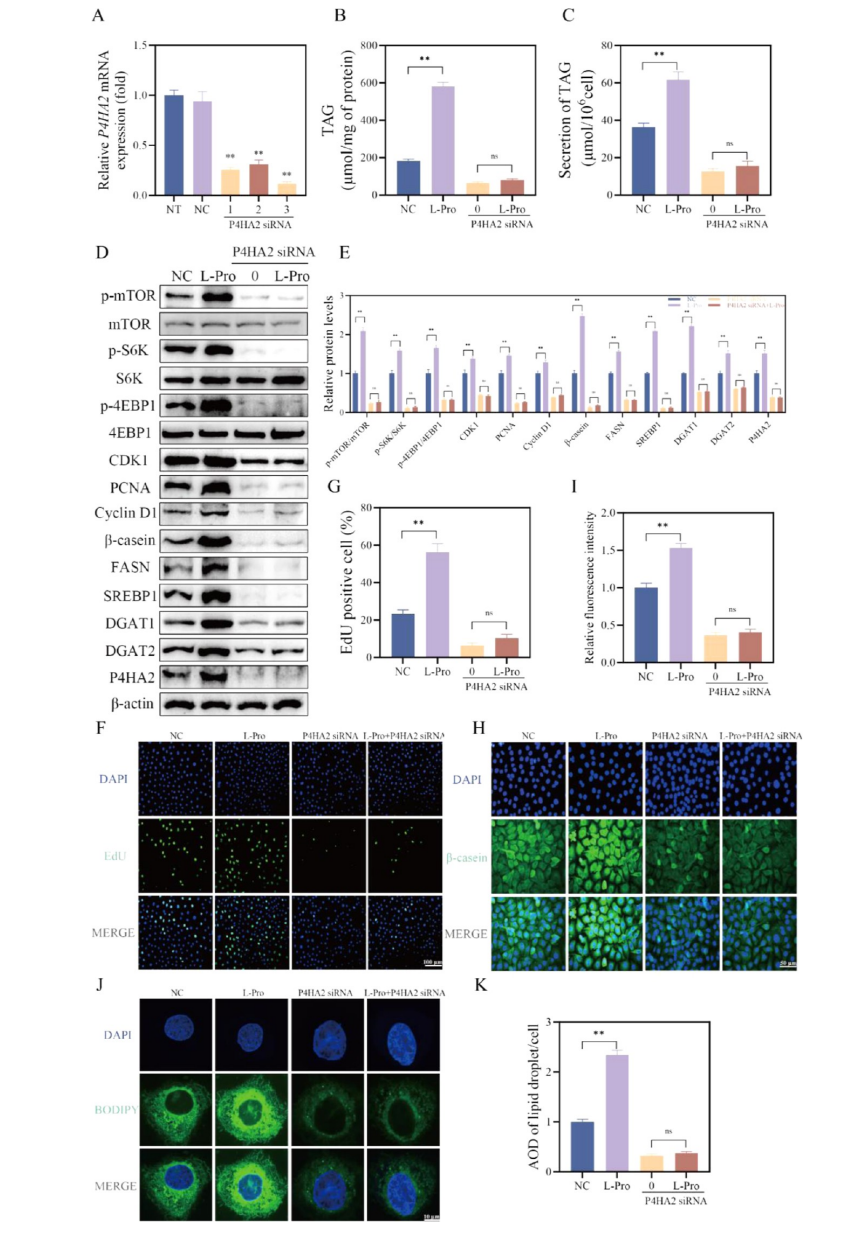 Figure 7. L?Proline promotes milk synthesis in BMECs via the P4HA2?mTOR signaling axis. (A) Validation of P4HA2 knockdown efficiency by qPCR after transfecting BMECs with three different siRNAs targeting P4HA2. BMECs were transfected with control siRNA (NC) or P4HA2? siRNA?3, followed by stimulation with 0.5 mM L?Pro or control for 24 h. (B) Intracellular TAG content analyzed using a detection kit. (C)TAG content analyzed in the culture medium using a detection kit. (D,E) Western blot analysis and quantification of phosphorylated/total mTOR pathway proteins (mTOR, S6K, 4E?BP1) and key milk synthesis?related proteins (CDK1, PCNA, Cyclin D1, β?casein, FASN, SREBP1, DGAT1, DGAT2). (F,G) Assessment of cell proliferation using an EdU incorporation assay. (H,I) Immunofluorescence staining for β?casein and quantification of relative fluorescence intensity per cell. (J,K) Confocal microscopy images of lipid droplets stained with BODIPY 493/503 and quantification of lipid droplet area and integrated density (AOD) per cell.
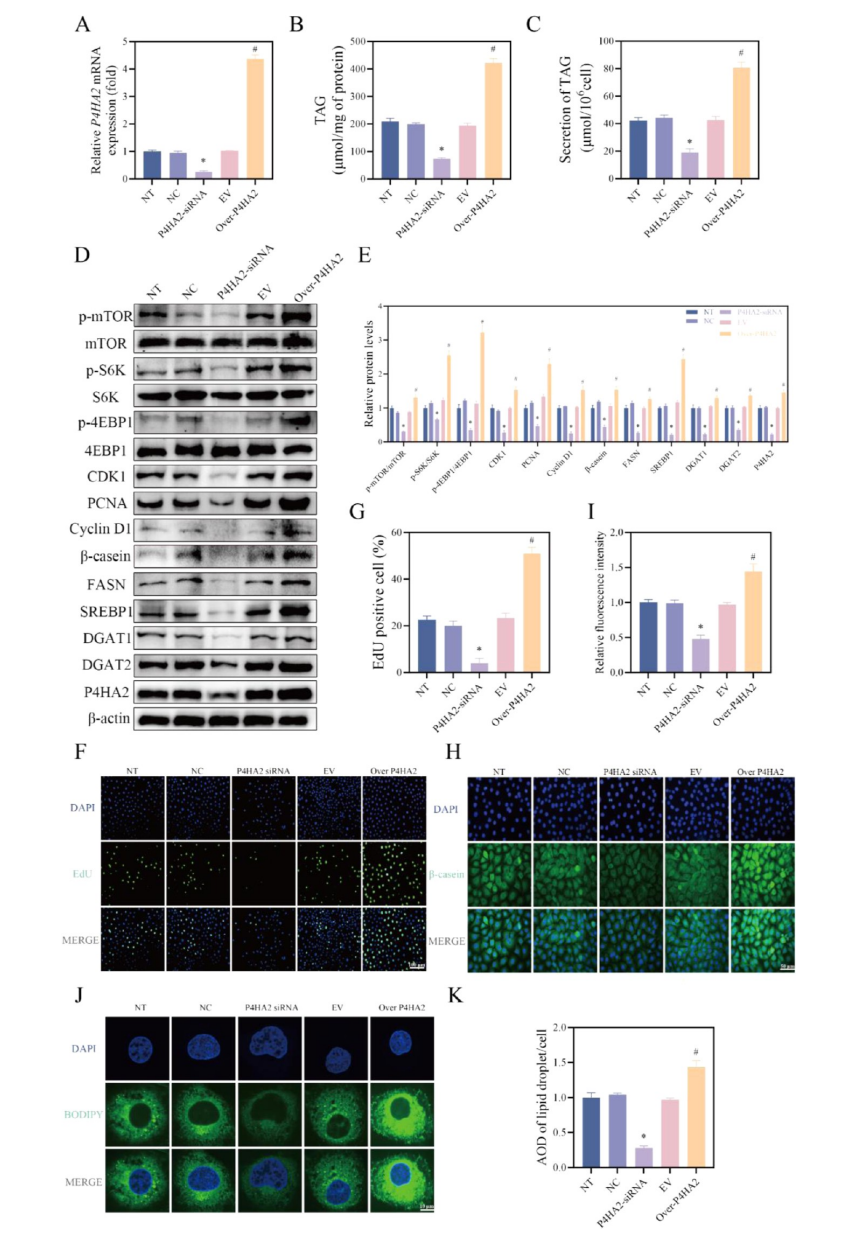 Figure 8. P4HA2 positively regulates milk synthesis in BMECs. BMECs were transfected with control siRNA (NC?siRNA), P4HA2?siRNA?3, empty vector (EV), or a P4HA2 overexpression plasmid (P4HA2?OE) for 24 h. (B) TAG content analyzed using a detection kit. (C) TAG content analyzed in the culture medium using a detection kit. (D,E) Western blot analysis and quantification of phosphorylated/total mTOR pathway proteins (mTOR, S6K, 4E?BP1) and key milk synthesis?related proteins (CDK1, PCNA, Cyclin D1, β?casein, FASN, SREBP1, DGAT1, DGAT2, P4HA2). (F,G) Assessment of cell proliferation using an EdU incorporation assay. (H,I) Immunofluorescence staining for β? casein and quantification of relative fluorescence intensity per cell. (J,K) Confocal microscopy images of lipid droplets stained with BODIPY 493/503 and quantification of lipid droplet area and integrated density (AOD) per cell.
模块 7:P4HA2 结合 mTOR 促进其溶酶体转位激活 mTORC1
1.研究逻辑:STRING 预测 P4HA2 与 mTOR 互作,蛋白对接、Co?IP、共定位荧光验证互作;mTOR 与溶酶体标记 LAMP2 共定位定量,证明 P4HA2 促进 mTOR 溶酶体招募;
2.核心实验:蛋白分子对接、Co?IP 互作验证、P4HA2/mTOR 共定位荧光、mTOR?LAMP2 双荧光共定位定量;
3.关键实验结果
4.Absin abs962 PBS 应用:双标免疫荧光染色全程用 abs962 PBS 梯度清洗,消除荧光交叉干扰。
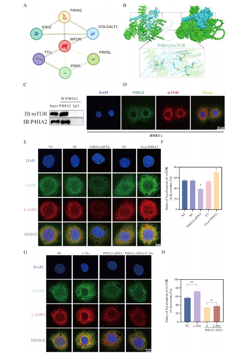 Figure 9. L?Proline promotes mTOR lysosomal localization and mTORC1 activation via P4HA2?mTOR interaction. (A) Predicted protein? protein interaction network for P4HA2 generated using the STRING database. (B) Optimal docked conformation resulting from protein? protein docking simulations between P4HA2 and mTOR. (C) Co?IP analysis demonstrating the interaction between P4HA2 and mTOR inBMECs. (D) Immunofluorescence staining showing colocalization of P4HA2 (green) and mTOR (red) in BMECs. (E,G) Representative immunofluorescence images showing subcellular localization of mTOR (green) and the lysosomal marker LAMP2 (red) under different experimental conditions. (F,H) Quantification of mTOR and LAMP2 colocalization using ImageJ software, corresponding to images in (E?G), respectively.
模块 8:体内饲喂过瘤胃 L?Pro 提升奶牛泌乳性能
1.研究逻辑:40 头泌乳中期奶牛分为对照、低 / 中 / 高剂量过瘤胃 L?Pro 组,9 周饲喂试验,动态监测产奶量、乳成分;
2.核心实验:日产奶量记录、每周混合奶样乳脂 / 乳蛋白 / 乳糖检测、3.5% 校正乳 FCM、能量校正乳 ECM 统计;
3.关键实验结果
4.Absin abs962 PBS 应用:奶牛乳腺活检组织离体后用 abs962 PBS 清洗,固定后用于后续组织蛋白提取。
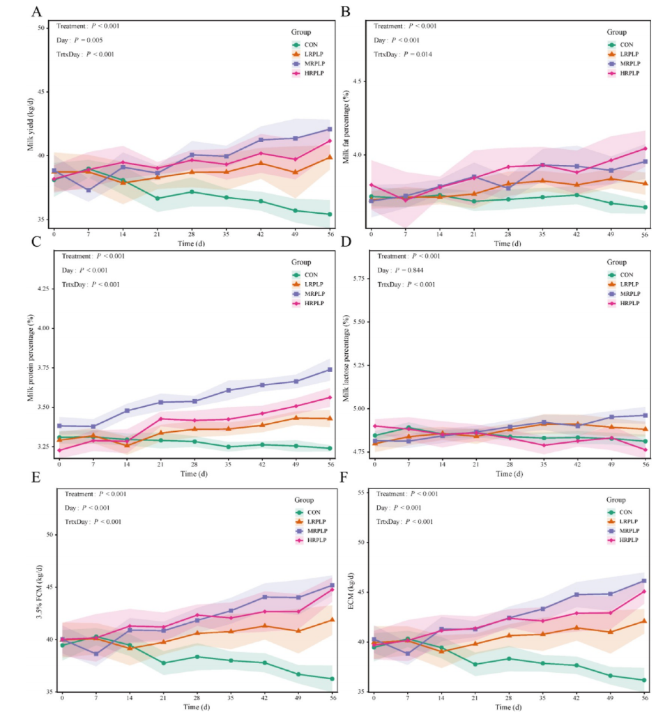 Figure 10. The effect of rumen?protected L?proline on dairy cows’ production performance. (A) Milk yield. (B) Milk fat percentage. (C) Milk protein percentage. (D) Lactose percentage. (E) 3.5% fat?corrected milk (FCM). (F) Energy?corrected milk (ECM). Treatments were the following: CON (Control, basal diet with no additives); LRPLP (basal diet supplemented with 25 g/d of L?proline); MRPLP (basal diet supplemented with 50 g/d of L?proline); and HRPLP (basal diet supplemented with 100 g/d of L?proline). Data are presented as least?squares means ± standard error of the mean (SEM; n = 10). P?values for the main effects of treatment (Trt), day of trial (Day), and their interaction (T × D) from repeated?measures analysis are shown in the upper right of each panel. Day 0 denotes the end of the 1 week adaptation period and the beginning of the 8 week formal experimental period.
五、爱必信 abs962 PBS 缓冲液产品整体作用总结
1.全流程基础缓冲支撑:本研究涵盖细胞培养、免疫荧光、蛋白印迹、组织样本前处理、分子互作多类实验,abs962 无菌等渗 PBS 缓冲液作为统一洗涤 / 重悬试剂,贯穿体外细胞与体内组织样本处理全部环节;
2.保障实验重复性:标准化离子配比维持细胞、蛋白天然生理状态,减少洗涤过程细胞凋亡、蛋白降解,降低荧光背景与 WB 杂带,提升定量结果稳定性;
3.适配多组学 + 分子机制高标准实验:代谢组、转录组、免疫共定位、CETSA 等高精度实验对缓冲液纯度要求高,abs962 无内毒素、无杂质污染,满足畜牧营养高分 SCI 论文细胞分子实验质控标准,是构建完整体外细胞? 体内动物验证体系不可或缺的基础通用试剂。

- 手机:13761418683