




- 手机:13761418683
联系人:爱必信
网址:http://www.absin.cn/
地 址:上海市浦东新区新浩路58号18号楼

文献解析:抑制 HO-1通过改善铁死亡介导的异常自噬缓解心肌纤维化
2026-07-24
文献信息
· 标题:Inhibition of HO1 alleviates cardiac fibrosis by improving ferroptosismediated aberrant autophagy
· 期刊:Free Radical Biology and Medicine(2026,IF≈11.7,氧化应激领域 Top)
· 核心结论:抑制 HO1 可通过阻断铁死亡、恢复异常自噬流,显著改善心肌梗死与 TGFβ1 诱导的心肌纤维化并保护心功能,为临床抗心肌纤维化提供全新靶点与机制。
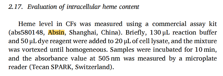
· 使用 Absin 产品:Heme Microplate Assay Kit(abs580148),高特异性、宽线性范围、微量样本即可准确定量,为 HO1 酶活与血红素代谢检测提供关键数据支撑。
一、领域背景与科学问题引出
1. 心肌纤维化临床困境
心肌纤维化是心梗、高血压心脏病共同病理通路,以心脏成纤维细胞过度活化、细胞外基质大量沉积为特征,直接导致心室僵硬、收缩舒张功能下降、心律失常甚至猝死。目前缺乏安全有效的靶向抗纤维化药物,核心瓶颈是机制不清。
2. 铁死亡与自噬在心脏疾病中的关键作用
铁死亡是铁依赖、脂质过氧化驱动的程序性死亡,在心肌损伤中被大量激活;自噬是细胞清除受损蛋白 / 细胞器的稳态机制,自噬流异常是纤维化重要驱动因素。二者交互调控,但在心肌纤维化中的上下游关系尚不明确。
3. HO1 的双重身份与争议
HO1(血红素氧合酶1)是经典抗氧化酶,可降解血红素生成 Fe2?、CO、胆绿素。适度激活具保护作用,但过度上调会导致铁过载、诱发铁死亡,在心脏病理中作用存在明显争议。
4. 本研究核心科学问题
HO1 是否通过铁死亡→异常自噬轴驱动心肌纤维化?抑制 HO1 能否成为抗心肌纤维化新策略?
二、文献综述
1. 心肌纤维化主流机制聚焦 TGFβ/Smad、炎症、氧化应激,但铁死亡与自噬交叉调控是近年新方向。
2. 多项研究证实铁死亡促进心肌纤维化,但上游调控靶点不明确。
3. HO1 可调控铁死亡,但在心肌纤维化中是否通过自噬发挥作用尚未报道。
4. 本研究首次建立 HO1–ferroptosis–autophagy–cardiac fibrosis完整调控轴,填补机制空白。
三、作者整体研究思路
构建体内 MI 小鼠模型 + 体外 TGFβ1 诱导 CFs 纤维化模型→验证 HO1 在纤维化中高表达→激活 / 抑制 HO1 观察铁死亡与纤维化表型→证实铁死亡介导自噬异常→阻断自噬回复实验→体内外验证 HO1 抑制剂 ZnPP 的治疗效应→提出靶向 HO1 的抗纤维化新策略。
四、分模块研究思路
模块 1:纤维化模型中 HO1 显著上调,激活 HO1 诱发铁死亡与纤维化
? 研究目的:确定 HO1 在心肌纤维化中的表达模式与功能表型。
? 实验设计:TGFβ1 刺激原代心脏成纤维细胞(CFs);小鼠 LAD 结扎构建 MI 模型;Hemin 激活 HO1。
? 检测指标:HO1 蛋白、Fe2?、ROS、脂质过氧化、MDA、αSMA、CollagenI、FN1。
? 关键结果:
1. TGFβ1 与 MI 均显著上调 HO1 表达。
2. Hemin 激活 HO1 后,铁死亡核心指标全面升高。
3. 纤维化标志物 αSMA、CollagenI、FN1 显著上调。
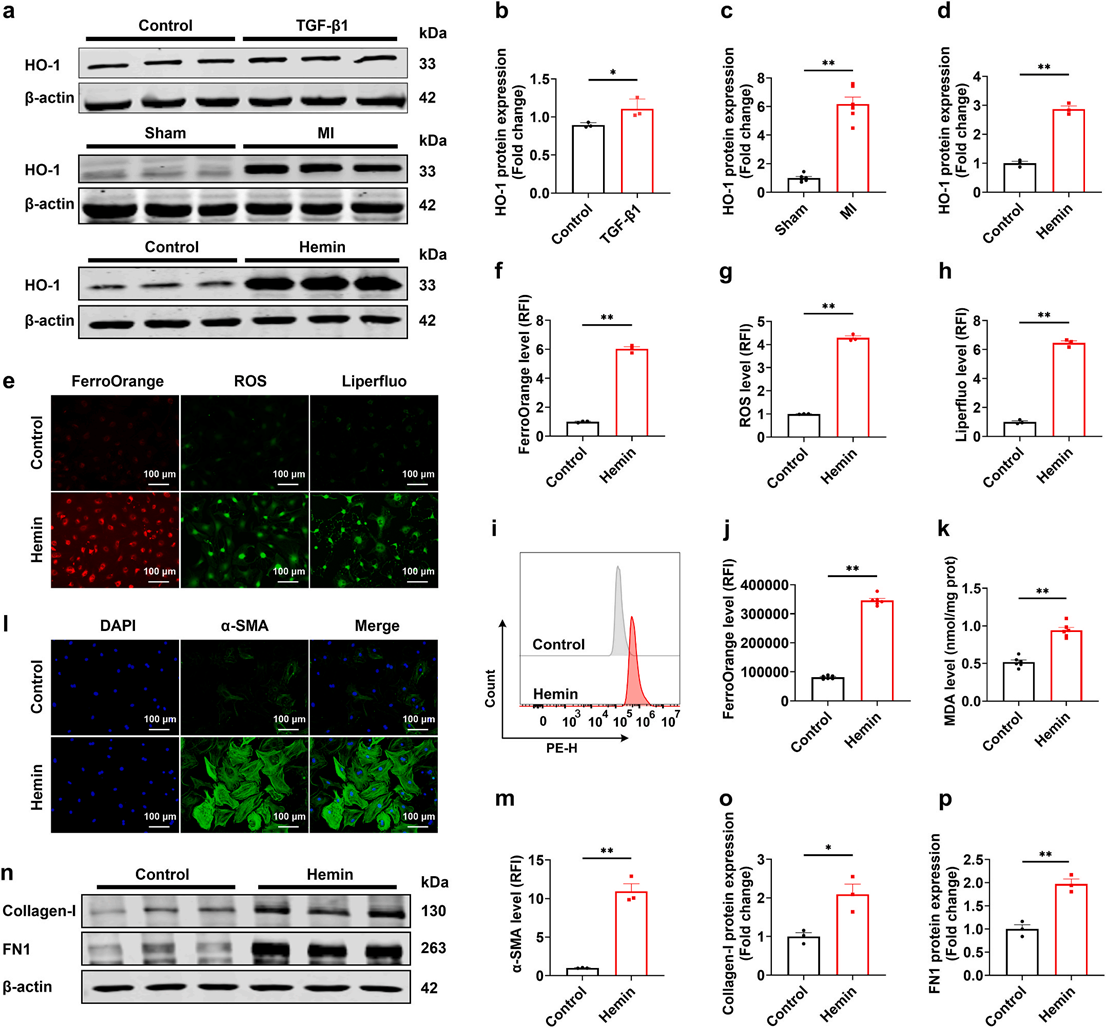
Fig. 1. Activation of HO-1 aggravates ferroptosis and fibrosis in CFs. a. Western blot assays for HO-1 protein expression in CFs and cardiac tissue of mice (n = 3 or n = 6). b-d. Densitometric quantification of HO-1 protein level (n = 3 or n = 6). e-h. Fluorescence detection and quantification of intracellular Fe2+ , ROS, and lipid peroxidation by FerroOrange, DCFH-DA, and Liperfluo, respectively, × 450 magnification, scale bar = 100 μm (n = 3). i, j. Flow cytometry analysis of intracellular Fe2+ levels (n = 6). k. Intracellular MDA levels (n = 6). l, m. Immunofluorescence staining of α-SMA at × 450 magnification, scale bar = 100 μm (n = 3). n-p. Western blot and densitometric analysis of Collagen-I and FN1 protein expression (n = 3). All data are presented as mean ± SEM. *P< 0.05, **P < 0.01.
模块 2:抑制铁死亡可缓解 TGFβ1 诱导的心肌纤维化
? 研究目的:证实铁死亡是心肌纤维化的关键介导环节。
? 实验设计:铁死亡抑制剂 Fer1 处理 TGFβ1 诱导的 CFs。
? 关键结果:
1. Fer1 显著降低 Fe2?、ROS、脂质过氧化、MDA 水平。
2. Fer1 明显抑制 αSMA、CollagenI、FN1 的上调。
3. 逆转 CFs 向肌成纤维细胞转化。
 Fig. 2. Inhibition of ferroptosis attenuates myocardial fibrosis in vitro. a. Viability of CFs exposed to IKE (0–30 μM) for 48 h (n = 6). b. CFs viability following 48 h treatment with IKE (25 μM) and Fer-1 (0–30 μM) for 48 h (n = 6). c, d. Intracellular Fe2+levels measured by flow cytometry (n = 5). e. Fluorescence images of intracellular Fe2+, ROS, and lipid peroxidation detected by FerroOrange, DCFH-DA, and Liperfluo, respectively, × 450 magnification, scale bar = 100 μm (n = 3). f-h. Quantification of intracellular Fe2+, ROS, and lipid peroxidation fluorescence intensity (n = 3). i. Intracellular MDA levels were measured using an assay kit (n = 4). j,Immunofluorescence staining of α-SMA at × 450 magnification, scale bar = 100 μm (n = 3). l. qRT-PCR analysis of ACTA2, COL1A1 and FN1 mRNA expression (n = 6). m. Western blot analysis of Collagen-I and FN1 protein expression (n = 3). n, o. Densitometric analysis of Collagen-I and FN1 protein levels (n = 3). All data are presented as mean ± SEM. *P< 0.05, **P < 0.01.
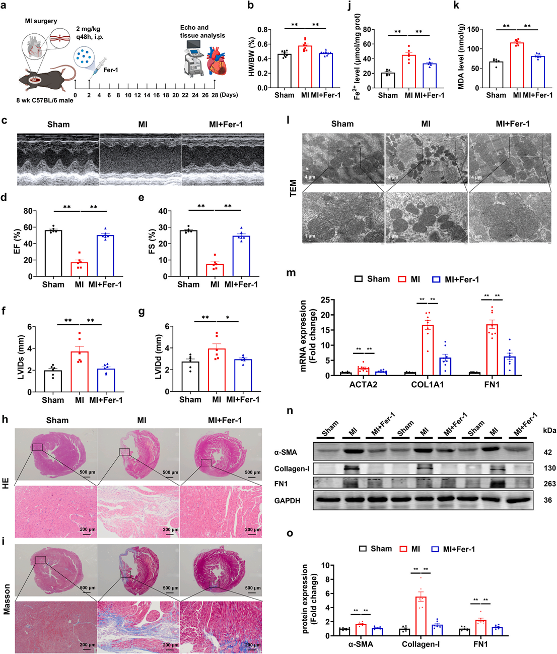
模块 3:体内抑制铁死亡改善 MI 小鼠心功能与心肌纤维化
? 研究目的:在动物水平验证铁死亡的促纤维化作用。
? 实验设计:MI 小鼠给予 Fer1 干预 4 周,超声、组织学、分子检测。
? 关键结果:
1. Fer1 显著提高 EF、FS,减小 LVIDs/LVIDd,改善心功能。
2. 减少胶原沉积、心肌损伤与线粒体结构破坏。
3. 降低心脏 Fe2?、MDA 及纤维化蛋白。
模块 4:铁死亡通过破坏自噬稳态驱动纤维化(不依赖 NCOA4 铁自噬)
? 研究目的:揭示铁死亡下游机制 —— 调控自噬流。
? 实验设计:检测自噬体、溶酶体、自噬流、LC3B/p62、NCOA4/FTH。
? 关键结果:
1. TGFβ1 导致自噬体蓄积、酸性溶酶体减少、自噬流阻断。
2. Fer1 恢复溶酶体功能、重启自噬流、降低 LC3B/p62。
3. NCOA4 无显著变化,不依赖铁自噬通路。
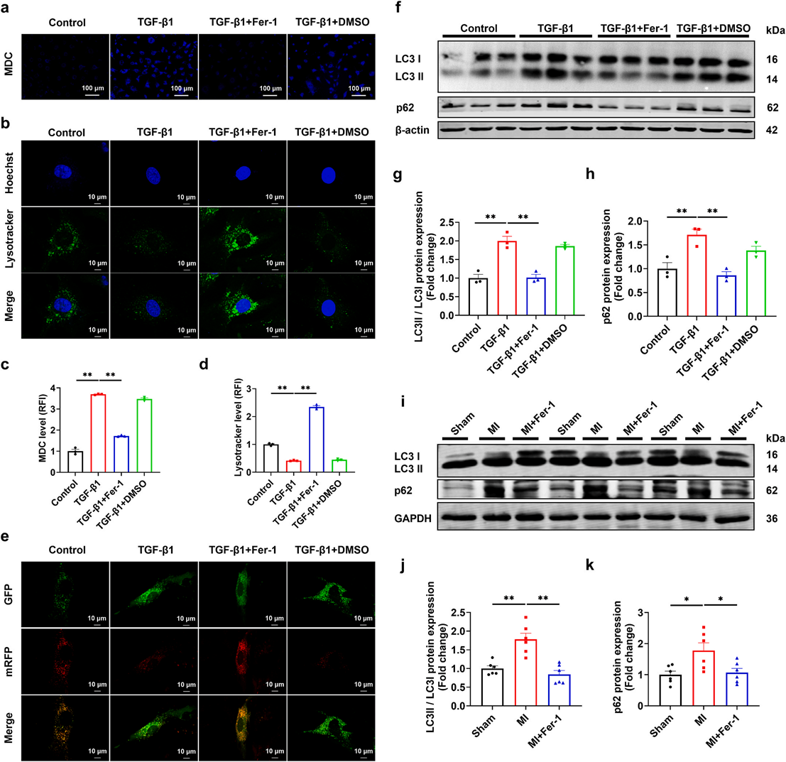
Fig. 4. Inhibition of ferroptosis restores dysregulated autophagy in TGF-β1-treated CFs and MI mice. a. MDC staining of autophagosomes in CFs at × 450 magni fication, scale bar = 100 μm (n = 3). b. Representative confocal microscope images of LysoTracker Green DND-26 staining at × 1000 magnification, scale bar = 10 μm (n = 3). c, d. Quantification of MDC and LysoTracker Green DND-26 fluorescence intensity (n = 3). e. Autophagic flux detected using pLVX-Puro-mRFP-GFP mLC3B, × 1000 magnification, scale bar = 10 μm (n = 3). f. Western blot analysis of LC3B and p62 protein expression in CFs (n = 3). g, h. Densitometric analysis of LC3B and p62 protein levels (n = 3). i. Western blot analysis of LC3B and p62 protein expression in cardiac tissues of mice (n = 6). j, k. Quantification of LC3B and p62 protein expression (n = 6). All data are presented as mean ± SEM. *P< 0.05, **P < 0.01. (For interpretation of the references to color in this figure legend, the reader is referred to the Web version of this article.)
模块 5:HO1 通过铁死亡介导自噬紊乱促进纤维化
? 研究目的:确立 HO1→铁死亡→自噬异常的级联通路。
? 实验设计:Hemin 激活 HO1+Fer1 处理 CFs。
? 关键结果:
1. Fer1 逆转 Hemin 诱导的铁死亡、溶酶体异常、自噬流阻滞。
2. Fer1 抑制 Hemin 诱导的纤维化激活。
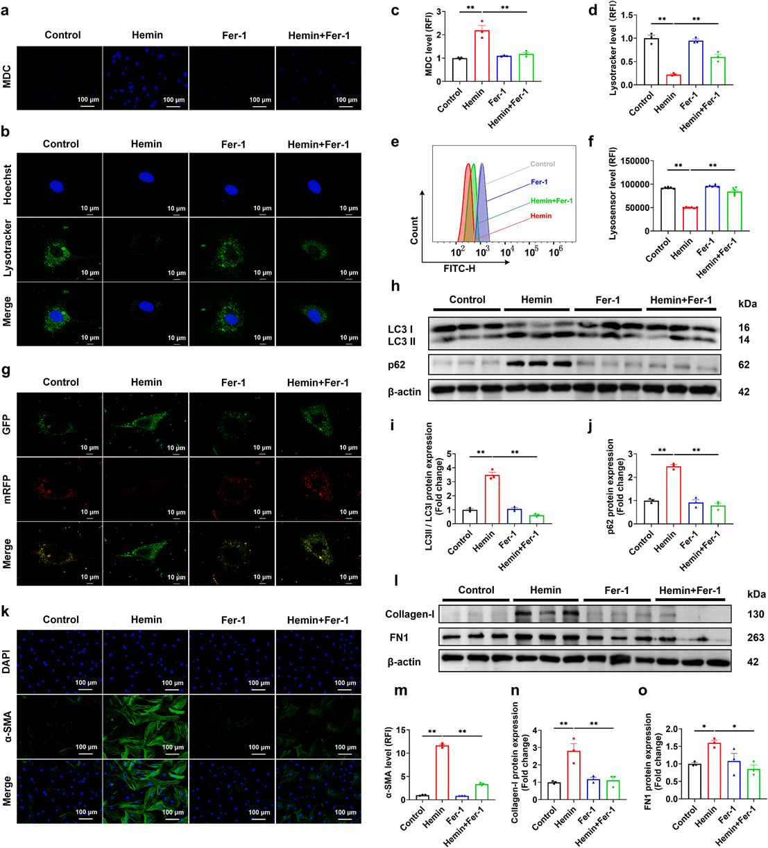
Fig. 5. Inhibition of ferroptosis restores autophagy and attenuates fibrosis in Hemin-induced CFs. a, b. MDC and LysoTracker Green DND-26 staining were used to assess the autophagosome level and the abundance of acidic lysosomes, respectively. × 450 and × 1000 magnification, scale bar = 100 μm and 10 μm (n = 3). c, d. Quantification of MDC and LysoTracker Green DND-26 fluorescence intensity (n = 3). e, f. Intracellular lysosomal pH measured using LysoSensor Green DND-189 (n = 6). g. Autophagic flux detected by pLVX-Puro-mRFP-GFP-mLC3B, × 1000 magnification, scale bar = 10 μm (n = 3). h-j. Western blot and densitometric analysis of LC3B and p62 protein expression (n = 3). k, m. Immunofluorescence staining of α-SMA in CFs at × 450 magnification, scale bar = 100 μm (n = 3). l-o. Western blot and densitometric analysis of Collagen-I and FN1 protein expression (n = 3). All data are presented as mean ± SEM. *P < 0.05, **P < 0.01. (For interpretation of the references to color in this figure legend, the reader is referred to the Web version of this article.)
模块 6:抑制 HO1(药物 / 基因)减轻铁死亡与纤维化
? 研究目的:验证 HO1 是抗纤维化有效靶点。
? 实验设计:ZnPP 抑制 HO1;siRNA 敲低 HO1。
? 关键结果:
1. 抑制 HO1 恢复胞内血红素水平(abs580148 检测)。
2. 显著降低铁死亡与纤维化指标。
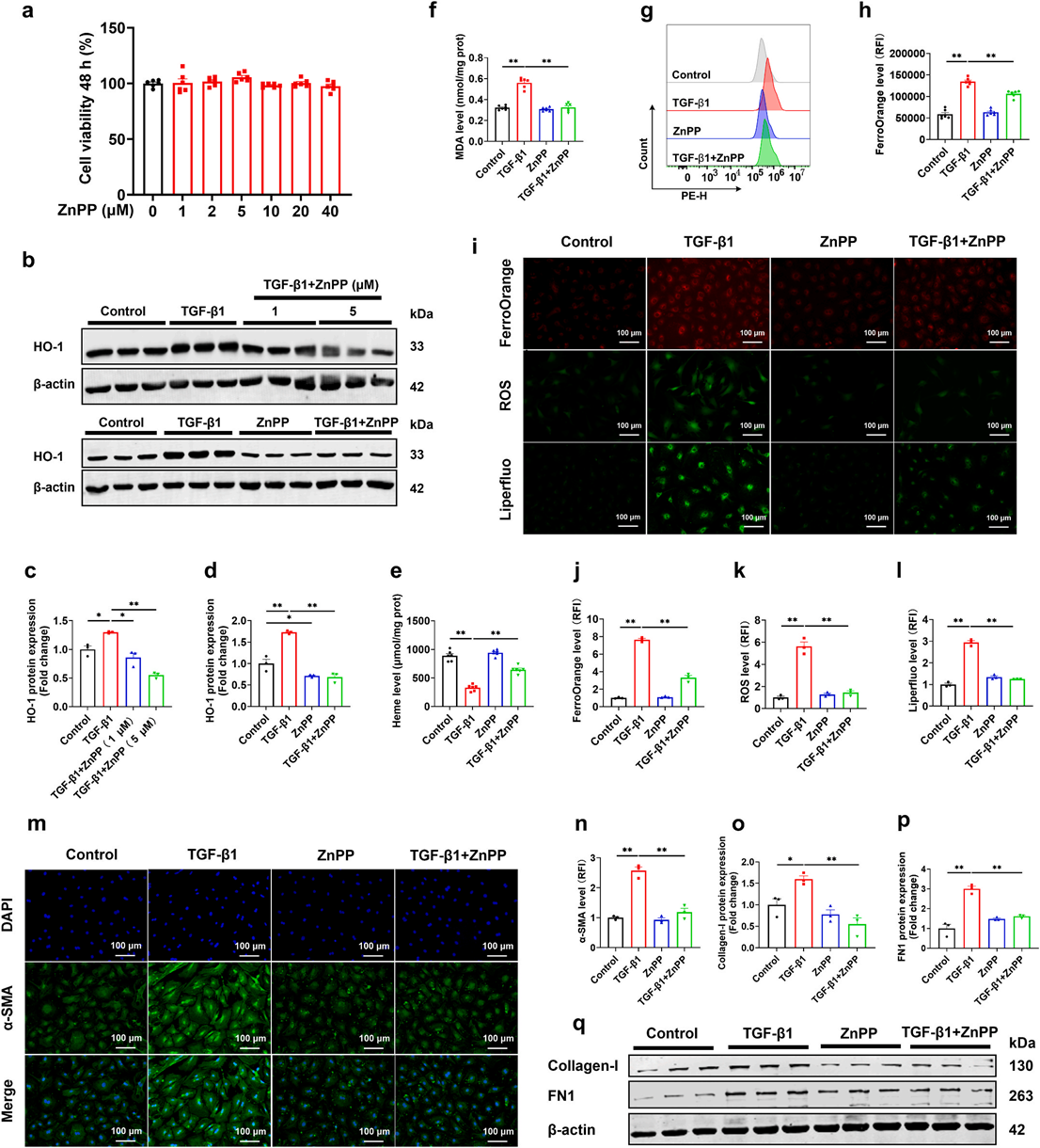
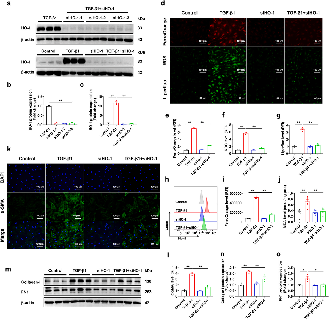
Fig. 6. Inhibition of HO-1 attenuates ferroptosis and fibrosis in CFs exposed to TGF-β1. a. Viability of CFs exposed to ZnPP (0–40 μM) for 48 h (n = 6). b. Western blot assays for HO-1 protein expression (n = 3). c, d. Quantification of HO-1 protein levels (n = 3). e. Intracellular heme levels (n = 6). f. Intracellular MDA level in CFs (n = 6). g, h. Flow cytometry analysis of intracellular Fe2+ level (n = 6). i. Fluorescence detection of intracellular Fe2+, ROS, and lipid peroxidation by FerroOrange, DCFH-DA, and Liperfluo, respectively, × 450 magnification, scale bar = 100 μm (n = 3). j-l. Quantification of intracellular Fe2+, ROS, and lipid peroxidation (n = 3). m, n. Immunofluorescence staining and quantification of α-SMA in CFs at × 450 magnification, scale bar = 100 μm (n = 3). o-q. Western blot analysis of Collagen-I and FN1 protein expression (n = 3). All data are presented as mean ± SEM. *P< 0.05, **P < 0.01.
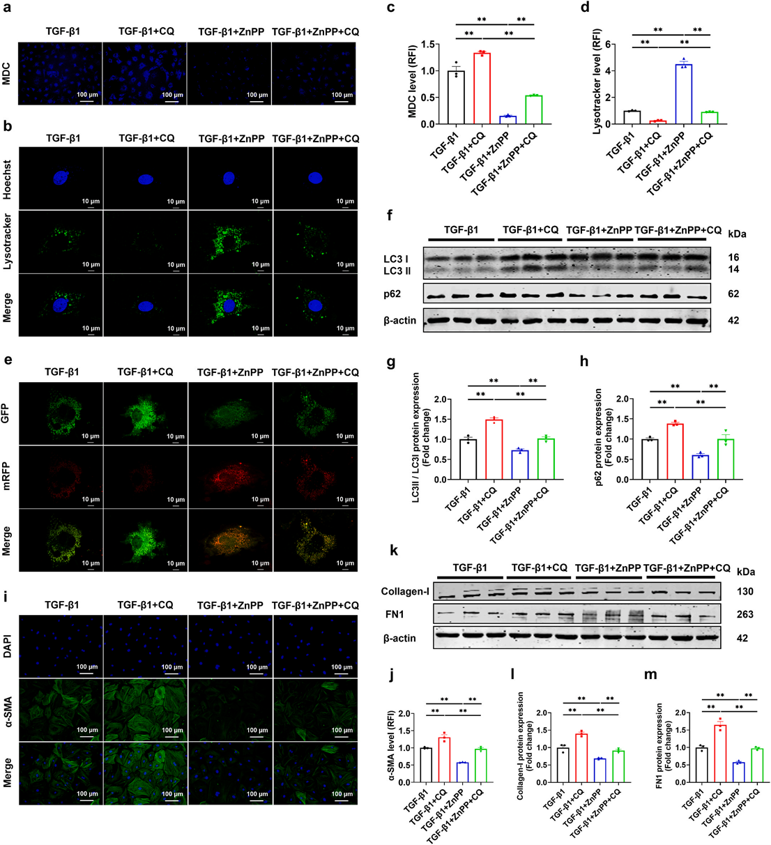 Fig. 7. HO-1 knockdown attenuates ferroptosis and fibrosis in TGF-β1-induced CFs. a. Representative Western blot images of HO-1 protein expression (n = 3). b, c. Quantitative analysis of HO-1 protein levels (n = 3). d. Fluorescence detection of intracellular Fe2+, ROS, and lipid peroxidation by FerroOrange, DCFH-DA, and Liperfluo, respectively, × 450 magnification, scale bar = 100 μm (n = 3). e-g. Quantification of intracellular Fe2+, ROS, and lipid peroxidation levels (n = 3). h, i. Flow cytometric analysis and quantification of intracellular Fe2+ level (n = 6). j. Intracellular MDA level in CFs (n = 6). k, l. Immunofluorescence staining and quantification of α-SMA in CFs at × 450 magnification, scale bar = 100 μm (n = 3). m-o. Western blot analysis and quantification of Collagen-I and FN1 protein expression (n = 3). All data are presented as mean ± SEM. *P< 0.05, **P < 0.01.
? 研究目的:机制闭环,证实保护效应依赖自噬。
? 实验设计:ZnPP+CQ(自噬抑制剂)联合处理。
? 关键结果:
1. CQ 逆转 ZnPP 对自噬流的挽救作用。
2. CQ 消除 ZnPP 的抗纤维化与心功能保护作用。
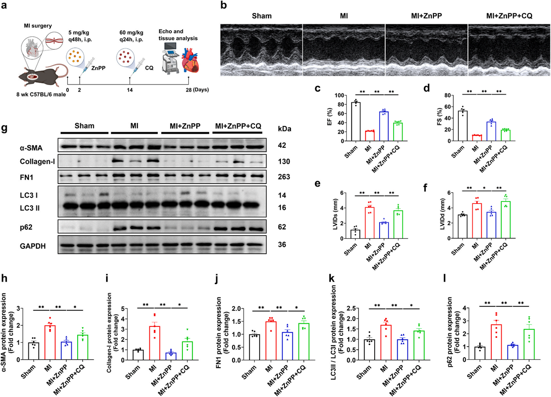 Fig. 8. Suppression of HO-1 alleviates myocardial fibrosis by restoring autophagy in TGF-β1-treated CFs. a, b. MDC and LysoTracker Green DND-26 staining, × 450 and × 1000 magnification, scale bar = 100 μm and 10 μm (n = 3). c, d. Quantification of MDC and LysoTracker Green DND-26 fluorescence intensity (n = 3). e. Autophagic flux was measured by pLVX-Puro-mRFP-GFP-mLC3B, × 1000 magnification, scale bar = 10 μm (n = 3). f-h. Western blot and densitometric analysis of LC3B and p62 protein expression (n = 3). i, j. Immunofluorescence staining of α-SMA in CFs at × 450 magnification, scale bar = 100 μm (n = 3). k-m. Western blot and densitometric analysis of Collagen-I and FN1 protein expression (n = 3). All data are presented as mean ± SEM. **P < 0.01. (For interpretation of the references to color in this figure legend, the reader is referred to the Web version of this article.)
五、Absin 产品 abs580148 应用与优势
1. 实验应用场景
本研究在原文 2.17 实验方法章节与原文 Fig.6e 结果图中使用Absin abs580148 血红素检测试剂盒检测心脏成纤维细胞内血红素含量,直接反映 HO1 的酶解活性:
1. HO1 激活→血红素被降解→胞内血红素下降;
2. ZnPP 抑制 HO1→血红素降解减少→胞内血红素回升;
3. 该数据是HO1 功能验证的核心生化证据。
2. 产品核心优势
1. 高特异性:专一识别血红素,不受样本中其他色素干扰。
2. 宽检测范围:1–100 μmol/L,完美覆盖细胞 / 组织生理浓度。
3. 操作简便:比色法,96 孔板高通量,酶标仪直接读数。
4. 样本适应性强:细胞裂解液、组织匀浆、全血、血清均可测。
5. 稳定性好:4℃保存,保质期 6 个月,蓝冰运输保障活性。
3. 对本研究的关键作用
1. 精准量化胞内血红素,直接反映 HO1 酶活性,为机制提供硬核数据。
2. 微量样本即可检测,适配原代细胞珍贵样本。
3. 结果稳定可靠,支撑HO1 作为治疗靶点的结论严谨性。
六、研究亮点与临床意义
1. 首次提出并证实 HO1→铁死亡→异常自噬→心肌纤维化的全新调控轴。
2. 澄清HO1 在心脏中的双重作用:过度激活促纤维化,抑制具保护作用。
3. 提供无创 / 药物可靶向的新靶点:HO1 抑制剂具临床转化潜力。
4. 拓展心肌纤维化治疗思路:联合靶向铁死亡 + 自噬,提高疗效。

- 手机:13761418683