




- 手机:13761418683
联系人:爱必信
网址:http://www.absin.cn/
地 址:上海市浦东新区新浩路58号18号楼

- 全基因组体外 + 体内 CRISPR 敲除筛选,联合 TCGA/GEO 预后数据交集锁定候选驱动基因 DEFB1;
- 单细胞测序、多重免疫组化、临床样本 WB/ELISA 验证 DEFB1 在肿瘤细胞特异性高表达,且是患者独立不良预后标志物;
- CRISPR 敲除 DEFB、回补过表达、外源蛋白回救三组细胞实验,明确 DEFB1 促进增殖、迁移、侵袭恶性表型;
- CoIP-MS 质谱筛选 DEFB 膜结合互作蛋白,锁定 PPL、MIF 两个核心靶蛋白;
- 分别验证 DEFB1-PPL-EMT 增殖轴、DEFB1-MIF-M2 巨噬细胞极化轴,双敲除 / 抑制剂回救实验确认通路必要性;
- 构建患者肿瘤类器官、裸鼠 CDX 移植瘤、人源化 Kras 自发肺癌三类体内模型,验证抗 DEFB1 单抗 mAb-5 抑瘤效果;
- 免疫健全人源化 DEFB 小鼠完成 4 周长期给药,评估 mAb-5 血液、脏器病理安全性,完整支撑临床转化潜力。
- 体外、体内 CRISPR 增殖筛选与预后不良基因交集共 15 个,DEFB1 因机制空白选为核心研究基因;
- 单细胞测序:DEFB1 仅在肿瘤细胞、髓系细胞高表达,正常上皮、免疫细胞几乎无表达;
- 临床配对组织:肿瘤 DEFB1 染色强度显著高于癌旁正常组织;细胞系、患者肿瘤组织 WB 证实肿瘤 DEFB1 蛋白上调;细胞上清 ELISA 证明 DEFB1 大量分泌;
- KM 生存、多因素 Cox:DEFB1 高表达患者无进展生存期、总生存期显著缩短,是独立预后危险因素;多癌 TCGA 队列提示 DEFB1 在肺鳞癌、肾透明细胞癌同步上调。
- WB/ELISA 验证 KO 细胞 DEFB1 几乎无表达,ROE 细胞可恢复蛋白分泌;
- 高内涵、Alamar Blue:DEFB1-KO 显著抑制细胞增殖,ROE、外源 DEFB1 (200pg/mL) 可完全回救增殖能力;
- Transwell 侵袭:DEFB1 敲除后穿膜细胞大幅减少,回补 DEFB1 逆转侵袭缺陷;
- 裸鼠皮下 CDX 模型:DEFB1-KO 体内成瘤重量显著降低,ROE 组肿瘤体积、重量显著升高。
- CoIP/WB 证实外源分泌 DEFB1 可结合细胞膜定位 PPL;免疫荧光显示 Flag-DEFB1 与细胞膜标记 ATP1A1、PPL 共定位;
- DEFB1-KO 上皮标志物 E-cad 上调,间质标志物 N-cad、Vimentin、Snail 下调,回补 DEFB1 逆转 EMT 表型;
- PPL 单独敲除直接激活 EMT、提升细胞恶性;同时敲除 PPL 完全消除 DEFB1 对增殖、侵袭的调控作用,证明 DEFB1 依赖 PPL 发挥促癌功能。
- 移植瘤组织:DEFB1 高表达组 M2 (CD206+) 巨噬浸润增多,M1 (iNOS+) 巨噬减少;
- 体外共培养:DEFB1 显著抑制 M1 极化、促进 M2 极化;添加 MIF 抑制剂 ISO-1 可完全阻断 DEFB1 诱导的 M2 极化;
- 体内荷瘤模型:单独敲除 PPL 或 ISO-1 仅部分抑制肿瘤生长;PPL 敲除联合 ISO-1 可彻底抵消 DEFB1 体内促瘤作用,证明 PPL、MIF 两条通路共同介导 DEFB1 致癌。
- 细胞水平:mAb-5 特异性阻断 DEFB1 促增殖、侵袭效应,抑制 EMT,逆转巨噬 M2 极化;DEFB1-KO 细胞中 mAb-5 无抑瘤效果,证明作用靶点专一;
- 3D 类器官:mAb-5 处理 7 天显著抑制患者肿瘤类器官活力、缩小球体体积;
- 异种移植瘤:mAb-5 腹腔给药显著降低肿瘤重量、抑制瘤内 EMT 与 M2 巨噬浸润;
- 免疫健全自发肺癌模型:mAb-5 持续给药缩小肺部肿瘤负荷,显著延长小鼠生存期。
- 体外侵袭模型核心耗材:abs9490 基质胶用于 Transwell 小室铺膜,模拟体内肿瘤细胞外基质屏障,精准量化 DEFB1、mAb-5 对肿瘤侵袭能力的调控,是体外转移功能验证的必备支撑;
- 患者肿瘤类器官三维培养支架:abs9490 提供仿生胞外基质微环境,支撑肺腺癌患者原代肿瘤细胞形成三维类器官,高度还原患者肿瘤异质性,为抗体 mAb-5 体外药物筛选、预临床药效评估提供更贴近临床的体外模型;
- 打通细胞 2D 实验与 3D 类器官转化证据链:abs9490 同时覆盖侵袭二维功能实验、3D 原代类器官药效筛选,从细胞、患者原代组织两个层面共同佐证 DEFB1 促癌与 mAb-5 抑瘤结论,完善本研究从基础机制到转化药物的完整证据链,是连接体外细胞实验与临床前转化研究关键试剂。
SCI 文献深度解析:揭示 DEFB1 作为肺腺癌全新驱动基因及极具潜力的治疗靶点
2026-08-13
一、文献基础综述
(一)期刊与基础信息
期刊:Cell Death and Disease,2026 年最新 IF=12.2,Springer Nature 开源期刊,中科院生物学 1 区;
DOI:10.1038/s41419-026-08748-4;卷期:Cell Death and Disease (2026) 17:519;
论文标题:Unveiling DEFB1 as a novel driver and promising therapeutic target in lung adenocarcinoma
(二)全文核心结论
本研究通过体内外全基因组 CRISPR 筛选联合多组临床数据库,首次证实分泌型抗菌肽 DEFB1 是肺腺癌全新致癌驱动基因;机制上 DEFB1 存在双重促癌通路:①结合膜蛋白 PPL 解除其对 EMT 的抑制,直接促进肿瘤增殖、侵袭转移;②结合 MIF 细胞因子诱导巨噬细胞向促瘤 M2 型极化,构建免疫抑制微环境;研究自主筛选得到中和单克隆抗体 mAb-5,在细胞、患者类器官、异种移植瘤、人源化 Kras 自发肺癌四大模型中均显著抑制肿瘤进展,且在免疫健全小鼠体内无明显脏器、血液毒性,证明 DEFB1 及 mAb-5 具备临床转化价值。
(三)Absin(爱必信 abs9490 基质胶)产品作用
本研究 Transwell 侵袭实验采用 abs9490 基质胶构建体外基底膜屏障,精准模拟肿瘤细胞穿透胞外基质的侵袭过程;同时利用 abs9490 构建患者来源肺腺癌类器官三维培养支架,还原肿瘤原生三维结构用于抗体药效评估。
二、研究领域背景介绍
肺癌为全球发病率、死亡率双高恶性肿瘤,肺腺癌占新发肺癌 60% 以上,具备增殖快、侵袭转移强、早期隐匿的特征。现阶段手术、放化疗、靶向、免疫治疗仅能小幅延长晚期无靶点突变患者生存期,临床亟需全新驱动基因与靶向药物。
β 防御素 DEFB1 是上皮细胞分泌的天然抗菌肽,经典功能为黏膜抗感染;既往少量研究发现 DEFB1 在肺腺癌组织高表达,但完全不清楚其促癌分子机制,且不同肿瘤中 DEFB 功能存在矛盾(口腔 / 乳腺癌中抑癌,肺癌中促癌),分子通路空白。同时现有靶向药物多针对胞内突变蛋白,分泌型胞外靶点开发空间巨大,中和单克隆抗体具备特异性强、副作用低的优势。
基于上述临床与基础研究空白,本研究依托 CRISPR 全基因组筛选、单细胞测序、多组临床队列、多层次体内外模型,完整解析 DEFB1 在肺腺癌的致癌功能、双重分子调控轴,并开发靶向中和抗体 mAb-5,填补该领域机制与转化空白。
三、作者整体递进研究思路总结
全文遵循临床队列生信挖掘→临床组织样本验证→体外细胞功能验证→互作蛋白质谱筛选→双通路分子机制解析→多重体内模型药效验证→抗体安全性评估完整转化医学逻辑链:
四、分模块详细研究思路、实验结果、对应原文图片
模块 1:生信 + 临床样本证实 DEFB1 高表达提示肺腺癌不良预后
1.研究逻辑:CRISPR 筛选与预后基因取交集锁定 DEFB1;单细胞、组织染色、多临床队列验证表达差异与预后关联;
2.核心实验:GeCKO v2 全基因组 CRISPR 筛选;单细胞转录组 UMAP 聚类;118 对患者组织多重免疫组化;TCGA/GEO 生存分析;单 / 多因素 Cox 回归;WB、细胞上清 ELISA;
3.关键实验结果
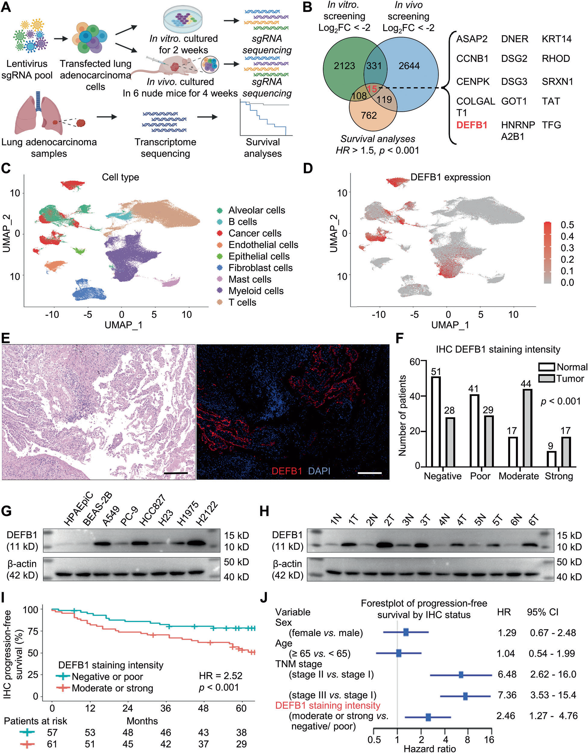 Fig. 1 DEFB1 exhibits elevated expression and indicates unfavorable survival in lung adenocarcinoma. A, B Intersection of genes that might promote lung adenocarcinoma proliferation obtained from in vitro and in vivo CRISPR/Cas9 genome-wide knockout experiments and genes associated with worse prognosis of lung adenocarcinoma; C, D UMAP clustering of single-cell transcriptome sequencing data for cell clustering and DEFB1 expression levels in each single cell; E, F DEFB1 expression in 118 pairs of lung adenocarcinoma tumor tissues and adjacent normal lung tissues by multiple immunohistochemical staining (red) (scale bar: 200 μm); G, H Western blot results of DEFB1 expression in normal lung epithelial cell lines, lung adenocarcinoma cell lines, as well as six pairs of tumor tissues and adjacent normal tissues obtained from patients with lung adenocarcinoma; I, J Kaplan–Meier and Cox survival analyses between tumor DEFB1 expression and progression-free survival in lung adenocarcinoma patients based on multiple immunohistochemical staining results. Statistical analysis: F Mann–Whitney U-test, and G Log-rank test.
模块 2:体外细胞实验证实 DEFB1 驱动肺腺癌增殖、迁移、侵袭(Absin abs9490 基质胶应用点)
1.研究逻辑:DEFB1 敲除 (DEFB1-KO)、敲除后回补过表达 (ROE)、敲除 + 外源 DEFB1 蛋白回救 (KO+EXO) 三组平行,从增殖、侵袭多维度验证促癌表型;
2.核心实验:CRISPR 稳转细胞株构建;高内涵活细胞成像、Alamar Blue 增殖实验;Transwell 迁移 / 侵袭实验(使用 Absin abs9490 基质胶);
3.Absin 产品实验步骤说明:Transwell 侵袭实验中,用无血清培养基 1:5 稀释 abs9490 基质胶,均匀铺于小室上室膜,37℃凝固形成胞外基质屏障,用于模拟体内肿瘤侵袭过程,量化细胞穿透基质能力;
4.关键实验结果
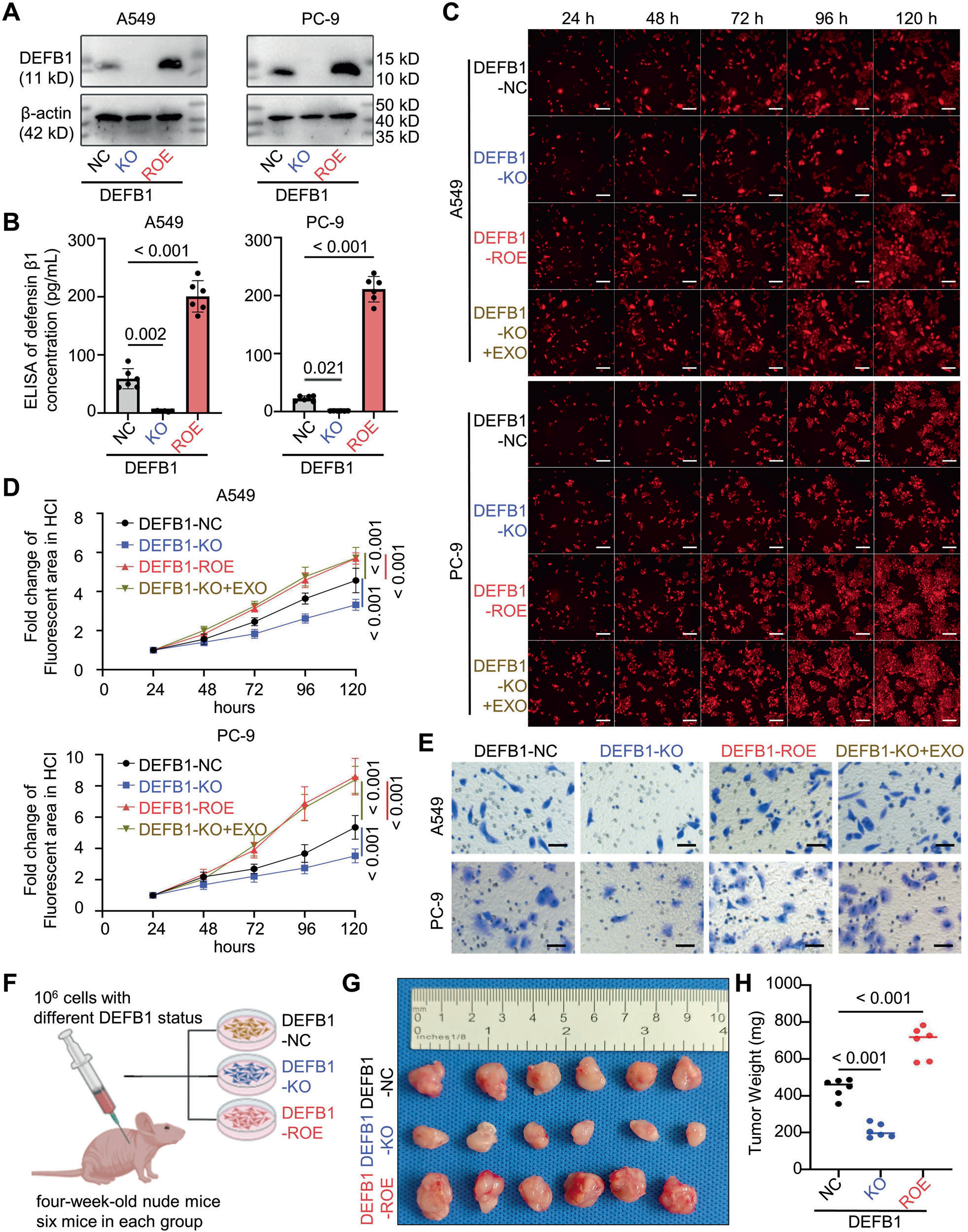 Fig. 2 DEFB1 enhances lung adenocarcinoma proliferation, migration, and invasion in vitro and tumorigenesis in vivo. A, B Western blot and ELISA for DEFB1 protein expression in A549/PC-9 DEFB1-NC/KO/ROE cells; C, D High-content imaging (HCI) for the proliferation of A549/ PC-9 DEFB1-NC/KO/ROE/KO + EXO cells (scale bar: 200 μm); E Transwell assay for the invasion capacity of A549/PC-9 DEFB1-NC/KO/ROE/ KO + EXO cells (scale bar: 100 μm); F Schematic diagram of for the xenograft experimental process; G, H In vivo proliferation and final tumor weights of A549 DEFB1-NC/KO/ROE cells. Statistical analysis: B, H One-way ANOVA and Bonferroni correction; D Two-way ANOVA test (n = 6) and Bonferroni correction.
模块 3:机制一:DEFB1 结合 PPL 调控 EMT 通路促进增殖侵袭
1.研究逻辑:CoIP-MS 筛选膜互作蛋白,锁定抑癌蛋白 PPL;免疫荧光、CoIP 验证二者结合;PPL 敲除回救实验确认 PPL 为 DEFB1 功能必需介质;
2.核心实验:3×Flag-DEFB1 CoIP 联合质谱;AlphaFold 蛋白结合预测;细胞膜共定位免疫荧光;EMT 标志物 WB;PPL CRISPR 敲除回救增殖、侵袭实验;
3.关键实验结果
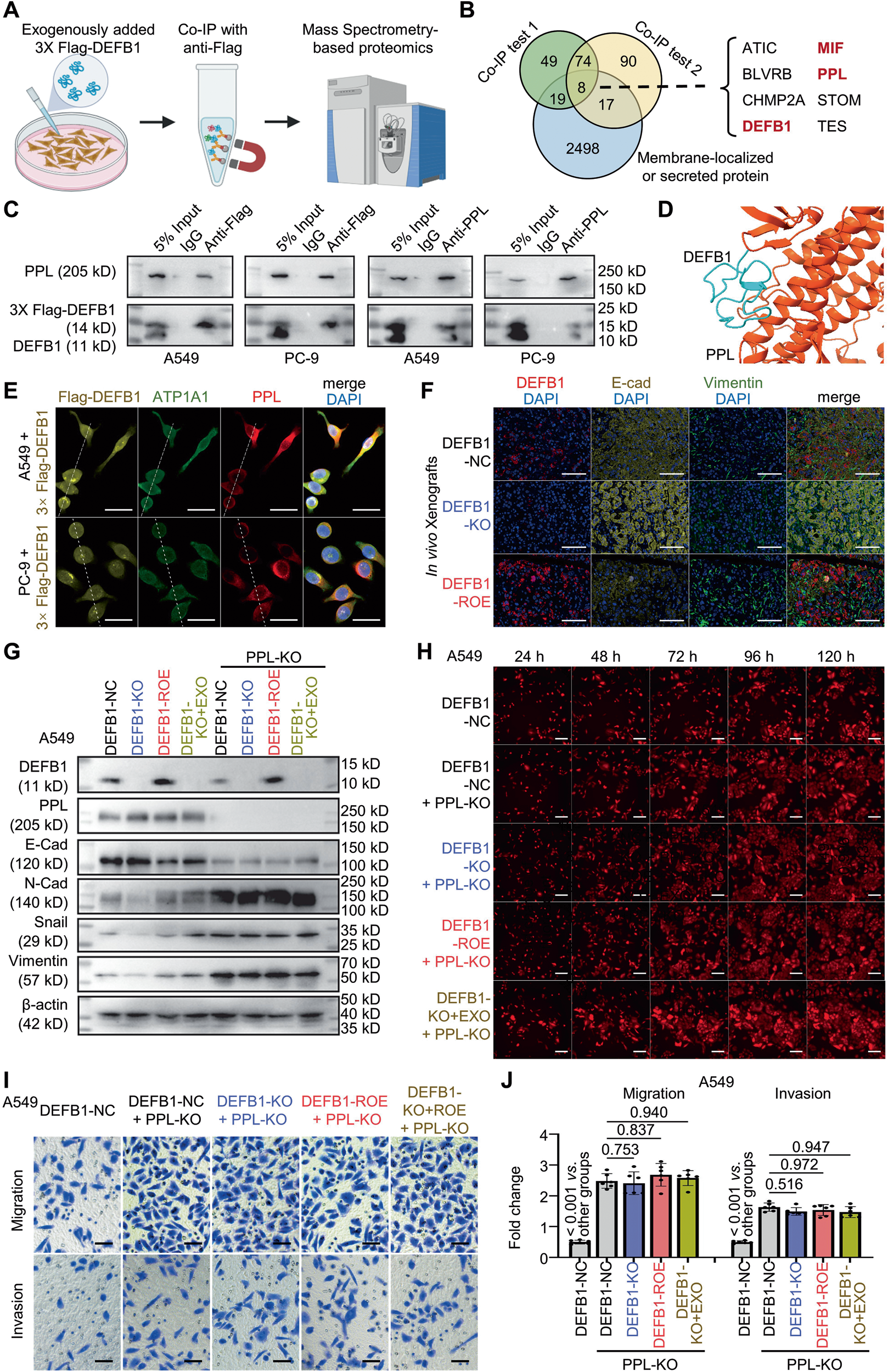 Fig. 3 DEFB1 interacts with Periplakin (PPL) to promote epithelial-to-mesenchymal transition (EMT) and proliferation in lung adenocarcinoma cells. A, B Flowchart and results for screening interacting membrane-localized and secreted proteins using coimmunoprecipitation (CoIP) combined with mass spectrometry and anti-Flag in A549 cells treated with exogenously added 3× Flag-tagged DEFB1 (200 pg/mL); C CoIP verification of the interaction between DEFB1 and PPL in A549/PC-9 cells treated with exogenously added 3× Flag tagged DEFB1 (200 pg/mL); D AlphaFold 3 prediction of the interaction mode between DEFB1 and PPL; E Immunofluorescence assay for detecting the cellular distribution of Flag-DEFB1, ATP1A1 (plasma membrane marker), and PPL in A549/PC-9 cells treated with exogenously added 3× Flag-tagged DEFB1 (200pg/mL)(scale bar: 30 μm); White dashed lines indicate the regions used for fluorescence intensity profile analysis; F Epithelial-mesenchymal transition (EMT) status of nude-mouse xenografts derived from A549 DEFB1-NC/KO/ROE cells (scale bar: 100 μm); G Western blot detection of the expression of EMT-related proteins in A549 DEFB1-NC/KO/ROE/KO + EXO cells with or without PPL knockout (PPL-KO); H High-content imaging (HCI) for the proliferation of A549 DEFB1-NC, and DEFB1-NC/KO/ROE/KO + EXO cells with PPL-KO (scale bar: 200 μm); I, J Transwell assays of the migration and invasion capacities of A549 DEFB1-NC, and DEFB1-NC/KO/ROE/KO + EXO cells with PPL-KO (scale bar: 100 μm). Statistical analysis: J One-way ANOVA test and Bonferroni correction.
模块 4:机制二:DEFB1 结合 MIF 诱导巨噬细胞 M2 促瘤极化
1.研究逻辑:CoIP-MS 发现 DEFB1 与 MIF 互作;肿瘤组织免疫荧光验证 DEFB1 调控巨噬分型;体外肿瘤 - 巨噬共培养 + MIF 抑制剂 ISO-1 回救实验验证通路;
2.核心实验:CoIP 验证 DEFB1-MIF 结合;THP-1 单核诱导巨噬共培养;流式 CD86 (M1)/CD206 (M2) 分型;qRT-PCR 巨噬标志物;裸鼠移植瘤巨噬多重免疫荧光;ISO-1 MIF 抑制剂体内回救;
3.关键实验结果
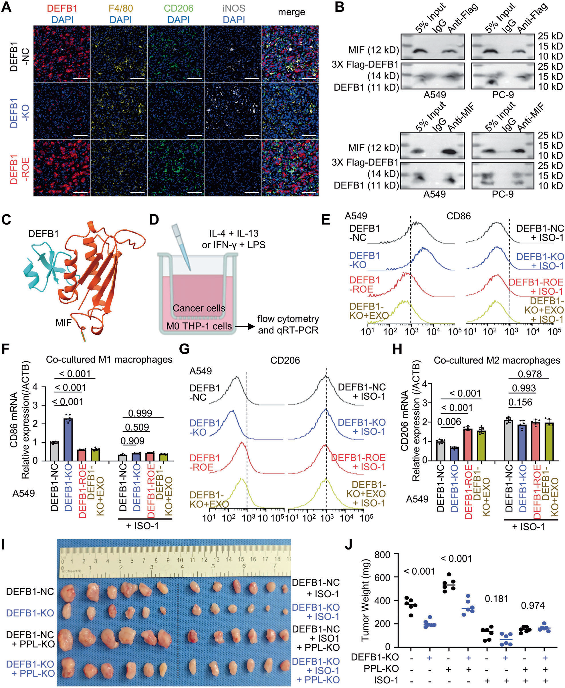 Fig. 4 DEFB1 interacts with macrophage Migration Inhibitory Factor (MIF) to promote M2 polarization of macrophages in vitro and in vivo. A Infiltrating status of M1 (marked by F4/80 and INOS) and M2 (marked by F4/80 and CD206) macrophages in nude-mouse xenografts derived from A549 DEFB1-NC/KO/ROE cells (scale bar: 200 μm); B CoIP verification of the interaction between DEFB1 and MIF in A549/PC-9 cells treated with exogenously added 3× Flag-tagged DEFB1 (200 pg/mL); C AlphaFold 3 prediction of the interaction mode between DEFB1 and MIF; D Schematic diagram of an in vitro co-culture model of lung adenocarcinoma cells and THP-1 derived macrophages; E, F Flow Cytometry and qRT-PCR for evaluating the effects of DEFB1 and/or MIF inhibitor (ISO-1) on M1 polarization of THP-1 M0 cells co-cultured with A549 cells; G, H Flow Cytometry and qRT-PCR for evaluating the effects of DEFB1 and/or ISO-1 on M2 polarization of THP-1 M0 cells co cultured with A549 cells; I, J Effects of DEFB1-KO, PPL-KO, and/or ISO-1 on the in vivo proliferative capacity in nude-mouse CDX model. Statistical analysis: (F, H, and G) One-way ANOVA test and Bonferroni correction.
模块 5:转化研究:抗 DEFB1 中和单抗 mAb-5 体内外抗肿瘤药效(Absin abs9490 基质胶应用点)
1.研究逻辑:构建 6 株抗 DEFB1 兔单克隆抗体,细胞筛选最优 mAb-5;细胞、患者肿瘤类器官、裸鼠移植瘤、人源化自发肺癌四重模型验证药效;检测 mAb-5 对 EMT、巨噬极化的逆转作用;
2.核心实验:抗体高内涵增殖筛选;Transwell 侵袭;EMT 蛋白 WB;肿瘤 - 巨噬共培养流式;患者 PDO 类器官培养;裸鼠 CDX 给药;Kras G12D 人源化 DEFB1 自发肺癌模型 CT 监测、生存分析;
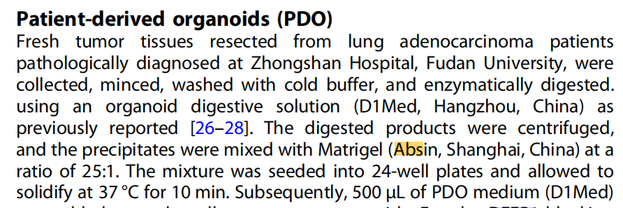
3.Absin 产品实验步骤说明:患者来源肺腺癌类器官培养环节,肿瘤消化沉淀与 abs9490 基质胶按 1:25 比例混合铺板,37℃凝固形成 3D 支撑支架,用于 mAb-5 药物体外药效筛选;
4.关键实验结果
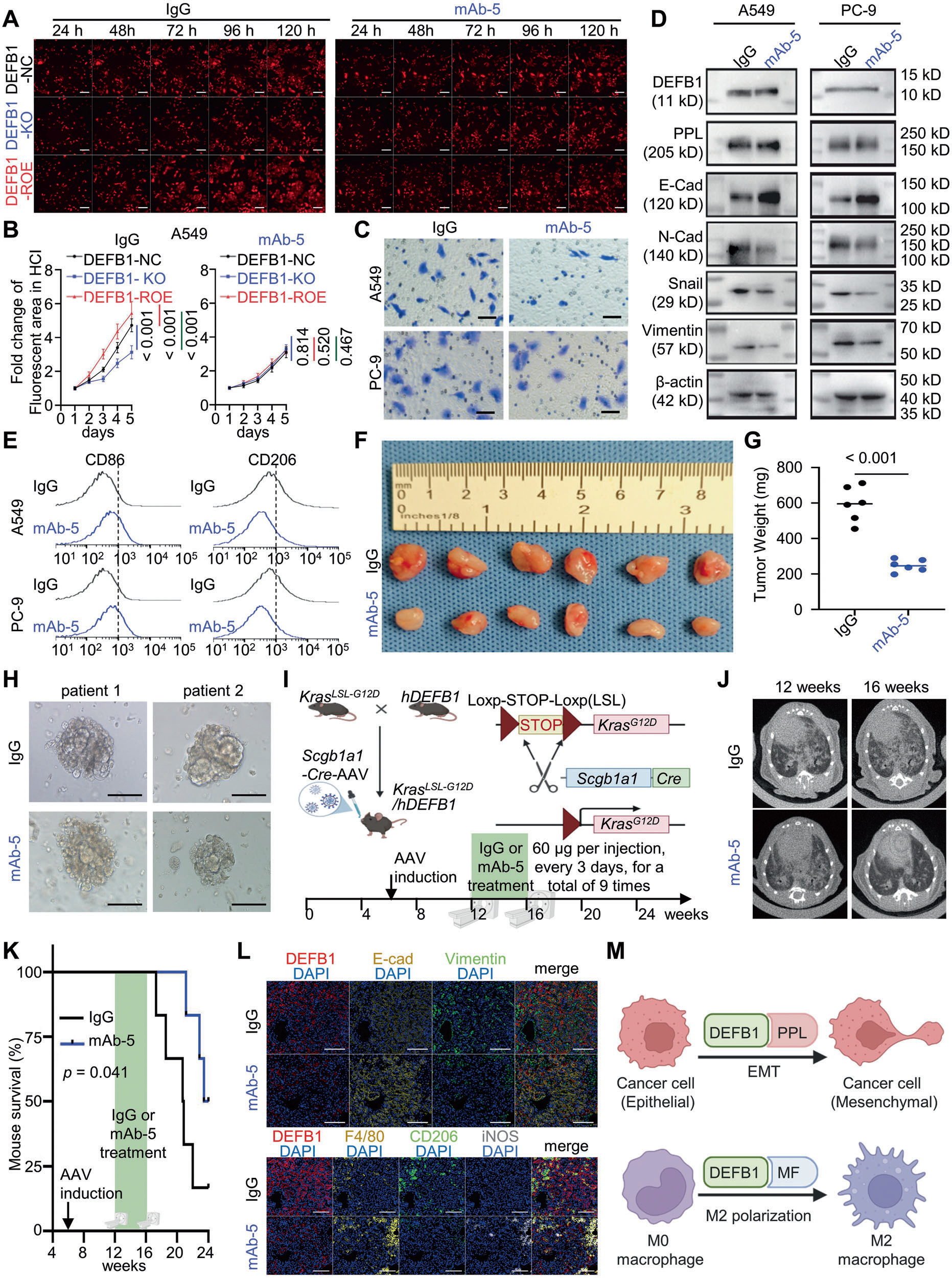 Fig. 5 Anti-DEFB1 monoclonal antibody as a promising therapeutic agent against lung adenocarcinoma. A, B High-content imaging for the proliferation of A549 DEFB1-NC/KO/ROE cells treated with 1 μg/mL IgG or anti-DEFB1 monoclonal antibody mAb-5 (scale bar: 200 μm); C Transwell assay for the invasion capacity of A549/PC-9 cells treated with 1 μg/mL IgG or mAb-5 (scale bar: 100 μm); D Western blot detection of the expression of epithelial-mesenchymal transition (EMT)-related proteins in A549/PC-9 cells treated with 1 μg/mL IgG or mAb-5; E Flow Cytometry for evaluating the M1/M2 polarization statues of THP-1 M0 cells co-cultured with A549/PC-9 cells treated with 1 μg/mL IgG or mAb-5; F–H In vivo proliferative capacity in nude mice CDX model and in human lung adenocarcinoma organoids (scale bar: 100 μm) treated with IgG or mAb-5 (60 μg per injection, every 3 days, for a total of 9 times); I Schematic description of KrasG12D-driven spontaneous lung tumor mice model expressing humanized DEFB1 and the treatment mode; J, K Representative images of chest computed tomography before and after the treatments of IgG or mAb-5 (60 μg per injection, every 3 days, for a total of 9 times) and survival analysis in spontaneous lung cancer model; L Epithelial-mesenchymal transition (EMT) and macrophage infiltrating statuses in spontaneous lung cancer model treated with IgG or mAb-5 (scale bar: 200 μm); M Mechanistic schema of DEFB1 in lung adenocarcinoma. Statistical analysis: B Two-way ANOVA test (n = 6) and Bonferroni correction; G Student’s t-test; K Log-rank test (n = 6).
模块 6:mAb-5 体内系统性安全性评估
1.研究逻辑:人源化 DEFB 免疫健全 C57BL 小鼠 4 周长期给药,从体重、行为、血液生化、主要脏器病理多角度评估毒性;
2.核心实验:每 3 天腹腔注射 mAb-5 共 9 次;每周监测体重;终点心脏穿刺采血生化检测;心 / 肝 / 脾 / 肺 / 肾 HE 病理染色;
3.关键实验结果:mAb-5 给药组小鼠体重、活动行为无异常;血液生化指标无显著改变;五大主要脏器 HE 切片无炎症、坏死、损伤病变,证实 mAb-5 体内安全性良好。
五、Absin(abs9490 基质胶)产品整体作用总结

- 手机:13761418683