




- 手机:13761418683
联系人:爱必信
网址:http://www.absin.cn/
地 址:上海市浦东新区新浩路58号18号楼

顶刊文献深度解析|姜黄源纳米囊泡负载 ROS 响应水凝胶重编程铁代谢促进微骨折术后软骨修复
2026-07-24
文献基础信息
● 标题:Curcuma-derived nanovesicle-loaded ROS-responsive hydrogels reprogram iron metabolism to enhance cartilage regeneration after microfracture
● 期刊:Bioactive Materials(2026, 64:744–762,生物材料领域顶刊,IF≈15+)
● DOI:https://doi.org/10.1016/j.bioactmat.2026.05.026
● 核心结论:首次证实微骨折术后术区铁过载诱发 BMSCs 铁死亡,是干细胞偏向纤维软骨分化、修复失败的核心诱因;从姜黄根茎提取 CDEVs(姜黄来源胞外囊泡),其核心载荷 Pvu-miR-159 靶向抑制 PTPN12、激活 ERK1/2-ATF4-GPX4/SLC7A11 抗氧化通路、阻断铁死亡;构建HAMA-APBA ROS 响应型可注射水凝胶实现病灶原位按需释放 CDEVs,在大鼠模型实现高质量透明软骨再生,为临床微骨折术后软骨修复提供植物源生物材料转化新策略。
● 使用 Absin 产品:OrganoGel 低因子无酚红基质胶(货号:abs9495),用于BMSCs 三维软骨类器官体外诱导培养,搭建 3D 体外软骨分化模型,验证 CDEVs 抑制纤维化、促进透明软骨形成的体外功能。
一、领域背景与科学问题引出
1. 临床痛点:微骨折(MF)软骨修复远期疗效差
微骨折是临床关节软骨缺损首选微创术式,通过打通软骨下骨、募集骨髓间充质干细胞(BMSCs)原位修复软骨,但临床随访显示30% 患者 5~10 年需二次手术,修复组织以力学性能差的纤维软骨为主,难以形成天然透明软骨,3~5 年渐进退变;目前纤维软骨分化的内在分子、微环境机制不明,缺乏靶向干预方案。
2. 前沿研究缺口
? 微环境机制空白:微骨折术中关节腔出血溶血,血红蛋白降解释放大量游离铁,局部铁过载微环境对 BMSCs 分化命运的调控机制尚无单细胞水平系统性研究,铁死亡在软骨修复中的调控地位未被阐明;
? 姜黄源胞外囊泡(CDEVs)应用短板:姜黄提取物已证实抗氧化、抗骨关节炎效果,但既往研究聚焦姜黄素小分子,植物源 miRNA 跨物种调控 BMSCs 铁死亡的机制未知,游离 CDEVs 体内易被体液快速清除,缺少靶向递送载体;
? 递送材料缺陷:常规水凝胶无病灶微环境响应能力,无法匹配术后 ROS 动态变化实现精准释药,难以在铁死亡关键窗口期(术后 7~14 天)持续起效。
3. 本研究核心科学问题
① 微骨折后局部铁过载是否通过铁死亡调控 BMSCs 分化分叉(透明软骨 / 纤维软骨)?
② CDEVs 能否通过内含植物 miR 抑制铁死亡、重编程 BMSCs 分化走向透明软骨?
③ 构建 ROS 响应可注射水凝胶能否实现 CDEVs 时序可控释放,体内提升软骨修复质量?
二、文献综述(研究领域脉络梳理)
1. 微骨折与 BMSCs 分化研究进展
BMSCs 是微骨折修复唯一功能细胞,局部炎症、氧化应激异常会诱导干细胞纤维化分化;既往研究仅聚焦炎症因子,铁代谢紊乱、铁死亡尚未被纳入修复失败关键诱因;单细胞测序技术逐步用于软骨异质性解析,但缺少术后全周期 BMSCs 分化轨迹图谱。
2. 铁死亡与软骨再生研究现状
铁死亡由铁依赖脂质过氧化驱动,GPX4/SLC7A11/GSH 是核心抗氧化轴;ATF4 直接调控 SLC7A11 转录、维持谷胱甘肽稳态;近年证实铁死亡参与骨关节炎软骨退变,但铁死亡调控 BMSCs 分化命运、决定微骨折修复表型是全新研究方向。
3. 植物胞外囊泡(PDEVs)与水凝胶递送
姜黄是传统药用植物,其来源胞外囊泡(CDEVs)富含 miRNA、脂质、小分子代谢物,低免疫原、高生物相容性,兼具抗氧化抗炎;但游离 CDEVs 半衰期短、关节腔留存差;苯硼酸(APBA)修饰透明质酸甲基丙烯酸(HAMA)可构建 ROS 响应水凝胶,高 ROS 环境下硼酸酯键断裂、凝胶降解释药,适配损伤高 ROS 病理微环境,是 EVs 理想递送平台。
4. 本研究创新点总结
? 机制创新:单细胞 + 时序病理首次明确「术后铁过载→BMSCs 铁死亡→分化偏向纤维软骨」完整病理轴,锁定分化关键节点 Node3;
? 靶点创新:首次发现 CDEVs 中 Pvu-miR-15 为核心功能分子,阐明跨物种靶向 PTPN12-ERK1/2-ATF4-GPX4 抗铁死亡通路;
? 材料创新:HAMA-APBA 智能水凝胶匹配术后 ROS 时序变化,在铁死亡窗口期(7~14d)按需释药,解决游离 CDEVs 体内快速清除难题;
? 模型创新:依托 Absin abs9495 基质胶搭建 3D 软骨类器官,体外高效模拟体内软骨分化,大幅缩短药效验证周期。
三、整体研究思路
第一步:病理机制挖掘:构建 SD 大鼠微骨折动物模型→多时间点组织取样 + 单细胞 RNA 测序 + 时序铁 / ROS 定量→证实铁死亡是 BMSCs 分化分叉关键开关;
第二步:体外药效验证:分离纯化 CDEVs→构建 Fe3?诱导 BMSCs 铁死亡体外模型→2D 细胞功能 +abs9495 基质胶 3D 软骨类器官培养,验证 CDEVs 抑制铁死亡、纠正异常分化;
第三步:分子机理深挖:多组学(转录组 + 代谢组 + miRNA 测序)+ 双荧光素酶 + WB 验证→锁定 Pvu-miR-159/PTPN12/ERK1/2/ATF4/GPX4 核心通路;
第四步:生物材料制备与表征:合成 HAMA-APBA 水凝胶、负载 CDEVs→流变 / 力学 / 体外释药 / 生物相容性检测,验证 ROS 响应释药性能;
第五步:体内动物疗效:大鼠关节原位注射给药(MF 组、空水凝胶组、游离 CDEVs 组、复合水凝胶组)→6w/12w 大体、Micro-CT、多染色组织学评分验证体内软骨修复效果。
四、分模块研究思路、实验结果与配套 Absin 产品应用
模块 1:体内病理探究 —— 微骨折后铁过载诱发铁死亡,驱动 BMSCs 纤维化分化
研究目的:阐明微骨折术后修复不良的本源病理,定位 BMSCs 分化关键调控因素
实验设计:SD 大鼠随机分假手术、微骨折组;术后 3/7/14/28d 取材,组织病理染色、单细胞测序、组织铁 / ROS 定量、基因表达检测
关键实验方法:
1. 大鼠膝关节微骨折造模:滑车沟全层软骨缺损 + 克氏针打孔穿透软骨下骨;
2. SOG 番红固绿染色、IHC(GPX4)、scRNA-seq(49095 个单细胞)、Pseudotime 拟时序分析、KEGG 富集;
3. 试剂盒定量组织总铁、DCFH-DA 测组织 ROS 水平。
核心实验结果(对应原文 Fig1、Fig2)
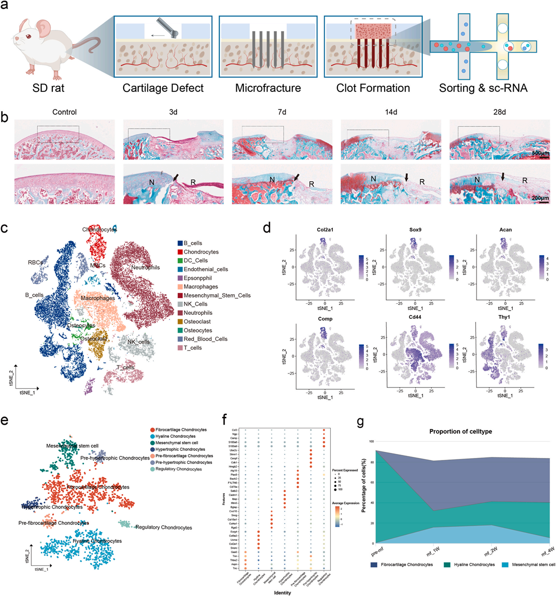
Fig. 1. Abnormal cartilage differentiation and single-cell sequencing of microfracture surgery. a) Modeling and single-cell sequencing of microfractures in SD rats. b) SOG staining of regenerated cartilage at different time points (Control、3d、7d、14d、28d) following microfracture surgery. c) t-distributed stochastic neighbor embedding (t-SNE) visualization of 49,095 cells using scRNA-seq. d) Visualization of the characteristic genes of the Chondrocytes cluster (Col2a1, SOX9, Acan, Comp) and Mesenchymal stem cells cluster (Cd44,Thy1) on the t-SNE map. e) t-distributed stochastic neighbor embedding (t-SNE) visualization of Chondrocytes cluster and Mesenchymal stem cells cluster reclustering using scRNA-seq. f) Dot plot of the top 4 marker genes in each cell subcluster. g) Proportion of each cell subcluster in different groups.
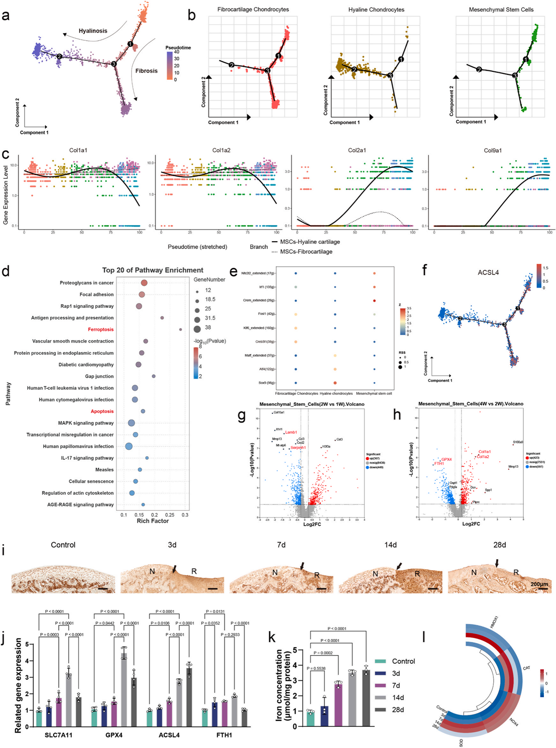
Fig. 2. Pseudotime analyses reveal ferroptosis as the core factor inducing the differentiation of MSCs trajectories after microfracture surgery. a) Pseudotime ordering (Node1,2,3-key nodes in the differentiation process, branch lines-differentiation trajectory) on the mean subclusters (Fibrocartilage Chondrocytes, Hyaline Chon drocytes, Mesenchymal stem cell) arranged them into a major trajectory (Hyalinosis and Fibrosis). b) The distribution of each subcluster at the Pseudotime map (Fibrocartilage Chondrocytes-red, Hyaline Chondrocytes-brown, Mesenchymal stem cell-green). c) Distribution of characteristic genes in the differentiation tra jectory(MSCs to Fibrocartilage- Col1a1,Col1a2; MSCs to Hyaline cartilage- Col2a1,Col9a1) (Color of point-each differentiation time state). d) The top 20 pathways after KEGG enrichment analysis of the differentiated gene set at Node 3.(Ferroptosis, Apoptosis). e) The top 3 transcription factors of each sucluscter revealed by the SCHENIC transcription factor regulatory network. f) The distribution of the gene ACSL4 in the Pseudotime trajectory. g-h) Volcano maps of differentiated genes of BMSCs in different groups after microfracture surgery, 2W vs 1W(G) and 4W vs 2W(H). i) Immunohistochemical staining of GPX4 between different groups after microfracture surgery of SD rats. j) Ferroptosis-related gene expression of rat's regenerated cartilage after microfracture surgery. k) Iron content of regenerated cartilage tissue in different groups. l) Heat map of ROS-related genes expression. Data are represented as mean ± SD. Statistical significance was determined by a two-sided Student's t-test.
4. SOG 染色:微骨折组随时间推移软骨基质糖胺聚糖持续丢失,缺损区大量纤维样瘢痕组织生成;
5. 单细胞分群:BMSCs 分化为透明软骨、纤维软骨两大谱系,Node3 是分化关键分叉位点;KEGG 富集提示铁死亡、MAPK 是分叉最高富集通路;
6. 时序检测:术后 0~14d 局部铁、ROS 持续升高,GPX4、SLC7A11(抗铁死亡基因)先代偿升高后骤降,纤维化标记 Col1A1 持续上调、透明软骨 Col2A1 显著下调;
7. ACSL4(铁死亡标志基因)沿 BMSC→纤维软骨分化轨迹高表达,直接证实铁死亡诱导纤维化分化。
模块 2:CDEVs 分离鉴定 + 体外 2D 细胞抗铁死亡功能验证
研究目的:表征姜黄源囊泡理化性质,体外验证 CDEVs 逆转铁死亡、恢复 BMSCs 增殖迁移和成软骨潜能
实验设计:姜黄根茎凝胶过滤层析分离 CDEVs,TEM/NTA 表征;FeCl?(100μM) 构建 BMSCs 铁死亡模型,分组(Control/Fe/Fe+CDEVs),优选 25μg/mL 给药浓度
关键实验方法:CCK8、EdU 增殖、划痕愈合、Transwell 迁移、FerroOrange 胞内铁染色、JC-1 线粒体膜电位、BODIPY 脂质过氧化、TEM 线粒体超微结构、qPCR 检测成软骨 / 纤维化 / 铁死亡基因。
核心结果(原文 Fig3、Fig5、Fig6)
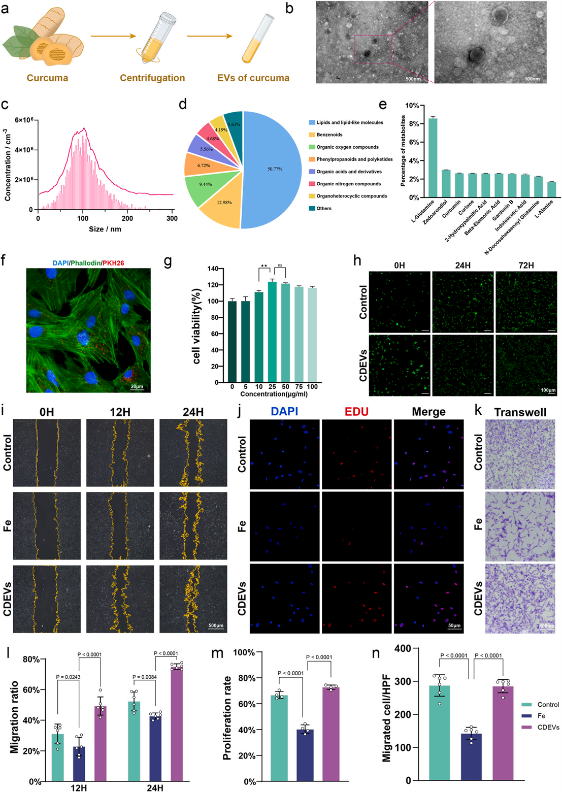
Fig. 3. Isolation, characterization, and biocompatibility of CDEVs. a) Schematic illustration of CDEVs collection isolation from Curcuma longa (turmeric). b) Representative TEM images revealing nanovesicular morphology. c) Size distributions analysis of CDEVs. d) Pie charts showing the biomolecular compositions of DEVs. e) The top 10 metabolites identified in CDEVs. f) Cellular uptake of PKH26-labeled CDEVs (red) by BMSCs (nuclei: DAPI, blue). g) BMSCs viability after 24 h exposure to increasing CDEVs concentrations (0, 5, 10, 25, 50, 75, and 100 μg/mL, respectively) for 24 h (n = 4). h) BMSCs treated with PBS or CDEVs(100 μg/mL) for different time periods(0H,24H; 72H) and stained with calcein-AM (living cells, green) and propidium iodide (dead cells, red). i) Wound healing capacity of BMSCs measured using a scratch wound healing test in different groups (Control:PBS; Fe:FeCl3-100μM; CDEVs: FeCl3-100μM, CDEVs-25 μg/mL). j) EDU proliferation assay (red) of BMSCs in different groups. Nuclei: DAPI (blue). k) Migration capacity of BMSCs measured using the transwell assay. l-n) Quantified scratch wound healing rate (l), proliferation rate of EDU staining (m) and migrated cell count/HPF in Transwell (n) (n = 6).
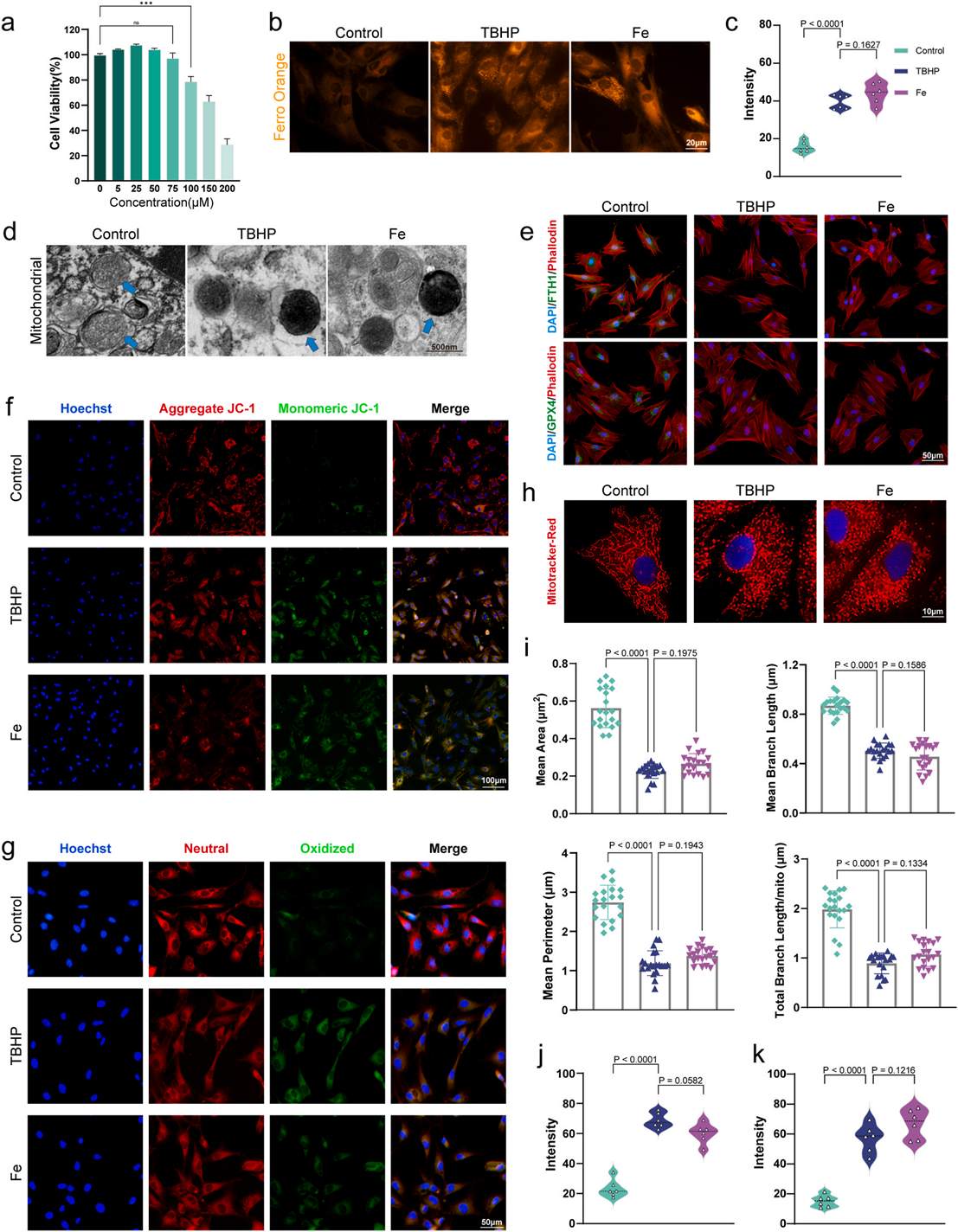
Fig. 5. Ferroptosis model constructed by high iron concentration in vitro. a) BMSCs activity under different iron concentration(0 μM,5 μM,25 μM,50 μM,75 μM,100 μM,150 μM,200 μM) detected by CCK8 assay. b-c) Ferro Orange staining of BMSCs in different groups(Control, TBHP:100 μM, Fe:100 μM)(b) and semi-quantitative visual analysis(c). d) Representative TEM images of mitochondria (blue arrows) in BMSCs treated by different groups. e) Immunofluorescence staining images of ferroptosis markers (GPX4, FTH1) of BMSCs. The nuclei were stained with DAPI, and the cytoskeletons were stained with TRITC Phalloidin. f) JC-1 staining images (DAPI-blue, JC-1 Aggregate-red, JC-1 Monomeric-green) of BMSCs in different groups. g) Bodipy staining images(DAPI-blue, Neutral-red, Oxidized-green) of BMSCs in different groups. h-i) Mito tracker red staining images of BMSCs in different groups (h) and quantitative analysis of mitochondrial morphology (i). j-k) Quantitative analysis of JC-1 staining (j) and Bodipy staining (k).Data are represented as mean ± SD. Statistical significance was determined by a two-sided Student's t-test (*p <0.05, **p < 0.01, ***p < 0.001).
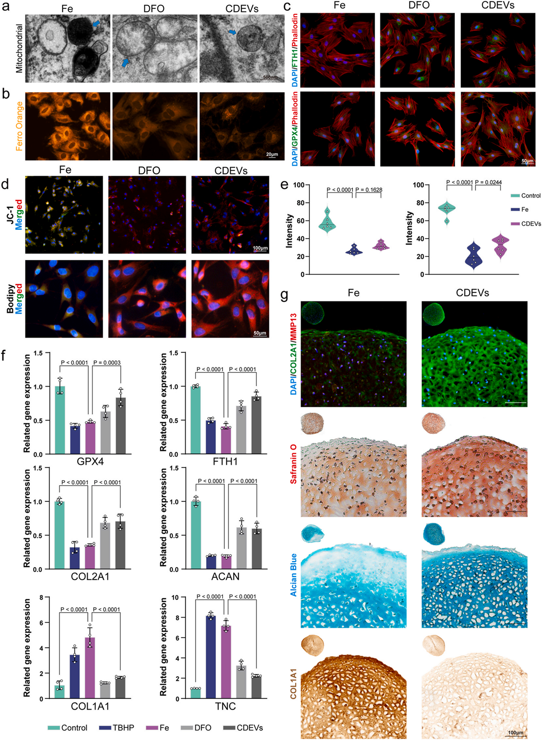
Fig. 6. Antagonistic effect of CDEVs on ferroptosis in vitro. a) Representative TEM images of mitochondria (blue arrows) in BMSCs treated by different groups(Fe:FeCl3-100μM; TBHP: FeCl3-100μM, DFO-10μM; CDEVs: FeCl3-100μM, CDEVs-25 μg/mL). b) Ferro Orange staining of BMSCs in different groups. c) Immunofluo rescence staining images of ferroptosis markers (GPX4, FTH1) of BMSCs. The nuclei were stained with DAPI, and the cytoskeletons were stained with TRITC Phalloidin. d-e) JC-1 and Bodipy staining images(merged) of BMSCs in different groups (d) and their quantitative analysis (e). f) qRT-PCR analysis of ferroptosis markers (GPX4, FTH1), hyalinosis markers (COL2A1, ACAN) and fibrosis markers (COL1A1, TNC) (n = 4). g) Immunofluorescence and pathological staining of 3D cultured cartilage organoids in different groups: Immunofluorescence staining (DAPI-blue, Col2a1-green, Mmp13-red); Safranin O staining; Alcian blue staining and Immunohistochemical staining of Col1a1. Data are represented as mean ± SD. Statistical significance was determined by a two-sided Student's t-test.
1. CDEVs 粒径 50~200nm(中位 107.6nm),球形囊泡结构,富集谷氨酰胺、姜黄素等抗氧化代谢物,4h 即可被 BMSCs 高效摄取;
2. Fe 诱导组:胞内铁超载、线粒体皱缩嵴缺失、膜电位崩塌、脂质过氧化飙升,Col2A/ACAN(透明软骨基因)下调,Col1A1/TNC(纤维化基因)上调;
3. CDEVs 干预:显著降低胞内 Fe2?、修复线粒体形态、上调 GPX4/SLC7A11,逆转分化异常,恢复 BMSCs 增殖与迁移能力。
模块 3:3D 软骨类器官培养
研究目的:突破 2D 细胞局限,3D 体外模拟体内软骨微环境,直观验证 CDEVs 调控软骨分化、抑制纤维化,使用 Absin OrganoGel 基质胶(货号 abs9495,无酚红低因子)
产品信息
产品全称:OrganoGel 基质胶(低因子、无酚红),产品特点:小鼠肿瘤基底膜提取,主要成分为层粘连蛋白、IV 型胶原;4℃液态、37℃成胶,无酚红不干扰荧光染色,适配软骨类器官长期 3D 培养,支撑干细胞自组装成软骨球。
实验方案 BMSCs+OrganoGel(abs9495)混合铺板,分组:空白对照组、Fe 损伤组、Fe+CDEVs 给药组;添加成软骨诱导培养基,连续培养 21d 收获类器官,COL2/ MMP13 免疫荧光、SOG、阿尔新蓝、COL1A1 免疫组化染色。
核心结果
1. Fe 损伤类器官:基质降解严重,COL2(透明软骨标志)极低表达、COL1(纤维化标志)大量沉积,糖胺聚糖(SOG / 阿尔新蓝染色)几乎缺失;
2. CDEVs 处理组:类器官结构完整,COL2 大面积阳性、COL1 表达显著下降,基质糖胺聚糖含量显著回升,证实 CDEVs 在 3D 微环境下有效重编程 BMSCs 向透明软骨分化。
应用价值:abs9495 基质胶完美复刻胞外基质支架,无需动物体内预实验即可快速筛选 CDEVs 药效,大幅节约动物实验成本。
模块 4:多组学 + 分子机制解析:Pvu-miR-159/PTPN12/ERK1/2/ATF4/GPX4 通路
研究目的:定位 CDEVs 中核心功能分子,完整阐明抗铁死亡分子通路
实验设计:CDEVs 小 RNA 测序、BMSCs 转录组 + 代谢组、5 种数据库靶基因预测、双荧光素酶报告基因、si-PTPN12 敲低、WB、GSH/GSSG 定量
核心结果
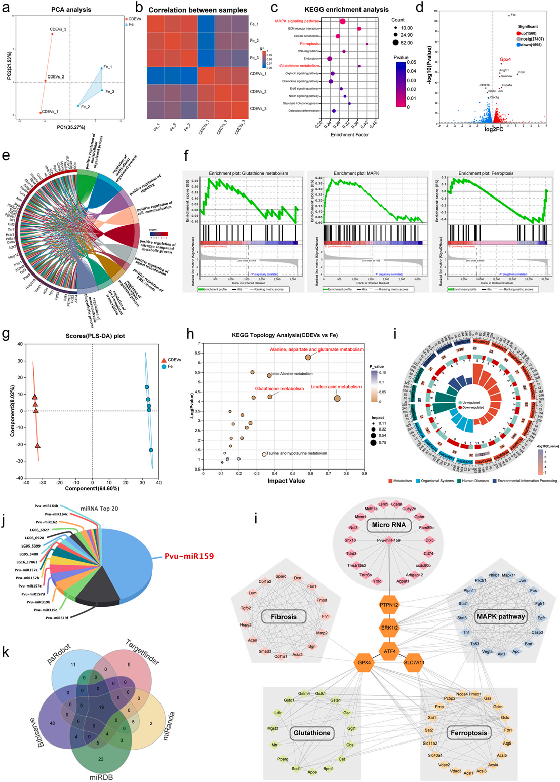 Fig. 7. CDEVs reverse high Fe level-induced ferroptosis in BMSCs through Pvu-mir159 by regulating the MAPK pathway and reshape Glutathione metabolism. a-b)PLS-DA score plot (a) and Pearson correlation (b) between different groups (Fe-treated BMSCs ± CDEVs). c) KEGG enrichment of DEGs, which are mainly associated with MAPK, Ferroptosis, and Glutathione metabolism. d) Volcano maps of BMSCs in different groups. e) Analysis of functional clustering enrichment for positive regulation of multicellular organismal process, positive regulation of signal transduction, positive regulation of ERK cascade, and regulation of DNA-templated transcription between Fe and CDEVs groups. f) GSEA of the MAPK pathway, Ferroptosis, and Glutathione metabolism. g) PLS-DA score plot of differentially abundant metabolites in Fe-treated BMSCs ± CDEVs. (h) KEGG topology analysis, which is mainly associated with Linoleic acid metabolism, Alanine, aspartate,glutamate metabolism, and Glutathione metabolism. (i) Multi-dimensional enrichment circle map of metabolites. (j) Distribution rate of the top 20 miRNAs in CDEVs. k) Venn analysis of all predicted genes for Pvu-miR-159 potential targets. l) miRNA-mRNA interaction of Pvu-miR-159 and the following pathway network on gene sets of Fibrosis, Glutathione metabolism, MAPK pathway, and Ferroptosis constructed by Cytoscape software.
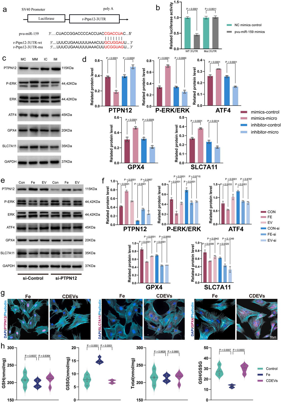 Fig. 8. Pvu-mir159 in CDEVs inhibits ferroptosis by binding to PTPN12 and regulating the downstream ERK-ATF4-GPX4 (SLC7A11) axis. a) Sequence of PTPN12 binding sites of wild type (WT) and mutant (Mut) Pvu-miR-159. b) Quantitative analysis of luciferase activity. c-d) Western blot analysis and quantitative analysis of PTPN12, P-ERK, ERK, ATF4, GPX4 and SLC7A11 in BMSCs transfected with Pvu-miR-159 mimics and inhibitor (MC: mimics-control, MM: mimics-micro, IC:inhibitor-control, IM: inhibitor-micro) (n = 4). e-f) Western blot analysis and quantitative analysis of PTPN12, P-ERK, ERK, ATF4, GPX4, and SLC7A11 in BMSCs which is transfected with si-PTPN12 or not (n = 4). g) Immunofluorescence staining images of PTPN12, GPX4, and SLC7A11, the nuclei were stained with DAPI, and the cytoskeletons were stained with phalloidin. h) Glutathione metabolism level identified by the concentration of GSH and GSSG in different groups. Data are represented as mean ± SD. Statistical significance was determined by a two-sided Student's t-test.
1. CDEVs 内 Pvu-miR-159 占总 miRNA 丰度 44.1%,是最优势功能性小 RNA;
2. 双荧光素酶证实:Pvu-miR-159 特异性结合 PTPN12 基因 3'UTR,抑制 PTPN12 蛋白表达;
3. PTPN12 为磷酸酶,被抑制后 ERK1/2 磷酸化水平上升,上调转录因子 ATF4,ATF 入核激活 SLC7A11 与 GPX4 转录;提升 GSH 含量、抑制脂质过氧化、阻断铁死亡;
4. PTPN12 敲低后,CDEVs 的保护效应大幅消失,明确Pvu-miR-159→PTPN12 是 CDEVs 发挥作用的必需靶点。
模块 5:HAMA-APBA ROS 响应水凝胶合成、理化与体外释药表征
研究目的:构建可注射智能载体,实现高 ROS 病灶按需缓释 CDEVs,解决游离囊泡体内快速清除痛点
实验设计:HA→甲基化改性 HAMA→APBA 酰胺接枝得到 HAMA-APBA,混合 CDEVs 制备复合水凝胶;核磁、SEM、流变、压缩力学、ROS(H?O?)诱导降解、体外 CDEVs 释放检测
核心结果
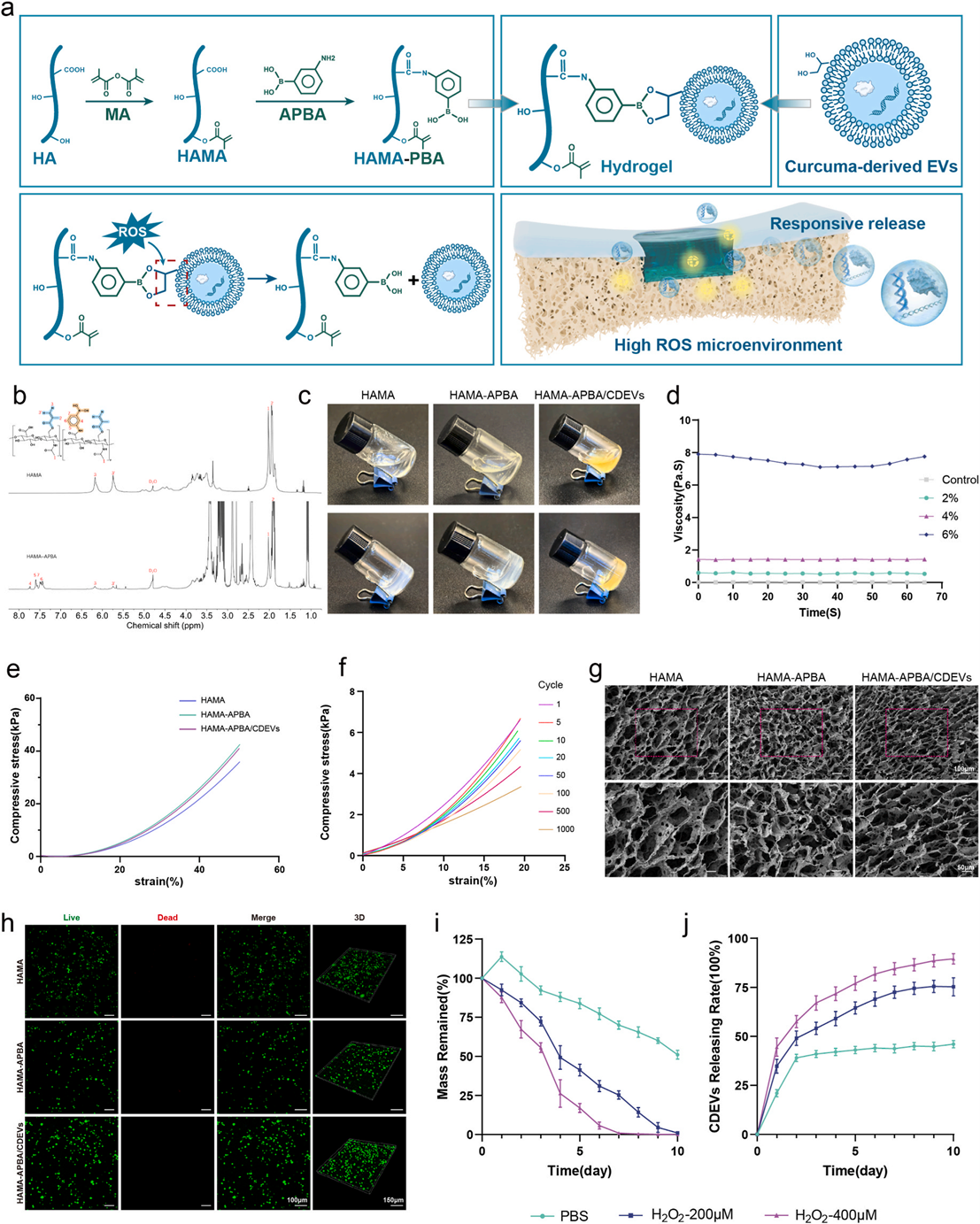
Fig. 4. Synthesis and Characterization of HAMA-APBA/CDEVs. a) Illustration of the stepwise preparation of HAMA-APBA/CDEVs. b) Nuclear magnetic resonance hydrogen spectrum ( 1 H NMR) of HAMA modified with APAB group or not. c) Morphology of hydrogels in different groups (HAMA, HAMA-APBA, HAMA-APBA/CDEVs). d) Viscosity test of hydrogels in different concentrations (Control, 2%, 4%, 6%). e) Representative tensile stress-strain curves of hydrogels in different groups at < 50% strain. f) The axial compressive cyclic stress-strain curves of hydrogels under 1000 loading-unloading cycles (1st, 5th, 10th,20th, 50th, 100th, 500th, and 1000th cycles) at 20% strain per time. g) Inner structure of hydrogels in different groups by Scanning Electron Microscope (SEM). h) BMSCs loaded with hydrogels in different groups and stained with calcein-AM (living cells, Green) and propidium iodide (dead cells, red). i) Degradation of hydrogels of different groups in the ROS environment. j) Release rates of exosomes from different scaffold groups.
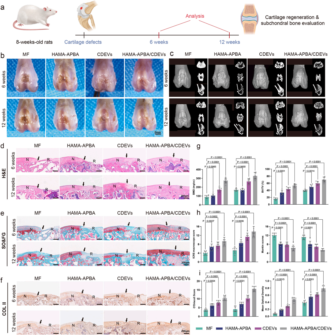
Fig. 9. HAMA-APBA/CDEVs facilitated the original differentiation of regenerated cartilage after microfracture surgery. a) Schematic diagram of the rat micro fracture surgery model and treatment with HAMA-APBA/CDEVs. b) Macroscopic observation of cartilage defects with different implants at 6 and 12 weeks post operation. c) Micro-CT images showing 2D and 3D reconstruction of the repaired cartilage at 6 and 12 weeks postoperation. d) H&E staining, e) Safranin O/Fast Green staining, and f) Type II collagen immunohistochemical staining of the repaired tissue from different groups at 6 and 12 weeks postoperation. g) BMD and BV/TV in Micro-CT 3D reconstruction, h) ICRS histological total score and Mankin histological total score, i) O'Driscoll histological total score and the mean optical density of type II collagen immunohistochemical staining. The data are presented as the mean ± SD (n = 8).
1. 1H-NMR 验证 APBA 成功接枝,优选 4% 水凝胶配方(兼顾注射器可注射性与体内结构稳定性);
2. 水凝胶多孔结构,H?O?浓度越高降解越快:400μM H?O?条件 7d 完全降解,CDEVs 累积释放 88.3%;200μM 组 10d 降解、释放 73.3%,完美匹配术后 7d ROS 峰值释药;
3. 活死染色证实 HAMA-APBA 无细胞毒性,生物相容性优异。
模块 6:大鼠体内原位修复药效(MF 动物最终验证)
研究目的:体内验证 HAMA-APBA/CDEVs 复合水凝胶的软骨修复效果,分组:MF 空白组、HAMA-APBA 空胶组、游离 CDEVs 组、HAMA-APBA/CDEVs 复合组
实验方案:大鼠股骨滑车全层软骨缺损 + 微骨折造模,术区原位注射对应制剂;术后 6w、12w 取材,大体观察、Micro-CT(BMD/BV/TV)、H&E、SOG、COL2 免疫组化,ICRS/Mankin/O'Driscoll 组织学评分;心肝肾脾肺 HE 毒理染色。
核心结果(原文 Fig9)
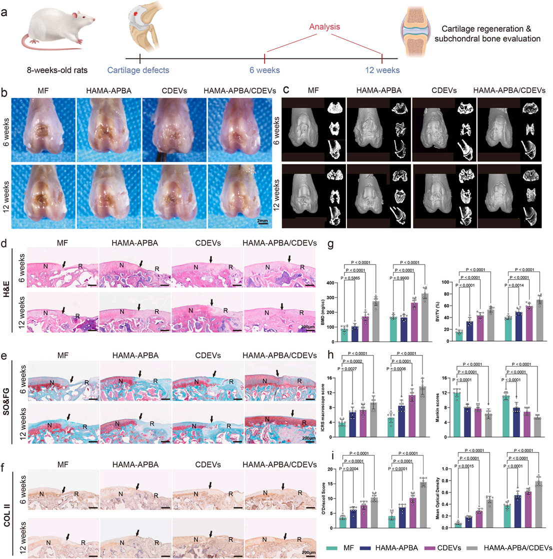
Fig. 9. HAMA-APBA/CDEVs facilitated the original differentiation of regenerated cartilage after microfracture surgery. a) Schematic diagram of the rat micro fracture surgery model and treatment with HAMA-APBA/CDEVs. b) Macroscopic observation of cartilage defects with different implants at 6 and 12 weeks post operation. c) Micro-CT images showing 2D and 3D reconstruction of the repaired cartilage at 6 and 12 weeks postoperation. d) H&E staining, e) Safranin O/Fast Green staining, and f) Type II collagen immunohistochemical staining of the repaired tissue from different groups at 6 and 12 weeks postoperation. g) BMD and BV/TV in Micro-CT 3D reconstruction, h) ICRS histological total score and Mankin histological total score, i) O'Driscoll histological total score and the mean optical density of type II collagen immunohistochemical staining. The data are presented as the mean ± SD (n = 8).
1. 大体:MF 组 12w 仍大面积缺损、纤维瘢痕;复合水凝胶组缺损近乎完全闭合,新生软骨平滑,与宿主软骨无缝整合;
2. Micro-CT:复合组软骨下骨 BMD、BV/TV 显著高于其余三组,骨重建最优;
3. 组织染色:复合组 SOG 强阳性、COL2 大面积富集,纤维化 COL1 极低,Mankin 评分显著降低;
4. 脏器病理无异常,证实材料与 CDEVs 体内安全性佳。
五、机制总结(完整作用链条)
1. 病理上游:微骨折术中关节腔出血溶血→局部大量游离铁蓄积→Fenton 反应催生高 ROS→BMSCs 铁死亡激活→ATF4 下调→SLC7A11/GPX4 下降→GSH 耗竭、脂质过氧化→BMSCs 偏离透明软骨分化、大量生成纤维软骨;
2. CDEVs 分子干预:HAMA-APBA 水凝胶在术后 7d(ROS 峰值)硼酸酯键断裂、原位释放 CDEVs→囊泡内化递送 Pvu-miR-159→靶向沉默 PTPN12→ERK1/2 磷酸化升高→ATF4 激活→SLC7A11/GPX4 上调→GSH 代谢修复、铁死亡被抑制→BMSCs 回归透明软骨分化轨迹;
3. 材料赋能:ROS 响应可控释药,在术后 7~14d 铁死亡关键窗口期持续给药,实现长效软骨保护。
六、研究亮点与临床转化价值
1. 科学亮点
? 机制原创:从单细胞水平阐明微骨折修复失败的铁死亡新机制,填补软骨再生领域铁代谢调控空白;
? 中药现代化:传统姜黄从粗提物升级为植物源纳米囊泡制剂,依托内源 miRNA 实现跨物种靶向调控;
? 材料创新:ROS 响应透明质酸水凝胶实现时序精准给药,打通植物 EVs 临床递送瓶颈;
? 体外模型标准化:采用 Absin abs9495 基质胶建立标准化 3D 软骨类器官筛选平台,缩短候选药物开发周期。
2. 临床转化价值
? 手术联用便捷:HAMA-APBA 为可注射水凝胶,微骨折术中关节腔原位注射即可,无需额外手术;
? 原料优势:姜黄全球量产、成本低廉,CDEVs 规模化提取可行性高,远优于动物源外泌体;
? 适用拓展:方案可延伸至骨关节炎、软骨缺损等多种软骨损伤疾病,具备从动物实验走向临床试验的潜力。
七、研究局限性与未来展望
局限
1. 仅大鼠体内验证,缺少非人灵长类大动物实验,长期反复给药的免疫原性仍需系统评价;
2. CDEVs 批量生产工艺尚未标准化,不同批次 Pvu-miR-159 含量波动待优化;
3. HAMA-APBA 水凝胶机械强度仍需优化,适配高强度负重关节。
未来方向
1. 优化 CDEVs 规模化工业制备工艺,固定活性 miRNA 含量;
2. 结合 abs9495 类器官高通量筛选体系,联用其他天然植物囊泡协同增效;
3. 大动物(兔 / 猴)体内长期毒理与药效评价,推进临床前申报。
【免责声明】原文版权归Bioactive Materials及原作者所有,本文为科研解析整理;文中 Absin 产品信息来源于爱必信官网公示资料。

- 手机:13761418683