




- 手机:13761418683
联系人:爱必信
网址:http://www.absin.cn/
地 址:上海市浦东新区新浩路58号18号楼

SCI 文献深度解析:高分文献解读|m?A-lncRNA MIAT 调控结直肠癌失巢凋亡抵抗与腹膜转移 Absin 助力患者来源类器官三维侵袭模型构建
2026-09-07
一、文献基础综述
期刊与基础信息
期刊:International Immunopharmacology,2026 IF=5.6,中科院医学 2 区,JCR Q1 区;
DOI:10.1016/j.intimp.2026.117132;卷期:2026, 186:117132;
论文标题:m?A?modified lncRNA MIAT promotes anoikis resistance and peritoneal metastasis of colorectal cancer through miR?181a?5p/MCL1 signaling axis。
全文核心结论:lncRNA?MIAT 在结直肠癌腹膜转移(CRCPM)组织与抗失巢凋亡(AR)结直肠癌细胞中显著上调,提示不良预后;METTL3 介导的 m?A 修饰以 IGF2BP2 依赖的方式提升 MIAT RNA 稳定性;MIAT 作为 ceRNA 海绵吸附 miR?181a?5p,上调抗凋亡蛋白 MCL1,促进结直肠癌细胞增殖、侵袭、失巢凋亡抵抗以及体内腹膜转移;该研究揭示m?A?MIAT/miR?181a?5p/MCL1全新信号轴,MIAT 可作为结直肠癌腹膜转移潜在预后标志物和治疗靶点。
爱必信产品应用简述:本研究使用abs47014921 I 型胶原蛋白(鼠尾来源)构建三维胶原凝胶体系,用于患者来源类器官(PDO)侵袭实验。该胶原凝胶模拟体内细胞外基质微环境,实现类器官侵袭表型的直观观察与评估,为类器官层面验证 MIAT 促侵袭功能提供三维体外模型支撑。
二、研究领域背景介绍
结直肠癌是全球高发恶性肿瘤,腹膜转移是结直肠癌恶性程度极高的转移亚型,7?15% 患者初诊即伴随腹膜转移,未经干预患者生存期仅 3?6 个月,临床治疗手段有限、预后极差。肿瘤细胞脱离原发灶后,会失去与胞外基质的粘附,触发失巢凋亡;失巢凋亡抵抗是肿瘤细胞完成腹膜播散、定植的核心先决条件,但结直肠癌腹膜转移中失巢凋亡抵抗的分子调控网络尚不清晰。
lncRNA 广泛参与肿瘤增殖、转移、凋亡抵抗等恶性表型;m?A 作为真核 RNA 最主要表观转录修饰,通过 “写、擦、读” 蛋白调控 RNA 的稳定性、剪接、翻译;m?A 修饰可调控 lncRNA 表达,在消化道肿瘤进展发挥关键作用,但 m?A 修饰调控 lncRNA 介导结直肠癌腹膜转移、失巢凋亡抵抗的机制报道较少。 患者来源类器官(PDO)高度保留原发肿瘤生物学特征,相比传统二维细胞更贴近肿瘤真实病理状态,三维胶原侵袭模型是评估肿瘤侵袭能力的重要工具。基于上述背景,该文聚焦 m?A 修饰调控的 lncRNA MIAT,系统探究其介导结直肠癌失巢凋亡抵抗与腹膜转移的完整分子通路。
三、作者整体研究思路总结
全文遵循临床样本挖掘筛选靶点→临床样本验证表达与预后→体外二维细胞功能验证→患者来源类器官(PDO)三维模型验证→体内动物模型验证→ceRNA 机制挖掘与回救实验→m?A 上游修饰机制探究→通路汇总完整转化医学逻辑链:
1. 芯片测序筛选结直肠癌腹膜转移差异 lncRNA,锁定在抗失巢凋亡细胞中高表达的 MIAT;82 对临床结直肠癌样本验证 MIAT 表达,结合生存分析明确 MIAT 和患者不良预后、腹膜转移的相关性;亚细胞定位实验提示 MIAT 同时定位于胞核与胞质,具备 ceRNA 作用基础。
2. 构建 MIAT 敲低稳转细胞株,体外开展增殖、克隆、凋亡、划痕、Transwell 侵袭实验,明确 MIAT 促进结直肠癌增殖、侵袭、失巢凋亡抵抗;同时在患者来源类器官模型验证 MIAT 促癌表型。
3. 构建裸鼠皮下异种移植、腹腔腹膜转移模型,体内验证 MIAT 促进肿瘤生长与腹膜转移。
4. 生物信息预测结合双荧光素酶、RNA?FISH、RIP 验证 MIAT 作为 ceRNA 靶向结合 miR?181a?5p;miR?181a?5p 抑制剂回救实验确认 miR?181a?5p 介导 MIAT 的促癌功能。
5. 预测并证实 MCL1 为 miR?181a?5p 直接下游靶基因;MCL1 过表达回救实验,体外细胞、类器官、体内动物多层次证明 MIAT 依赖 MCL1 发挥促癌作用。
6. 探究 MIAT 上调上游机制:MeRIP、RNA 稳定性实验证实 METTL3 介导 m?A 修饰,通过阅读器 IGF2BP2 增强 MIAT 的 RNA 稳定性,最终完成完整调控轴的论证。
四、分模块详细研究思路、实验结果、对应图片
模块 1:MIAT 高表达与结直肠癌腹膜转移及不良预后相关
研究逻辑:利用芯片测序筛选腹膜转移相关 lncRNA,构建抗失巢凋亡 CRC 细胞模型;临床组织样本检测 MIAT 表达,生存分析、临床病理关联分析 MIAT 的临床意义;亚细胞定位明确 MIAT 分布。
核心实验:lncRNA 芯片、GSEA/KEGG 富集分析;抗失巢凋亡(AR)细胞构建;RT?qPCR;RNA?ISH;Kaplan?Meier 生存分析;核质分离;RNA?FISH。
关键实验结果:① GSEA 提示失巢凋亡抵抗通路在结直肠癌腹膜转移组织显著富集;芯片筛选得到差异 lncRNA,MIAT 在 AR 细胞上调最显著; ② MIAT 在结直肠癌组织高于癌旁,腹膜转移灶表达高于原发灶;高 MIAT 表达对应患者更差总生存期;MIAT 表达与肿瘤大小、远处转移正相关; ③ MIAT 同时分布于细胞核、细胞质,具备 ceRNA 作用基础。
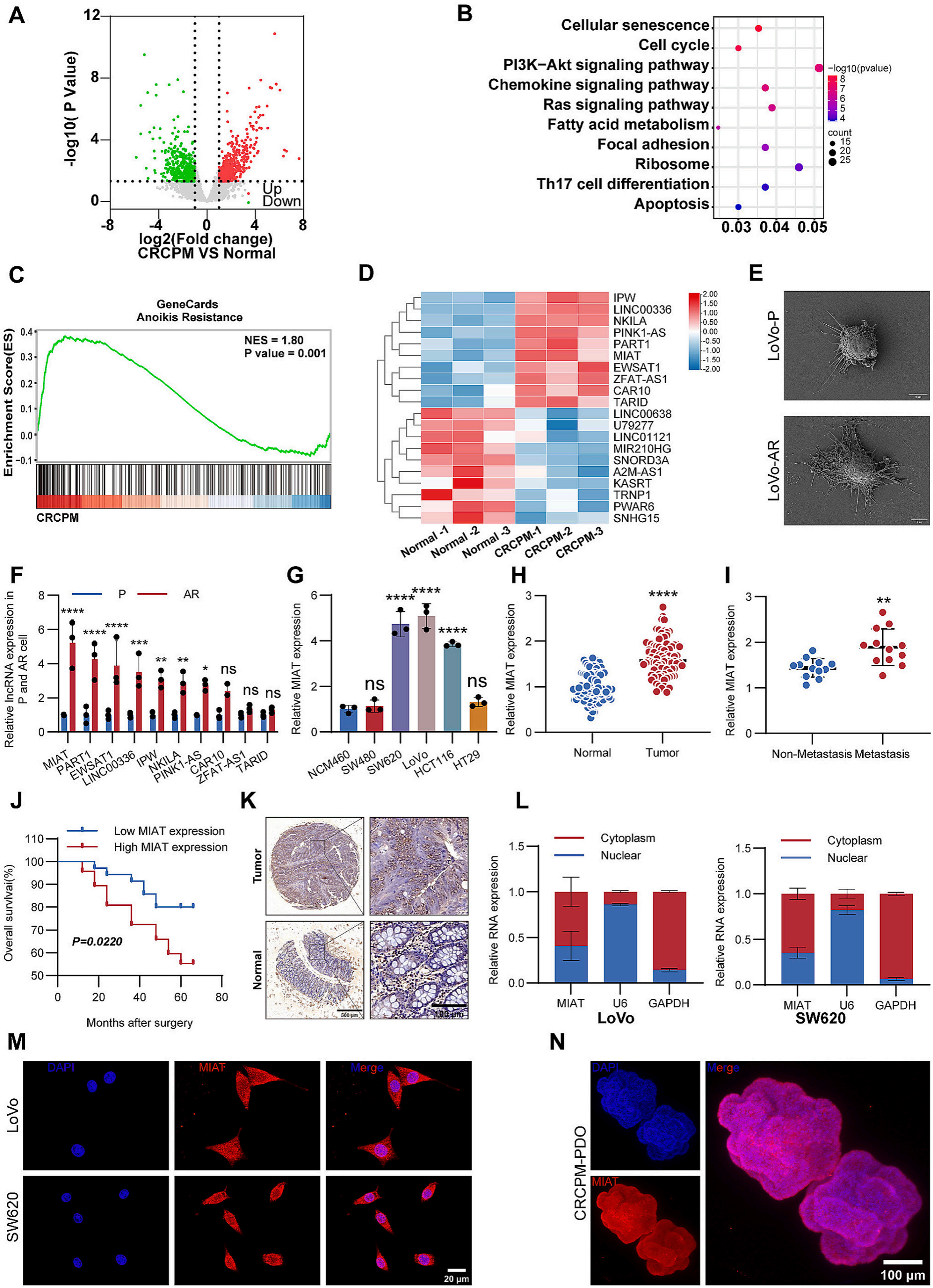 Fig. 1. Elevated MIAT expression is associated with CRCPM A Volcano plot showing differentially expressed genes between CRCPM tissues and matched normal colon tissues. B KEGG enrichment in CRCPM tissues as compared to normal colon tissues. C GSEA showing enrichment of anoikis resistance-related gene signatures in CRCPM tissues compared with normal colon tissues. D Heatmap showing the top differentially expressed lncRNAs in CRCPM tissues compared with normal colon tissues. E Ultrastructural differences between AR LoVo cells and parental cells. F Relative expression levels of the top 10 candidate lncRNAs in AR LoVo cells were determined by RT–qPCR. G Relative MIAT expression in the human normal colorectal epithelial cell line (NCM460) and CRC cell lines (SW480, SW620, LoVo, HCT116 and HT29). H MIAT expression in CRC tissues compared with adjacent normal tissues. I MIAT expression in CRCPM tissues compared with matched primary CRC tissues. J Kaplan–Meier analysis of overall survival in CRC patients stratified by MIAT expression. K Representative RNA-ISH images showing MIAT expression in CRC tissues and adjacent normal tissues. L Subcellular distribution of MIAT in CRC cells was analyzed by nuclear–cytoplasmic fractionation. M?N Representative fluorescence in situ hybridization (FISH) images showing MIAT localization in CRC cells (M) and organoids (N). (Data are presented as mean ± SD;* P < 0.05, **P < 0.01, ***P < 0.001, ****P < 0.0001).
模块 2:MIAT 体外促进结直肠癌细胞增殖、失巢凋亡抵抗、侵袭;PDO 类器官验证表型
研究逻辑:MIAT 敲低,多组学体外功能实验评估细胞恶性表型;使用患者来源结直肠癌类器官模拟体内肿瘤特征,验证 MIAT 功能。
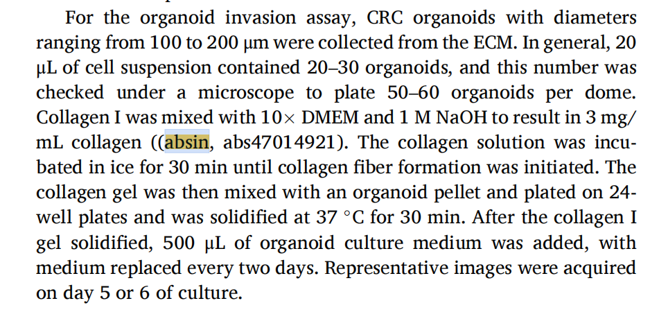
?爱必信产品使用步骤:收集直径 100?200μm 的 CRC 类器官,使用abs47014921 I 型胶原蛋白(鼠尾来源)配制 3?mg/mL 胶原工作胶,将类器官与胶原凝胶混合铺板,37℃凝固后培养 5?6 天,显微镜拍照评估类器官向外侵袭突起能力。
核心实验:慢病毒敲低 MIAT;CCK?8 增殖、克隆形成;悬浮 / 贴壁条件下 Annexin?V 流式凋亡;划痕愈合、Transwell 迁移侵袭;Western blot 凋亡、EMT 相关蛋白;患者来源类器官培养、类器官侵袭实验。
关键实验结果:① MIAT 敲低显著抑制 AR?LoVo、AR?SW620 细胞增殖、克隆形成;悬浮(失巢凋亡诱导)条件下敲低 MIAT 诱导凋亡效应比贴壁条件更显著; ② MIAT 敲低抑制细胞迁移侵袭;促凋亡蛋白 BAX、Cleaved?caspase?3 上调,抗凋亡 Bcl?2 下调;EMT 被抑制(E?cadherin 上调,N?cadherin、Vimentin、Snail 下调); ③ PDO 模型中 MIAT 敲低抑制类器官形成,三维胶原侵袭体系下类器官向外侵袭突起显著减少。
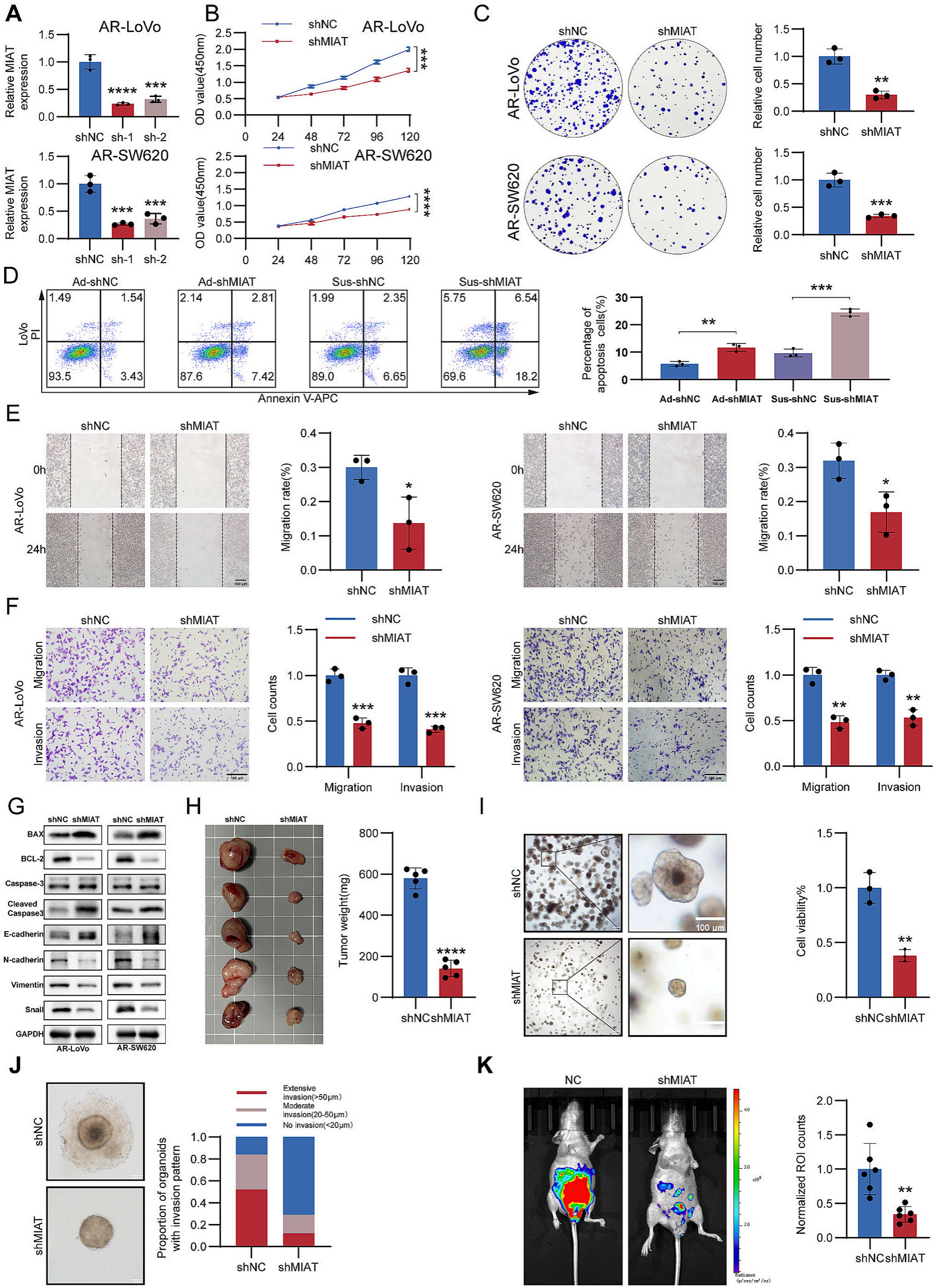 Fig. 2. MIAT promotes CRC proliferation, anoikis resistance, and invasion in vitro and in vivo A MIAT expression levels in AR CRC cells following lentiviral transduction were determined by RT–qPCR. B and C Cell proliferation was assessed using CCK-8 assays (B) and colony formation assays (C). D Flow cytometry was conducted to analyze the effects of MIAT knockdown on the Parental LoVo cells under either adherent or suspension conditions. E and F Effects of MIAT knockdown on the migration and invasion abilities of AR CRC cells, evaluated by Wound healing assay(E) and Transwell migration or invasion assay (F). G Changes in apoptosis?related proteins and EMT related proteins after MIAT knockdown were analyzed via western blot in AR CRC cells. H Representative images and weight analysis of subcutaneous xenograft tumors in nude mice(n = 5). I CRC patient?derived organoid was infected with MIAT knockdown or a control lentivirus, representative bright?field images and cell viability analysis are exhibited. J Representative image of CRC patient?derived organoid invasion. K Representative images of peritoneal metastasis model(n = 6). (Data are presented as mean ± SD; * P < 0.05, **P < 0.01, ***P < 0.001, ****P < 0.0001).
模块 3:MIAT 充当 ceRNA 分子海绵直接吸附 miR?181a?5p
研究逻辑:DIANA、starBase 数据库预测 MIAT 结合 miRNA;敲低 MIAT 筛选发生显著变化的 miRNA;双荧光素酶报告、RNA?FISH 共定位验证结合;临床样本分析表达相关性。
核心实验:生物信息预测;RT?qPCR;双荧光素酶报告基因;RNA?FISH 共定位;临床样本表达相关性分析。
关键实验结果:① MIAT 敲低后 miR?181a?5p 上调最显著;miR?181a?5p 在结直肠癌组织下调,与 MIAT 表达呈负相关; ② 双荧光素酶证实 MIAT 与 miR?181a?5p 直接结合;RNA?FISH 显示 MIAT 与 miR?181a?5p 在结直肠癌细胞及 PDO 类器官胞质共定位。
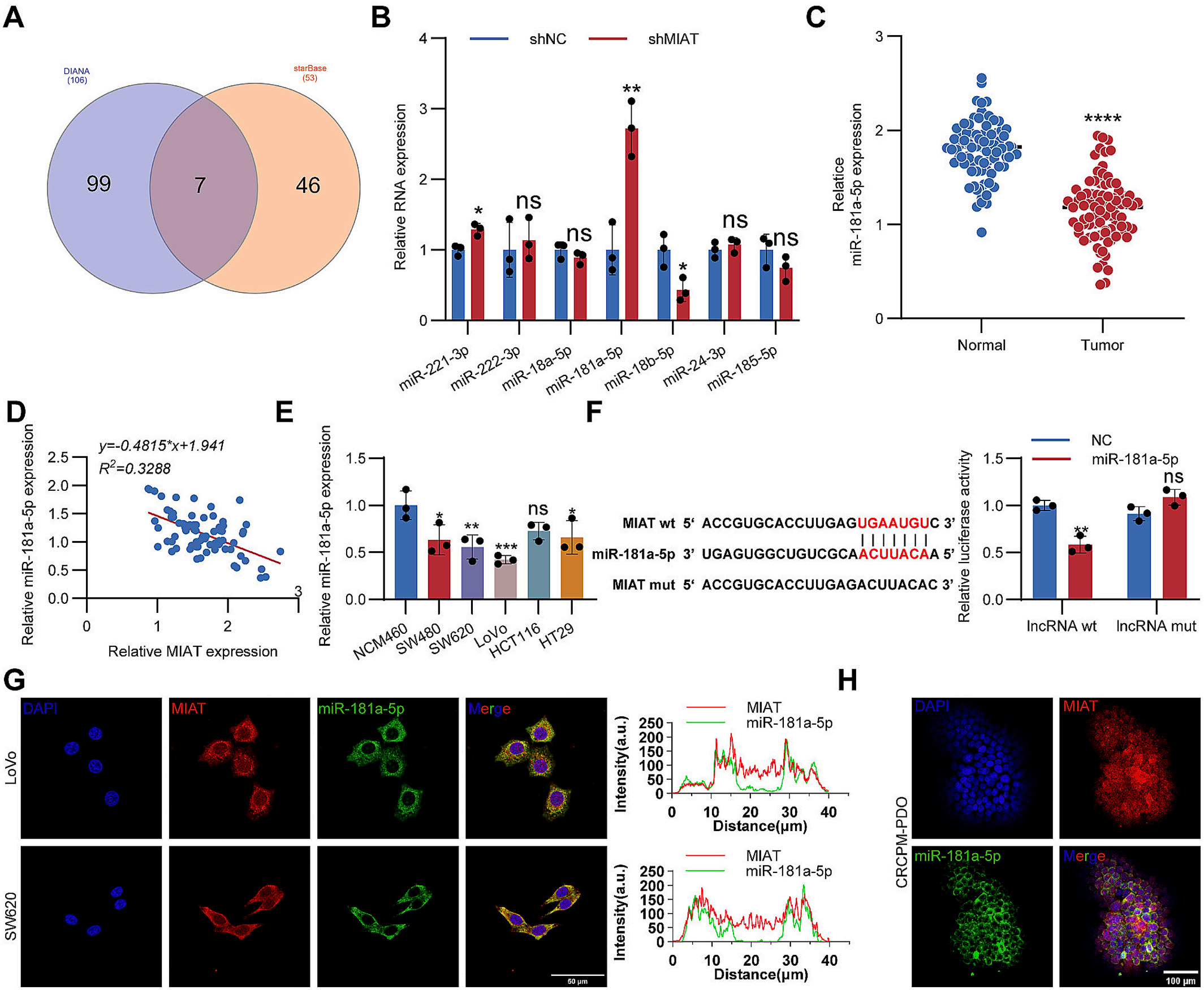 Fig. 3. MIAT functions as a miR-181a-5p sponge in colorectal cancer A Venn diagram showing candidate miRNAs predicted to bind MIAT based on DIANA and starBase databases. B Expression changes of candidate miRNAs following MIAT knockdown were determined by RT–qPCR. C miR-181a-5p expression in CRC tissues compared with normal tissues. D Correlation analysis between MIAT and miR-181a-5p expression in CRC tissues. E Relative expression of miR-181a-5p in CRC cell lines and normal colorectal epithelial cells. F Luciferase reporter assays confirming the direct interaction between MIAT and miR-181a-5p. G?H RNA?FISH assays showing colocalization of MIAT and miR?181a?5p in CRC cells (G) and organoids (H). (Data are presented as mean ± SD; * P < 0.05, **P < 0.01, ***P < 0.001, ****P < 0.0001).
模块 4:miR?181a?5p 介导 MIAT 的促癌生物学效应(回救实验)
研究逻辑:在 MIAT 敲低细胞中转染 miR?181a?5p 抑制剂,开展回救实验,证明 MIAT 促增殖、抗失巢凋亡、促侵袭功能依赖 miR?181a?5p。
核心实验:miR?181a?5p 抑制剂转染;CCK?8、克隆形成;悬浮条件流式凋亡;划痕、Transwell 迁移侵袭。
关键实验结果:抑制 miR?181a?5p 可以部分逆转 MIAT 敲低造成的增殖抑制、凋亡升高、迁移侵袭能力下降,证明 miR?181a?5p 是 MIAT 下游功能效应分子。
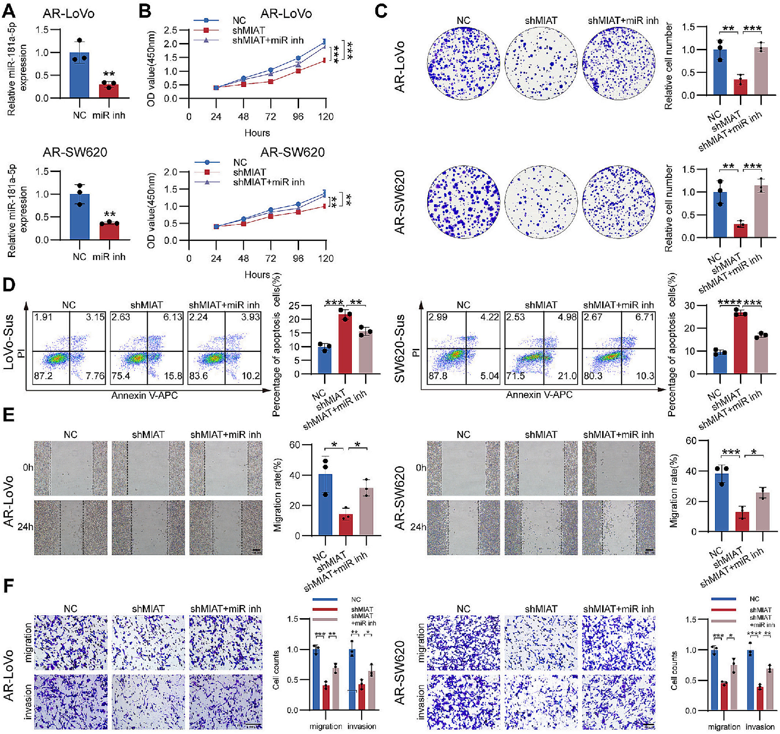 Fig. 4. miR-181a-5p mediates the oncogenic effects of MIAT in CRC cells A Transfection efficiency of miR-181a-5p inhibitor in AR CRC cells was confirmed by RT–qPCR. B and C Cell proliferation was evaluated using CCK-8 assays (B) and colony formation assays (C). D Flow cytometry was conducted to analyze the effects of miR-181a-5p inhibition on MIAT?mediated apoptosis under suspension conditions in LoVo and SW620 cells. E and F Cell migration and invasion were evaluated using Wound healing assays (E) and Transwell assays (F). (Data are presented as mean ± SD; * P < 0.05, **P < 0.01, ***P < 0.001, ****P < 0.0001).
模块 5:MIAT 通过 miR?181a?5p/MCL1 轴调控结直肠癌恶性表型
研究逻辑:靶基因预测筛选 miR?181a?5p 下游靶基因;双荧光素酶确认 MCL1 为直接靶基因;细胞、类器官层面 MCL1 过表达回救,验证 MCL1 介导 MIAT 的表型。
核心实验:靶基因预测;双荧光素酶报告;RT?qPCR、Western blot;CCK?8、克隆、流式凋亡;划痕、Transwell;PDO 类器官增殖与侵袭实验。
关键实验结果:① MCL1 在结直肠癌、AR 细胞高表达,和 MIAT 表达正相关;双荧光素酶证明 MCL1 是 miR?181a?5p 直接靶基因; ② MCL1 过表达能够部分逆转 MIAT 敲低诱导的增殖抑制、失巢凋亡增加、侵袭减弱,同时逆转凋亡、EMT 相关蛋白的表达改变;PDO 类器官中 MCL1 过表达恢复 MIAT 敲低带来的增殖与侵袭缺陷。
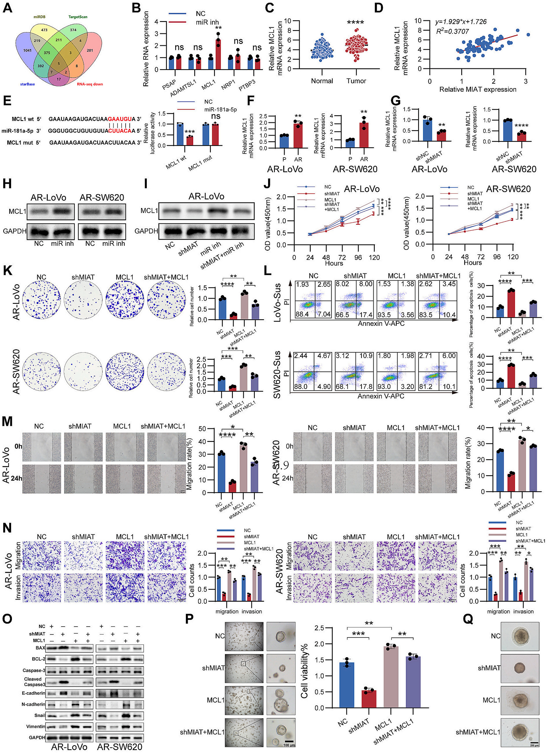 Fig. 5. MIAT regulates colorectal cancer progression through the miR-181a-5p/MCL1 axis A Venn diagram showing candidate downstream targets of miR- 181a-5p based on bioinformatic prediction and RNA sequencing analysis. B Expression changes of candidate target genes following miR-181a-5p inhibition. C MCL1 expression in CRC tissues compared with normal tissues. D Correlation analysis between MIAT and MCL1 expression in CRC tissues. E Luciferase reporter assays confirming the direct interaction between miR-181a-5p and MCL1. F Relative MCL1 mRNA expression in AR CRC cells. G MCL1 mRNA expression in AR CRC cells after MIAT knockdown. H MCL1 protein level in AR CRC cells after miR-181a-5p inhibited. I MCL1 protein level in MIAT knockdown AR?LoVo cells with miR? 181a?5p inhibited. J and K Cell proliferation was assessed using CCK-8 assays (J) and colony formation assays (K). L Flow cytometry was conducted to analyze the effects of MCL1 overexpression on MIAT?mediated apoptosis under suspension conditions in LoVo and SW620 cells. M Wound healing assay in AR CRC cells infected with MIAT knockdown or MCL1 overexpression lentivirus. N Transwell migration or invasion assay in AR CRC cells infected with MIAT knockdown or MCL1 overexpression lentivirus. O Changes in apoptosis related proteins and EMT related proteins in AR CRC cells infected with MIAT knockdown or MCL1 overexpression lentivirus. P Representative bright?field images and cell viability analysis of CRC organoids infected with MIAT knockdown or MCL1 overexpression lentivirus. Q Representative images of CRC organoid invasion. (Data are presented as mean ± SD; * P < 0.05, **P < 0.01, ***P < 0.001, ****P < 0.0001).
模块 6:体内动物模型验证 MIAT?MCL1 轴促进肿瘤生长、腹膜转移
研究逻辑:裸鼠皮下成瘤模型验证肿瘤增殖;腹腔注射构建腹膜转移模型,活体生物发光评估腹膜肿瘤负荷;IHC 检测肿瘤组织蛋白表达。
核心实验:裸鼠皮下异种移植;裸鼠腹腔腹膜转移模型;生物发光成像;肿瘤组织 IHC 染色(MCL1、Ki?67、BAX)。
关键实验结果:MIAT 敲低显著抑制皮下肿瘤生长,降低腹腔腹膜转移负荷;MCL1 过表达可以逆转 MIAT 敲低带来的体内抑瘤效果;IHC 结果显示 MIAT 敲低降低 MCL1、Ki?67,升高 BAX;MCL1 过表达逆转该变化。
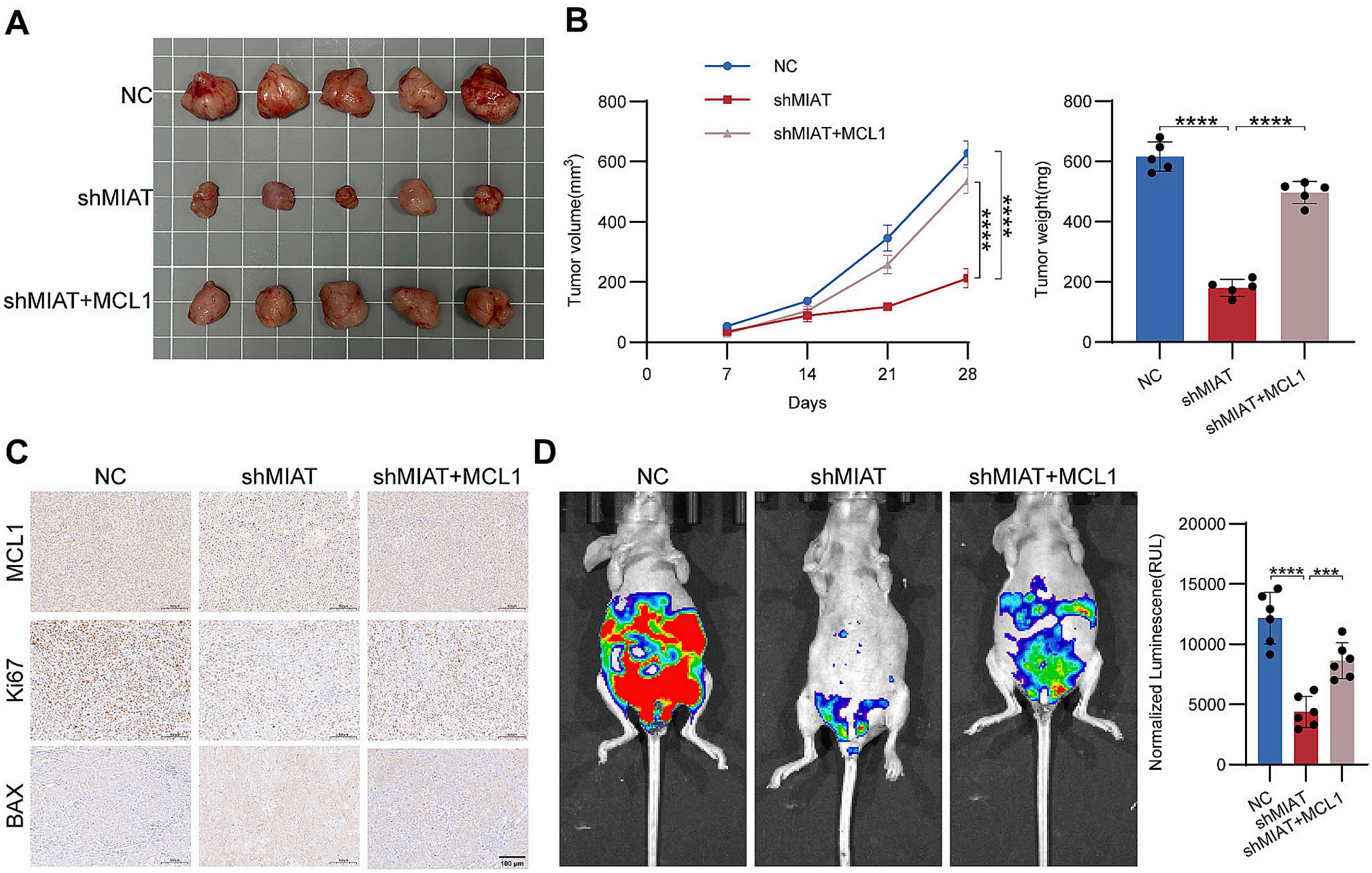 Fig. 6. MIAT drives the growth and peritoneal metastasis of colorectal cells in vivo A Representative image of dissected subcutaneous xenograft tumors in nude mice(n = 5). B Growth curve and weight analysis of xenograft tumors in nude mice. C Immunohistochemical staining of MCL1, Ki-67, and BAX in tumor tissues. D Representative images of intraperitoneal metastasis models(n = 6). (Data are presented as mean ± SD; * P < 0.05, **P < 0.01, ***P < 0.001, ****P < 0.0001).
模块 7:METTL3 介导 m?A 修饰,依赖 IGF2BP2 增强 MIAT RNA 稳定性
研究逻辑:SRAMP 预测 MIAT 的 m?A 修饰位点;临床样本分析 METTL3、IGF2BP2 与 MIAT 表达相关性;MeRIP?qPCR、RNA 半衰期、RIP 实验解析 m?A 调控 MIAT 分子机制。
核心实验:SRAMP m?A 位点预测;临床样本 qPCR、IHC+RNA?ISH;siRNA 敲低 METTL3/IGF2BP2;MeRIP?qPCR;放线菌素 D RNA 稳定性实验;RIP?qPCR;通路模式图绘制。
关键实验结果:① METTL3、IGF2BP2 在结直肠癌高表达,与 MIAT 表达正相关; ② METTL3 敲低降低 MIAT 的 m?A 修饰水平,缩短 MIAT 的 RNA 半衰期,MIAT 表达下调; ③ m?A 修饰的 MIAT 被阅读器 IGF2BP2 识别结合,IGF2BP2 敲低同样降低 MIAT 稳定性;METTL3 缺失会削弱 IGF2BP2 与 MIAT 相互作用;完整建立 m?A?METTL3?IGF2BP2?MIAT?miR?181a?5p?MCL1 调控模式。
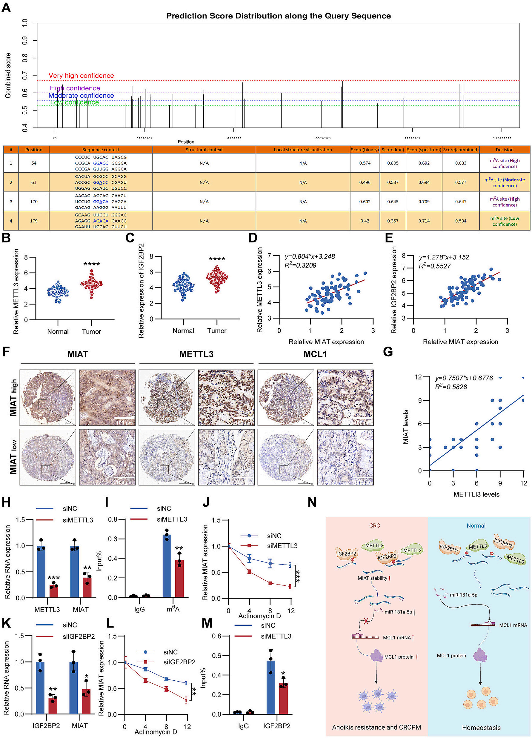 Fig. 7. m6 A modification contributes to MIAT upregulation in colorectal cancer A Predicted m6 A modification sites in MIAT based on SRAMP analysis. B and C The METTL3(B) and IGF2BP2(C) expression between CRC tissues and normal tissues. D?E Correlation between MIAT and METTL3(D) or IGF2BP2(E). F Representative images of MIAT, METTL3 and MCL1 analyzed by IHC and RNA?ISH staining in MIAT high expression and MIAT low?expression CRC tissues. G The correlation between MIAT and METTL3 was analyzed in CRC tissues using the IHC score and RNA?ISH score. H Relative expression of METTL3 and MIAT in LoVo cells after transfected with METTL3 siRNA. I MeRIP?qPCR analysis of the m6 A levels in MIAT in LoVo cells transfected with METTL3 siRNA. J RNA stability of MIAT following METTL3 knockdown. K Relative expression of IGF2BP2 and MIAT in LoVo cells after transfected with IGF2BP2 siRNA. L MIAT stability was determined by RT?qPCR after transfected with IGF2BP2 siRNA. M RIP?qPCR analysis showing the enrichment of IGF2BP2 on MIAT in LoVo cells after transfected with METTL3 siRNA. N Schematic diagram of the mechanism by which the m6 A modification?mediated MIAT facilitates peritoneal metastasis and growth in CRC. (Data are presented as mean ± SD; * P < 0.05, **P < 0.01, ***P < 0.001, ****P < 0.0001).
五、爱必信(Absin)产品整体作用总结
1. 构建肿瘤类器官三维侵袭体外模型:abs47014921 鼠尾 I 型胶原蛋白用于配制三维胶原凝胶,模拟体内细胞外基质生理微环境,实现结直肠癌患者来源类器官侵袭能力可视化观测,弥补传统二维 Transwell 无法还原类器官三维侵袭行为的缺陷。
2. 打通细胞?类器官表型证据链:二维细胞 Transwell 实验完成细胞侵袭评估,基于 Absin I 型胶原的 PDO 侵袭实验进一步在更贴近患者肿瘤真实特征的类器官模型上佐证 MIAT 促侵袭功能,提升体外实验的生理相关性。
3. 支撑靶点转化论证:该胶原产品为 PDO 功能验证提供关键基质材料,和细胞、动物实验结果互相印证,共同支撑 MIAT 作为结直肠癌腹膜转移潜在治疗靶点的结论,符合高分肿瘤类器官研究的三维模型质控要求。
相关产品
| 货号 | 品名 | 链接 |
| abs47014921 | I?Collagen, from Rat Tail(I型胶原蛋白,鼠尾来源) | https://www.absin.cn/ |

- 手机:13761418683