生物科技有限公司")


- 手机:13761418683
联系人:爱必信
网址:http://www.absin.cn/
地 址:上海市浦东新区新浩路58号18号楼

- 惰性前药无法肿瘤空间特异性激活,易引发全身毒副作用;
- 传统纳米酶催化效率低,ROS 生成不足,难以支撑强效治疗;
- 多数肿瘤因 GSDMD 表观沉默,焦亡启动困难,疗效受限。
- ? 以 π 共轭多酚骨架打造 "电子高速路",实现全颗粒高效电子穿梭,过氧化物酶活性拉满;
- ? 搭载 GSH "AND" H?O? 双响应逻辑门,全身循环惰性、肿瘤微环境精准 "开";
- ? 前药原位转化为胡桃醌,逆转 GSDMD 沉默 + 激活 NLRP3 炎症小体,双管齐下启动焦亡,同步叠加铜死亡、铁死亡,实现 ICD 免疫原性细胞死亡,激活全身抗肿瘤免疫。
- ● 以 L-半胱氨酸为调节剂,精准控制 Cu–DHN 粒径,最优比例 Cu:DHN:Cys=0.6:1:0.2,粒径约 50 nm,比表面积提升 27 倍,催化位点充分暴露;
- ● 形成 Cu?/Cu2? 混合价态与 Cu–O、Cu–S 混合配位壳层,电子传递效率飙升,实现高效·OH 生成 + DHN 向胡桃醌转化(转化率超 55%)。对应原文图:图 2b、2c、2h、2j、3a、3b
- ? 原位 4T1 乳腺癌模型:肿瘤生长抑制率 75%,显著延长生存期;
- ? 双侧肿瘤模型:触发远隔效应,抑制对侧肿瘤生长;
- ? 肺转移模型:肺转移结节减少 90% 以上,生存率大幅提升;
- ? 安全性:正常组织无激活,肝肾功、血小板无异常,完胜直接胡桃醌治疗。
STTT 重磅:Cu–DHN 纳米酶前药介导三通路细胞死亡,助力原位肿瘤疫苗构建
2026-06-15
近期,国际顶级期刊 Signal Transduction and Targeted Therapy 发表了突破性研究 —— 导电配位纳米酶前药 Cu–DHN,通过 GSH/H?O? 双响应逻辑门精准激活,同步触发细胞焦亡、铜死亡、铁死亡,将原位肿瘤变 "疫苗工厂",强效抑制原发与转移瘤,为肿瘤精准免疫治疗开辟全新路径。Absin 作为核心试剂供应商,以高灵敏、高稳定的 ELISA 试剂盒为关键实验保驾护航,助力成果顺利登顶顶刊!
文献标题:Conductive coordination nanozyme prodrugs precisely trigger pyroptosis, cuproptosis and ferroptosis for in situ cancer vaccination
发表期刊:Signal Transduction and Targeted Therapy (IF=52.7)
DOI:https://doi.org/10.1038/s41392-026-02607-6
使用 Absin 产品:Mouse TNF-α ELISA Kit(货号:abs520010)
![]()
一、研究核心思路:破解焦亡临床转化三大痛点
细胞焦亡是理想的原位肿瘤疫苗诱导方式,可快速释放肿瘤抗原与 DAMPs,激活系统性抗肿瘤免疫,但临床转化面临三大瓶颈:
为此,团队创新构建 Cu–DHN 导电配位纳米酶前药:
二、核心研究成果:三死协同,原位疫苗强效抑瘤
1. 结构设计:L-Cys 精准调控,催化性能拉满
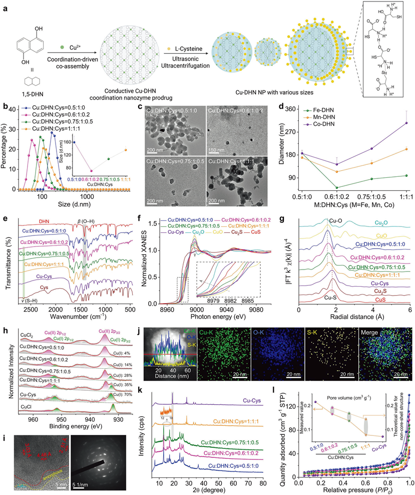
Engineering the architecture and coordination environment
a Synthesis scheme of Cu–DHN, illustrating the role of L-Cys as a coordination terminator and size regulator. The zoomed-in region illustrates the intermolecular hydrogen bonding between L-Cys molecules. b Hydrodynamic size distribution profiles (n = 3), and c TEM images of Cu–DHN with varying Cu:DHN:Cys feed molar ratios, revealing a non-monotonic size trend. d Variation profiles of hydrodynamic size for different M-DHN (M = Fe, Mn, Co) nanoparticles (n = 3) demonstrating the generalizability of Cys-mediated size control. e Fourier transform infrared (FT-IR) spectra confirming the involvement of phenolic –OH and thiol –SH groups; f normalized Cu K-edge XANES spectra, g Fourier transforms of k3-weighted Cu K-edge EXAFS spectra, and h high-resolution Cu 2p XPS spectra of Cu–DHN with varying Cu:DHN:Cys molar ratios, collectively evidencing the evolution of a mixed (O, S)-coordination shell and a rising Cu?/Cu2? ratio with increasing Cys content. i HR-TEM image and corresponding SAED pattern, and j elemental distribution mapping and corresponding line-scan profile of Cu–DHN at a Cu:DHN:Cys molar ratio of 0.6:1:0.2. k XRD patterns, and l Brunauer–Emmett–Teller (BET) N2 adsorption–desorption isotherms demonstrating the pore structure evolution of Cu–DHN with varying Cu:DHN:Cys molar ratios, confirming the progressive surface passivation that defines the core–shell pore structure. Data are represented as mean ± SD
2. 机制突破:单药触发三通路死亡,攻克 GSDMD 低表达困境
细胞焦亡:胡桃醌抑制 DNA 甲基转移酶,逆转 GSDMD 沉默;K?外流激活 NLRP3 炎症小体,Caspase-1 切割 GSDMD,形成焦亡孔;
铜死亡:纳米载体高效递送 Cu?,下调 FDX1、LIAS,触发铜死亡;
铁死亡:耗竭 GSH、抑制 GPX4,引发脂质过氧化;
三通路协同,即使 GSDMD 低表达肿瘤也能强效焦亡,释放 CRT、HMGB1、ATP,强力促 DC 成熟与 CD8?T 细胞浸润。
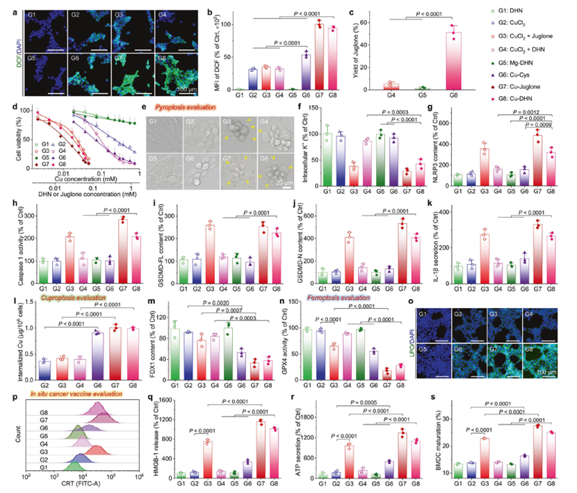
In vitro effects of Cu–DHN (Cu:DHN:Cys = 0.6:1:0.2) on 4T1 cells
a CLSM images and b mean fluorescence intensity (MFI) of 4T1 cells stained by DCFH-DA (ROS probe) after co-incubation with Cu–DHN for 6 h (n = 3). c Juglone yield in 4T1 cells after co-incubation with Cu–DHN for 12 h (n = 3). d Cell viability curves of 4T1 cells after co-incubation with Cu–DHN for 24 h (n = 6). e Bright-field microscopic images of 4T1 cells after co-incubation with Cu–DHN for 18 h (yellow arrows highlight membrane-bound vesicular protrusions indicative of pyroptotic morphology). f Intracellular K+ content in 4T1 cells after co-incubation with Cu–DHN for 6 h (n = 3). g NLRP3 content, h Caspase-1 activity, i GSDMD-FL content, j GSDMD-N content in 4T1 cells after co-incubation with Cu–DHN for 18 h (n = 3). k IL-1β secretion from 4T1 cells after co-incubation with Cu–DHN for 18 h (n = 3). l Internalized amounts of Cu by 4T1 cells after co-incubation with Cu–DHN for 6 h (n = 3). m FDX1 content in 4T1 cells after co-incubation with Cu–DHN for 18 h (n = 3). n GPX4 activity in 4T1 cells after co-incubation with Cu–DHN for 18 h (n = 3). o CLSM images of 4T1 cells stained by Liperfluo (LPO probe) after co-incubation with Cu–DHN for 12 h (n = 3). p Flow cytometry analysis of 4T1 cells labeled by AF488-CRT antibody after co-incubation with Cu–DHN for 18 h. Release of q HMGB-1, and r ATP from 4T1 cells after co-incubation with Cu–DHN for 18 h (n = 3). s BMDC maturation after co-incubation with Cu–DHN treated 4T1 cells for 24 h (n = 3). Cu concentration: 0.020 mM, DHN or juglone concentration: 0.033 mM. Data are represented as mean ± SD
3. 体内疗效:抑原发、控转移、安全无忧
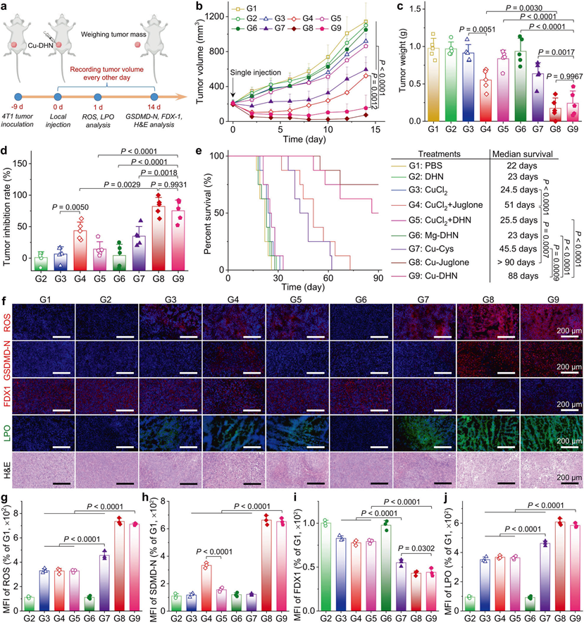
In vivo effects of Cu–DHN (Cu:DHN:Cys = 0.6:1:0.2) on 4T1 tumor
a Schematic illustration of inhibiting tumor growth via inducing pyroptosis, cuproptosis, and ferroptosis by a single intratumoral injection of Cu–DHN. b Longitudinal monitoring of tumor growth following single-dose Cu–DHN intratumoral administration (3.78 μmol kg?1 Cu, 6.24 μmol kg?1 DHN or juglone) (n = 5). c Weights of dissected tumors, and d tumor inhibition rate at 14 days post-treatment (n = 5). e Survival rate analysis of 4T1 tumor-bearing BALB/c mice treated with Cu–DHN (n = 8). f CLSM images and MFI of tumor slices labeled with g DHE (1 day post-treatment), h GSDMD-N antibody (14 days post-treatment), i FDX1 antibody (14 days post-treatment), j Liperfluo (1 day post-treatment), and H&E (14 days post-treatment) (n = 3). Data are represented as mean ± SD
三、Absin 硬核助力:关键试剂支撑顶刊数据
本研究中,Absin 小鼠肿瘤坏死因子-α(TNF-α)ELISA 试剂盒(abs520010-96T) 成为免疫激活验证的核心工具,在全身免疫应答、远隔效应、抗转移三大关键实验中发挥不可替代作用:
产品信息
| 项目 | 详情 |
|---|---|
| 产品名称 | 小鼠肿瘤坏死因子-α(TNF-α)ELISA 试剂盒 |
| 货号 | abs520010-96T |
| 品牌 | Absin |
核心作用
1. 精准定量血清 / 肿瘤组织 TNF-α
检测 Cu–DHN 治疗后细胞因子风暴水平,验证免疫激活强度与可控性;
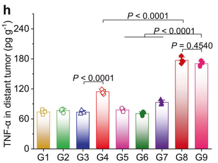
h TNF-α and i IFN-γ contents in distant tumors (n = 4).
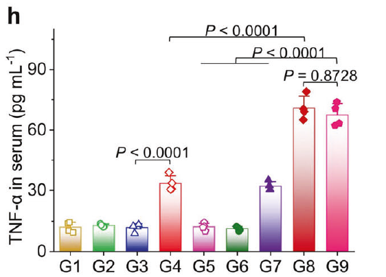
h TNF-α and (i) IFN-γ contents in serum from mice at 3 days post-treatment (n = 4).
2. 高灵敏、低背景
适配微量样本检测,准确反映 DC 成熟、CD8?T 细胞活化程度,支撑免疫机制结论;
3. 稳定可靠
批间差小、重复性强,保障体内疗效数据真实可重复,助力顶刊数据严谨性。
本研究构建的 Cu–DHN 导电配位纳米酶前药,实现肿瘤特异性激活 + 三死协同 + 原位疫苗三重突破,解决焦亡治疗靶向性、效率、安全性三大痛点,为纳米酶催化免疫治疗提供全新设计范式。Absin 以高品质试剂深度赋能前沿研究,未来将持续推出肿瘤免疫、细胞死亡、纳米材料领域优质产品,助力更多中国科研成果登顶国际顶刊!

- 手机:13761418683