




- 手机:13761418683
联系人:爱必信
网址:http://www.absin.cn/
地 址:上海市浦东新区新浩路58号18号楼

顶刊文献解读|神经干细胞命运调控分子机制的实验体系构建与深度解析
2026-07-15
文献标题:Single‐Chain Anti‐IL‐1β Antibody Carried by Outer Membrane Vesicles of Bacteroides fragilis Alleviates Tubular Inflammation in Chronic Kidney Disease
发表期刊:J Extracell Vesicles. (IF=14.5)
DOI:https://doi.org/10.1002/jev2.70234
使用 Absin 产品:Mouse IL-1β ELISA Kit(货号:abs520001)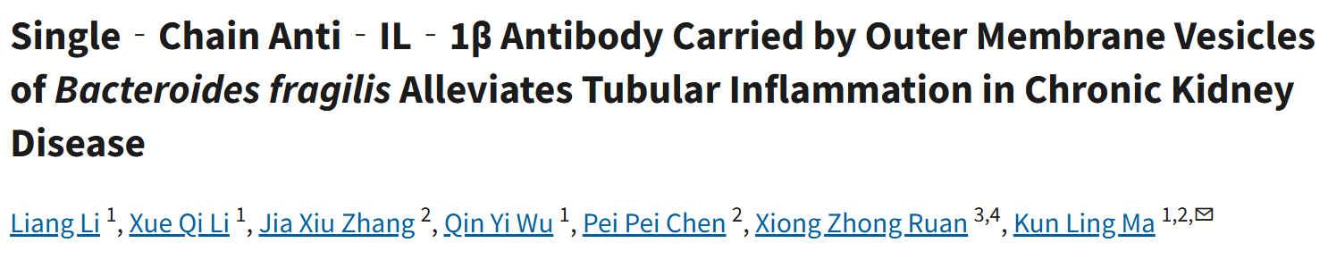

一、研究核心思路:锚定 PCM1,解码神经干细胞命运密码
本研究以中心体周质蛋白 1(PCM1)为核心靶点,围绕神经干细胞不对称分裂与细胞命运决定展开全链条验证,构建严谨科学逻辑:
1. 科学问题:脊椎动物放射状胶质祖细胞(RGP)通过不对称分裂平衡自我更新与分化,但中心体不对称如何调控子代细胞命运尚不明确;
2. 表型定位:明确 PCM1 在神经干细胞中心体上的不对称分布,鉴定其与 Par-3、动力蛋白的相互作用;
3. 机制深挖:解析 PCM1 通过调控内体极化运输(早期内体→循环内体转换),维持 Notch 信号、抑制神经元过度分化的分子通路;
4. 跨物种验证:在斑马鱼胚胎与人类大脑类器官中证实机制保守性,为神经发育与神经疾病研究提供新理论。
研究结合活体成像、超高分辨显微镜、类器官模型、基因敲除,系统揭示 PCM1 作为 "细胞 GPS" 调控神经干细胞命运的全新机制。
二、核心研究成果:PCM1—— 神经干细胞命运的 "核心开关"
1. PCM1 不对称定位决定中心体极性
PCM1 特异性富集于母中心体,呈不对称分布,是神经干细胞不对称分裂的标志性分子(对应原文Figure 1)。
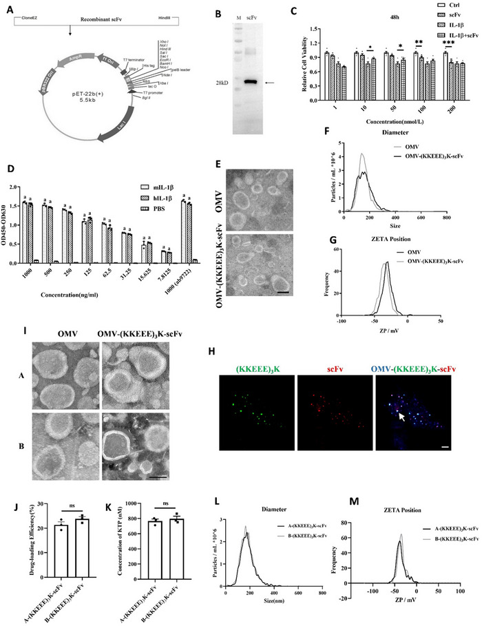
FIGURE 1.
Preparation of IL‐1β scFv and construction of OMV‐(KKEEE)3K‐scFv. (A) Schematic representation of recombinant scFv expression plasmid construction. (B) Western blotting (WB) assay of recombinant scFv from Escherichia coli T7 expression system lysate. Lane 1: marker. Lane 2: cultured medium lysate with IPTG induction. (C) CCK‐8 assay detected the neutralizing effect of various scFv concentrations (1, 10, 50, 100, and 200 nmol/L) on IL‐1β (n = 5). (D) ELISA detected the affinity of various scFv concentrations to human and murine IL‐1β (hIL‐1β and mIL‐1β). Commercial IL‐1β antibody was used as control (n = 3). “a” indicates that there is a significant difference between this group and the PBS group, with p ≤ 0.001. (E) TEM images of OMVs and constructed OMV‐(KKEEE)3K‐scFv (scale bar: 100 nm). (F, G) Size and zeta potential values of the OMVs and OMV‐(KKEEE)3K‐scFv were measured by ZetaView. (H) Representative confocal microscopy image of OMV‐(KKEEE)3K‐scFv. The white arrow indicates typical colocalization of FITC‐labelled (KKEEE)3K (green), DID (1,1'‐dioctadecyl‐3,3,3',3'‐tetramethylindodicarbocyanine, 4‐chlorobenzenesulfonate salt) ‐labelled OMVs (blue), and RB200‐labelled scFv (red). Scale bar: 1 μm. (I) TEM images of OMVs and OMV‐(KKEEE)3K‐scFv constructed from various batches (scale bar: 100 nm). (J) Drug loading efficiency statistics of OMV‐(KKEEE)3K‐scFv constructed from various batches (n = 3). (K) Concentrations of (KKEEE)3K in 1 mg/mL OMV‐(KKEEE)3K‐scFv samples from Batch A and Batch B (n = 3). (L, M) Size and zeta potential values of OMV‐(KKEEE)3K‐scFv constructed from various batches measured by ZetaView. Mean ± SEM was used for reporting the data. *P < 0.05, **P < 0.01, ***P < 0.001. Two groups were compared by independent Student's t‐test, and multiple groups were compared by one‐way ANOVA, followed by Tukey's Honest Significant Difference (HSD) test.
2. PCM1 调控内体极化运输与信号传递
PCM1 促进早期内体(Rab5b+)向循环内体(Rab11a+)转换,组装 Par-3 - 动力蛋白复合物,驱动内体后向极化运输,保障 Notch 配体高效传递(对应原文Figure 2、3)。
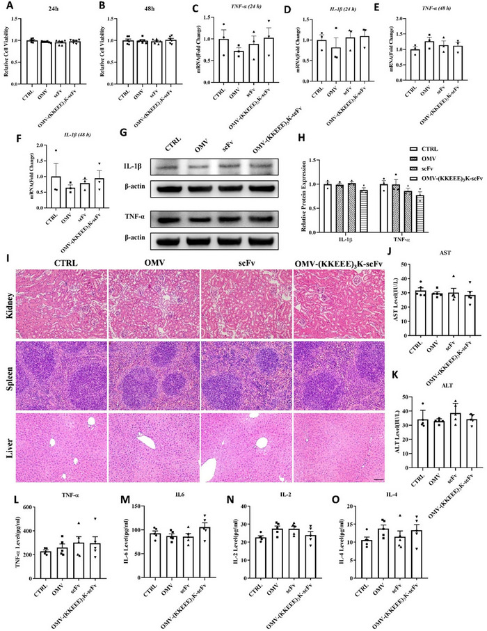
FIGURE 2.
Safety evaluation of OMV‐(KKEEE)3K‐scFv in vivo and in vitro. (A, B) CCK‐8 assay detected PTECs (Primary tubular epithelial cells) viability after drug treatment for 24 and 48 h (n = 6). (C‐F) Estimation of mRNA levels of TNF‐α and IL‐1β in PTECs after drug treatment for 24 and 48 h by RT‐qPCR (n = 3). Data are presented as fold changes relative to the CTRL group, which was set as 1‐fold. (G‐H) Representative WB assay images and summarized data displaying relative protein levels of IL‐1β and TNF‐α in PTECs after drug treatment for 48 h (n = 3). (I) Representative haematoxylin and eosin staining images of major organs in mice treated with various drugs for 2 months (scale bar: 50 μm). (J, K) Serum AST (aspartate transaminase) and ALT (alanine transaminase) levels in mice from various groups (n = 5). (L‐O) Estimation of serum levels of TNF‐α, IL‐6, IL‐2, and IL‐4 levels in mice from various groups (n = 5). Two groups were compared by independent Student's t‐test, and multiple groups were compared by one‐way ANOVA, followed by Tukey's HSD test.
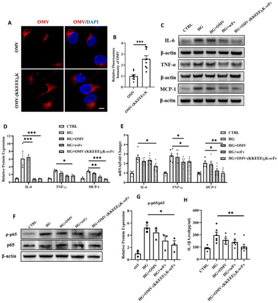
FIGURE 3.
OMV‐(KKEEE)3K‐scFv attenuates high glucose‐induced inflammation in primary tubular epithelial cells. (A, B) PTECs were incubated with DID‐conjugated OMVs and OMV‐(KKEEE)3K for 48 h. Representative confocal microscopy images of cellular uptake of vesicles (red: DID‐labelled OMVs; blue: DAPI; scale bar: 5 μm) and the quantitative analysis (n = 9) are presented. (C, D) Representative WB assay images and summarized data displaying the relative protein levels of MCP‐1, TNF‐α, and IL‐6 in PTECs after high glucose (30 mmol/L) and vesicle treatments for 48 h (n = 3). (E) RT‐qPCR analysis of MCP‐1, TNF‐α, and IL‐6 mRNA levels in PTECs after drug treatment for 48 h (n = 6). Data are presented as fold changes relative to the CTRL group, which was set as 1‐fold. (F, G) Representative WB assay images and summarized data displaying the relative protein levels of p‐p65 in PTECs after high glucose (30 mmol/L) and vesicle treatments for 48 h (n = 3). (H) Measurement of IL‐1β levels in cell culture medium supernatants from various groups (n = 5). Mean ± SEM was used for reporting the data. *P < 0.05, **P < 0.01, ***P < 0.001. Two groups were compared by independent Student's t‐test, and multiple groups were compared by one‐way ANOVA, followed by Tukey's HSD test.
3. 缺失 PCM1 破坏干细胞稳态
敲除 PCM1 导致内体运输紊乱,神经干细胞提前分化为神经元,祖细胞库耗竭,大脑皮层发育异常(对应原文Figure 4)。
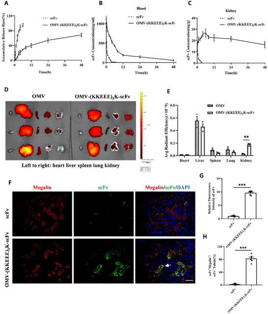
FIGURE 4.
Pharmacokinetic parameters and vesicle biodistribution in vivo. (A) Drug release profiles of scFv and OMV‐(KKEEE)3K‐scFv in PBS (pH 7.4) supplemented with 1% glycerol and 0.1% Tween 80. (B, C) Kidney and blood clearance of scFv and OMV‐(KKEEE)3K‐scFv in DN mice (n = 3). (D) Images of major organs were acquired with the Caliper IVIS Lumina II system at 6 h following dosing. (E) Fluorescence intensity in organs was quantified based on images shown in D. The fluorescence intensity was estimated as radiant efficiency by using Living Image 4.5 software (n = 3). (F‐H) Representative fluorescence images displaying the colocalization of scFv (labelled by anti‐HIS antibodies) and megalin‐positive cells in sections from DN kidneys (scale bar: 50 μm) and quantitative analysis (n = 6). The white arrow indicates typical colocalization of megalin and scFv. Mean ± SEM was used for reporting the data. **P < 0.01, ***P < 0.001. Two groups were compared by independent Student's t‐test, and multiple groups were compared by one‐way ANOVA, followed by Tukey's HSD test.
4. 人类大脑类器官验证机制保守
人源神经干细胞中 PCM1 同样呈现不对称分布,与 Par-3、Rab11a 共定位,证实该机制跨物种保守(对应原文Figure 5)。
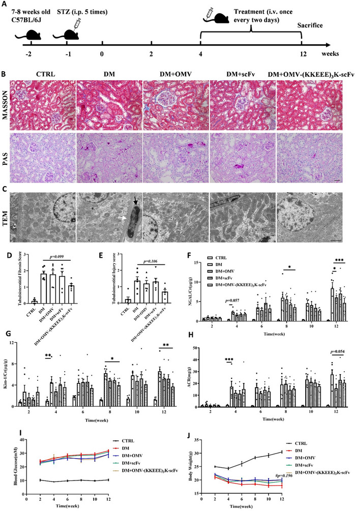
FIGURE 5.
Effects of OMV‐(KKEEE)3K‐scFv on renal injury in STZ‐induced diabetic mice. (A) Animal experiment procedure. (B) Representative kidney photomicrographs from various groups stained by PAS and Masson's trichrome stain (scale bar: 20 μm). (C) TEM images of kidney sections from various groups (scale bar: 2 μm). Black and white arrows indicate infiltrating immune cells in the renal tubulointerstitium and the thickened basement membrane of renal tubular epithelial cells, respectively. (D, E) Bar graphs denote tubulointerstitial injury and fibrosis scores based on Masson's trichrome staining and PAS staining (n = 6). (F‐H) Urinary Kim‐1/creatinine and NGAL/creatinine ratios and ACR (albumin/creatinine ratio) in mice from various groups evaluated at 2, 4, 6, 8, 10, and 12 weeks after the diabetic mouse model was successfully established (n = 6). (I, J) Trends in blood glucose and body weight during treatment in the mouse groups (n = 6). # represents a comparison between the DM and DM+OMV‐(KKEEE)3K‐scFv groups. Mean ± SEM was used for reporting the data. *P < 0.05, **P < 0.01, ***P < 0.001. Multiple groups were compared by one‐way ANOVA, followed by Tukey's HSD test.
三、Absin abs520001:精准支撑蛋白定位与功能验证
本研究中,Absin abs520001(高特异性 SALL2 多克隆抗体) 作为核心免疫检测试剂,全程支撑神经干细胞干性标志物检测、免疫荧光共定位、蛋白表达定量,对应原文Figure 1、Figure 5,为干细胞表型鉴定与机制验证提供精准数据支撑。
abs520001 在本文中的核心作用
? 适配原文 Figure 1:精准标记神经干细胞干性标志物 SALL2,清晰区分干细胞与分化神经元,验证 PCM1 在干性细胞中的特异性定位;
? 适配原文 Figure 5:在人类大脑类器官中检测 SALL2 表达,量化神经干细胞比例,证实 PCM1 调控人源干细胞命运的保守性;
? 高特异性:仅识别 SALL2 蛋白,无交叉杂带,免疫荧光信号清晰、背景极低;
? 高灵敏度:可检测低丰度内源蛋白,适配类器官等微量样本检测。
abs520001 核心优势
? ? 高特异性:经 WB/IF/IHC 多重验证,不与其他同源蛋白交叉反应;
? ? 多场景适配:完美支持 Western Blot、免疫荧光、免疫组化等实验;
? ? 批间稳定:严格质控确保批次一致性,实验结果可重复;
? ? 顶刊品质:助力多篇高分神经科学论文发表,数据可靠有保障。
四、Absin:神经发育与干细胞研究试剂伙伴
Absin 聚焦生命科学前沿需求,为神经干细胞、类器官、细胞分裂、神经发育研究提供一站式试剂方案:
? 干细胞干性检测:SALL2、SOX2、Nestin 等标志物抗体;
? 免疫荧光工具:高特异性一抗、荧光二抗、抗荧光淬灭封片剂;
? 类器官培养:神经类器官专用培养基、基质胶、消化试剂盒;
? 细胞分裂研究:中心体蛋白、内体标志物、信号通路抗体。
本次 abs520001 助力高分神经发育研究,再次印证 Absin 试剂的文献级品质。未来,Absin 将持续以高性能试剂赋能科研,助力神经疾病、干细胞治疗等领域突破创新!

- 手机:13761418683