
欢迎联系我们!




- 手机:13761418683
联系人:爱必信
网址:http://www.absin.cn/
地 址:上海市浦东新区新浩路58号18号楼
产品中心
联系我们

公司动态
IF14 + 高分期刊成果|eNOS 通路激活介导血管正常化,开创肿瘤免疫治疗全新策略
2026-07-13
近期,中山大学、北京协和医学院联合团队在Acta Pharmaceutica Sinica B发表重磅研究,创新性构建脂质纳米递送系统 MC@L,通过内皮转胞吞精准激活 eNOS、重塑肿瘤血管正常化,协同免疫原性细胞死亡(ICD)与肿瘤微环境(TME)重塑,显著提升 PD-L1 抑制剂疗效。Absin 明星产品abs42016331(DAR-1 NO 荧光探针) 全程支撑核心实验,为 NO 精准定位与血管正常化验证提供关键数据支撑!
文献标题:Exploiting eNOS activation to achieve tumor vascular normalization via endothelial transcytosis of lipid nanoparticles
发表期刊:Acta Pharm Sin B. (IF=14.6)
DOI:https://doi.org/10.1016/j.apsb.2025.11.040
使用 Absin 产品:DAR-1(货号:abs42016331)

一、研究痛点:肿瘤血管 "乱",免疫治疗 "难"
异常肿瘤血管结构紊乱、渗漏、高间质压,造就缺氧 / 酸性免疫抑制 TME,导致药物递送差、免疫细胞浸润低,PD-1/PD-L1 单抗单药效果有限。
传统外源性 NO 供体易弥散、定位不准,难以维持血管周 NO 梯度,无法稳定实现血管正常化。
二、核心研究思路:一石三鸟,精准破局
研究团队提出 "eNOS 激活 + 转胞吞递送 + TME 重塑" 一体化策略:
1. 递送载体:脂质纳米粒 MC@L 共包埋二甲双胍(Met)+CaO?
2. 血管正常化:内皮细胞中释放 Ca2?+Met,双通路激活 eNOS,原位生成 NO 并维持血管周梯度,稳定血管、改善灌注
3. 肿瘤杀伤:经内皮转胞吞进入肿瘤细胞,酸性溶酶体中 CaO?降解引发Ca2?超载 + 氧化应激,触发 ICD
4. 免疫唤醒:释氧改善缺氧、中和酸性;Met 抑制色氨酸摄取,逆转免疫抑制,协同 PD-L1 单抗强效抑瘤
三、关键机制与 Absin abs42016331 硬核支撑
1. eNOS 激活→NO 精准生成,Absin 探针 "看见" 关键信号
? 机制:Ca2?直接促进 eNOS 偶联;Ca2?+Met 激活AMPK→eNOS 磷酸化,双通路提升 NO 合成
? 实验支撑:采用Absin abs42016331(DAR-1 探针) 对 HUVEC 细胞内 NO 荧光成像,直接证实MC@L 组 NO 水平显著高于单药组,验证 eNOS 协同激活效果(原文图 2D)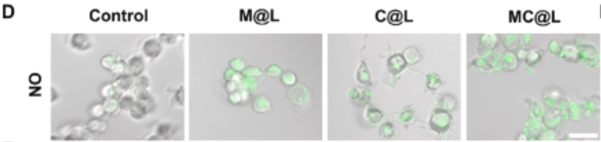
? 动物水平:肿瘤组织 CD31 与 abs42016331 共染色,清晰显示血管周 NO 梯度重建,为血管正常化提供直接可视化证据(原文图 4B)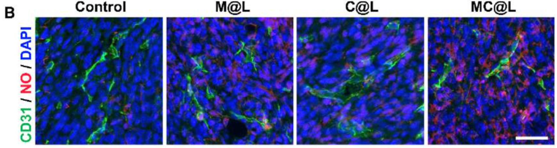
2. 内皮转胞吞:低渗透肿瘤 "主动递送" 新路径
针对 4T1 等低 EPR 效应肿瘤,团队证实 MC@L 主要经内皮转胞吞进入肿瘤实质:
? 流式与 TEM 直观显示:HUVEC 摄取 Cy5-MC@L 后外吐释放,完成跨内皮转运(原文图 2I-L)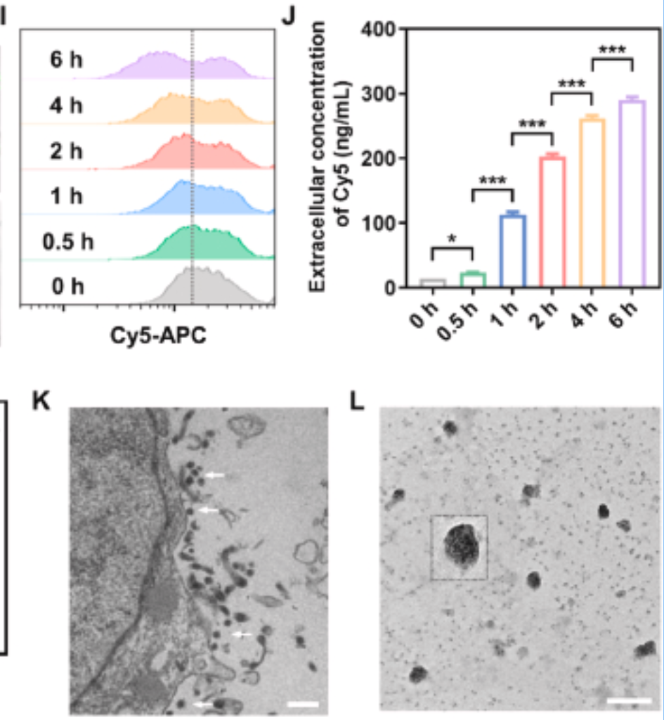
? 活体成像:Cy5-MC@L 在肿瘤富集显著高于游离 Cy5,24 h 仍高滞留(原文图 4E-G)
? 组织共定位:随时间延长,纳米粒从血管内转胞吞至血管外,实现深部递送(原文图 4I)
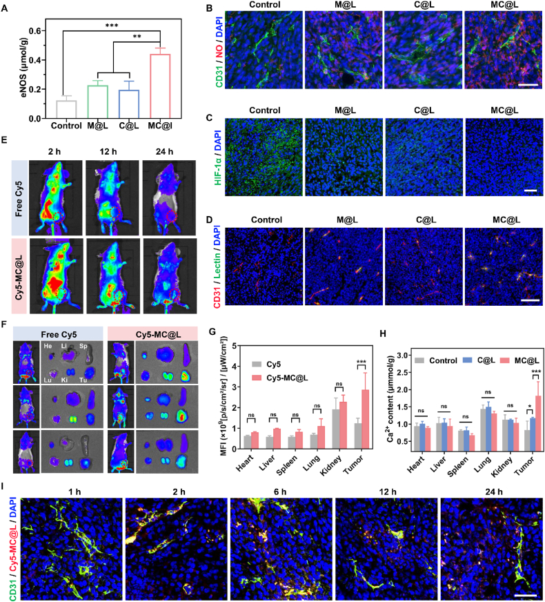
Figure 4. In vivo vascular normalization and biodistribution. (A) eNOS protein expression levels in tumor tissues with different treatments. (B) The distribution of NO (red) and tumor vessels (green) in tumor tissues with different treatments. Scale bar = 50 μm. (C) Representative immunofluorescent images of HIF-1α expression (green) in tumor tissues with different treatments. Scale bar = 50 μm. (D) The perfusion of tumor vessels after different treatments. Scale bar = 100 μm. (E) In vivo fluorescence images of 4T1 tumor-bearing mice at different time points after intravenous injection of free Cy5 or Cy5-MC@L. (F) In vivo fluorescence images of 4T1 tumor-bearing mice at 24 h post-injection and ex vivo fluorescence images of major organs and tumor tissues harvested at 24 h post-injection. (G) The corresponding quantification of mean fluorescence intensity (MFI) values of ex vivo major organs and tumor tissues (n = 3). (H) The content of Ca2+ in major organs and tumor tissues of 4T1 tumor-bearing mice at 24 h post-injection of C@L or MC@L. Data are presented as mean ± SD (n = 3). (I) The co-localization of Cy5-MC@L (red) and tumor vessels (green) at different times. Scale bar = 50 μm ?P < 0.05, ??P < 0.01, ???P < 0.001. ns, no significance.
3. 肿瘤内级联反应:ICD + 缺氧改善 + 酸性中和
? CaO?在酸性溶酶体降解:Ca2?超载 + H?O?积累触发 ICD,CRT 外翻、HMGB1/ATP 释放(原文图 3I-J)
? 原位产 O?缓解缺氧、消耗 H?中和酸性,改善免疫抑制微环境(原文图 3C-D)
? Met 下调 SLC7A5,抑制色氨酸摄取,减少犬尿氨酸生成,保护 T 细胞功能(原文图 3F-H)
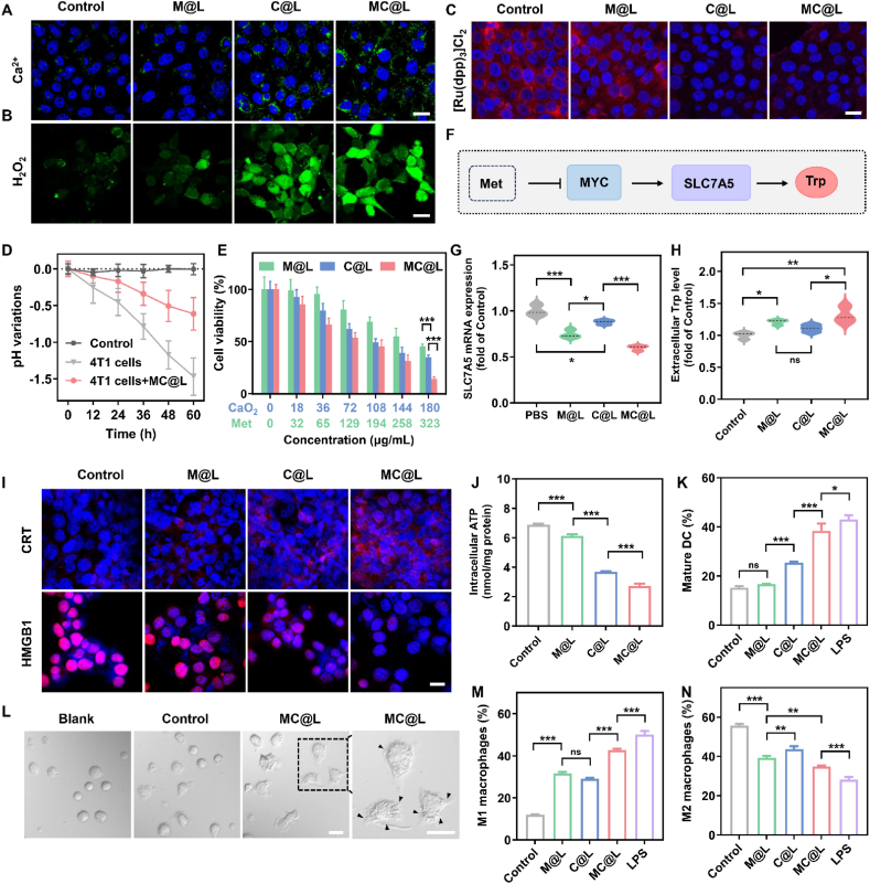 Figure 3. In vitro cytotoxicity and immune activation. (A, B) CLSM examination of intracellular Ca2+ concentration (A) and H2O2 level (B) in 4T1 cells with different treatments. Scale bar = 20 μm. (C) CLSM examination of intracellular O2 in 4T1 cells under hypoxic conditions with different treatments. Red fluorescence represents the hypoxic level. Scale bar = 20 μm. (D) Extracellular pH variations of 4T1 cells (n = 3). (E) 4T1 cell viability after 48 h incubation with different treatments (n = 5). (F) Schematic diagram illustrating that Met-mediated Trp uptake inhibition. (G) mRNA expressions of SLC7A5 in 4T1 cells with different treatments analyzed by qPCR (n = 3). (H) ELISA analysis of extracellular Trp levels in the culture medium of 4T1 cells treated with different formulations (n = 3). (I) CLSM examination of CRT exposure and HMGB1 release in 4T1 cells with different treatments. Scale bar = 20 μm. (J) Quantitative analysis of intracellular ATP concentration in 4T1 cells with different treatments (n = 3). (K) Quantitative analysis of in vitro DC maturation after incubation with 4T1 cells pretreated with different formulations (n = 3). (L) The optical images of DCs with different treatments. Scale bar = 20 μm. (M, N) Flow cytometry examination of M1 macrophages (CD11b+F4/80+CD86+) (M) and M2 macrophages (CD11b+F4/80+CD206+) (N) after different treatments. Data are presented as mean ± SD (n = 3). ?P < 0.05, ??P < 0.01, ???P < 0.001. ns, no significance.
4. 体内疗效:血管正常化 + 免疫激活,协同 PD-L1 "王炸"
? 血管正常化:eNOS 表达↑、NO 梯度重建、血管灌注↑、缺氧 / HIF-1α↓(原文图 4A-D)
? 免疫激活:DC 成熟↑、CD8?/CD4? T 细胞浸润↑、M2→M1 巨噬细胞极化、Treg/IL-10↓(原文图 6)
? 抑瘤效果:MC@L+αPD-L1组肿瘤生长近乎停滞,抑瘤率显著优于单药组(原文图 5)
? 安全性:体重稳定、血常规 / 生化无异常,心肝肾无损伤
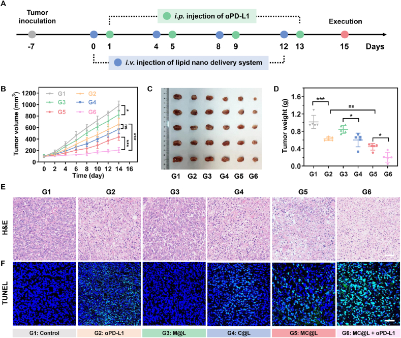 Figure 5. In vivo antitumor efficacy. (A) Treatment schedule for in vivo antitumor efficacy in the unilateral 4T1 tumor-bearing mice model. i.p., intraperitoneal. i.v., intravenous. (B) Tumor growth curves of the unilateral 4T1 tumor-bearing mice (G1: Control; G2: αPD-L1; G3: M@L; G4: C@L; G5: MC@L; G6: MC@L+αPD-L1) (n = 5). (C and D) Photograph (C) and the corresponding weights (D) of the resected tumors in different groups at the end of treatment. Data are presented as mean ± SD (n = 5). (E and F) H&E (E) and TUNEL (F) staining images of the tumor tissues in different groups. Scale bar = 50 μm (F). ?P < 0.05, ??P < 0.01, ???P < 0.001. ns, no significance.
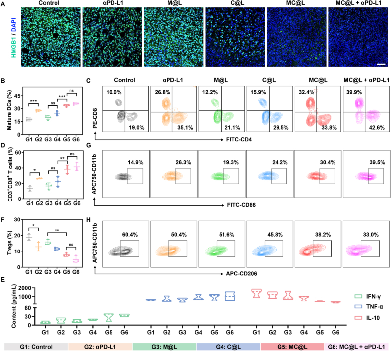 Figure 6. In vivo immune activation. (A) Representative immunofluorescent images of HMGB1 release in tumor tissues with different treatments. Scale bar = 50 μm. (B) Quantification of mature DCs in the TDLNs of 4T1 tumor-bearing mice post-treatments (G1: Control; G2: αPD-L1; G3: M@L; G4: C@L; G5: MC@L; G6: MC@L+αPD-L1) (n = 3). (C) Flow cytometry examination of intratumorally infiltrated CD4+ T cells (CD4+, gated on CD3+) and CD8+ T cells (CD8+, gated on CD3+) post-treatments. (D) Quantification of intratumorally infiltrated CD8+ T cells of 4T1 tumor-bearing mice post-treatments (n = 3). (E) The heatmaps of the content of IFN-γ, TNF-α, and IL-10 within the tumor tissues post-treatments (n = 3). (F) Quantification of intratumorally infiltrated Treg cells of 4T1 tumor-bearing mice post-treatments. Data are presented as mean ± SD (n = 3). (G, H) Flow cytometry examination of intratumorally infiltrated M1 macrophages (CD11b+CD86+, gated on CD11b+F4/80+) (G) and M2 macrophages (CD11b+CD206+, gated on CD11b+F4/80+) (H) post-treatments. ?P < 0.05, ??P < 0.01, ???P < 0.001. ns, no significance.
四、Absin abs42016331:NO 研究 "金标准",顶刊实证可靠
产品核心价值
? 货号:abs42016331
? 产品:DAR-1 一氧化氮荧光探针
? 核心作用:
1. 活细胞 / 组织高特异性标记 NO,荧光信号强、背景低
2. 与 CD31 共染,精准定位血管周 NO,直观验证血管正常化
3. 操作简便、适配 CLSM / 荧光显微镜,数据稳定可重复
? 本文贡献:支撑体外 NO 生成、体内血管周 NO 梯度两大关键结论,是机制验证的 "硬核工具"(原文图 2D、图 4B)
五、研究意义与转化前景
1. 策略创新:不依赖外源性 NO 供体,激活内源 eNOS实现持久、精准的血管正常化
2. 递送突破:立足ATR 效应 + 转胞吞,解决低渗透肿瘤纳米药物递送难题
3. 临床价值:MC@L 联合 PD-L1 单抗,为三阴性乳腺癌等冷肿瘤提供高效、安全的组合方案
4. 平台化:脂质纳米载体可拓展搭载多种药物,适配血管正常化 + 免疫治疗通用平台
六、Absin:高品质试剂,赋能高分科研
Absin 始终以高特异性、高稳定性、高性价比助力生命科学突破。本次abs42016331成功支撑 IF 14 + 顶刊研究,再次印证 Absin 试剂在血管生物学、肿瘤免疫、纳米药物领域的可靠价值。
Absin 提供NO 探针、ELISA 试剂盒、荧光抗体、细胞培养试剂等全系列科研工具,覆盖分子、细胞、动物水平研究,助力更多科研团队实现从基础发现到临床转化的跨越!
【免责声明】原文献《Acta Pharm Sin B.》(DOI:10.1016/j.apsb.2025.11.040),由 AI 解读整理;文中涉及的原文献图片、数据等知识产权归原期刊及研究团队所有。若存在侵权情形,敬请及时联系我方删除,我方将积极配合处理。
联系方式
- 手机:13761418683